3477
Diffusion Imaging for Mapping Hydraulic White Matter Parameters in Convection Enhanced Delivery1Medical Physics, University of Wisconsin at Madison, Madison, WI, United States, 2Waisman Center, University of Wisconsin at Madison, Madison, WI, United States, 3ImgGyd, LLC, Middleton, WI, United States, 4Therataxis, Baltimore, MD, United States, 5Biomedical Engineering, University of Wisconsin at Madison, Madison, WI, United States, 6Radiology, University of Wisconsin at Madison, Madison, WI, United States
Synopsis
Keywords: Simulation/Validation, Diffusion/other diffusion imaging techniques
Motivation: Intraparenchymal brain infusions using convection enhanced delivery (CED) require predictive modeling to plan catheter locations to visualize end distributions. Currently, diffusion MRI indirectly estimates extracellular volume fraction (ECVF) and creates hydraulic conductivity maps.
Goal(s): To directly measure ECVF parameters using advanced biophysical diffusion models.
Approach: Quantitative brain maps of ECVF from a pre-existing database were generated with diffusion model SANDI. These were compared with prior ECVF estimates determined via invasive physiological techniques.
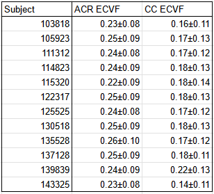
Results: SANDI modeling predicted ECVF ranges of 0.14-0.26 in white matter across 12 cases, consistent with consensus values around 0.20 estimated from physiology.
Impact: SANDI can quantitatively measure extracellular volume fractions in white matter. Previous methods estimated these parameters indirectly. Direct parametrization could be used for more accurate estimates of end-state biologic distributions in proposed trials of monogenic pediatric neurodegenerative gene therapy trials.
Introduction
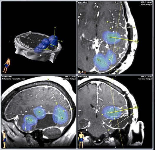
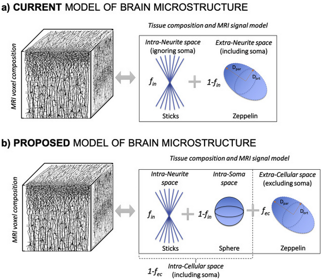
Gene therapies for fatal rare neurodegenerative pediatric lysosomal storage diseases are now capable of correcting cellular function but remain limited by drug delivery methods1,2. End distributions of gene therapies using convection enhanced delivery (CED), which uses intra-parenchymal catheter placements to slowly infuse therapeutic agents directly into the brain, are difficult to predict due to heterogeneous hydraulic attributes influencing flow across differing tissues, such as infusate’s preferential flow along white matter tracts2,3. Moving from palliative strategies to one-time cures will require advances in predictive modeling of end-state CED distributions before pharmaceutical companies begin new clinical trials.Software to predictively model CED distributions and overlay them on anatomical MRI studies has been integrated into presurgical planning tools, as shown in Figure 14 (Brainlab, Munich, Germany). T1, T2, and diffusion tensor imaging (DTI) have been used to segment the adult brain and estimate key determinants of flow, including ECVF and from it, hydraulic conductivity4,5. The extracellular volume fractions (ECVF), the space in which the infusion distributes, is often treated as uniform across all brain tissue using an accepted physiological estimate6. However, ECVF is known to vary across brain regions, though this has not yet been extensively investigated7. Previous attempts to quantitatively measure ECVF with noninvasive imaging were limited by a lack of diffusion imaging techniques8. Soma And Neurite Density Imaging (SANDI), a recent modeling technique, can map ECVF in a manner more relevant to CED due to its ability to delineate cell soma from extracellular space (Figure 2)9. Presented here are measurements made using SANDI to compare its spatially-varying ECVF measurements against prior standards.
Materials and Methods
SANDI:Many diffusion models, like NODDI, prioritize measuring the orientation and density of neurite axons to map white matter tracts10. This information is less useful in CED where infusates diffuse through and largely remain in the extracellular spaces between neurites. NODDI’s prioritization of axonal information disregards neurite cell bodies, leading to inaccuracies in measurements of extracellular volumes9,10. Alternatively, SANDI uses a different compartment model to account for the intrasomal volumes, providing more relevant measures of the spaces at play in CED, as shown in Figure 2.
Dataset Source:
A set of 12 diffusion MRI (dMRI) acquisitions were sourced from the publicly available Human Connectome Database11. Multishell diffusion-weighted acquisitions with b-values of 1000, 2000, and 3000 s/mm2, and 90 directions were obtained on a Siemens 3T scanner with 1.25mm isotropic resolution.
Data Analysis:
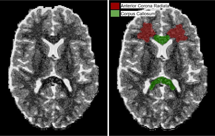
Previous CED planning methodologies have estimated the extracellular volume fraction (ECVF) of regular brain tissue as 20%, based on physiological experimentation5,6. Some regions, like the corpus callosum (CC), are expected to have differing ECVF, though a quantitative estimation is not yet established. Segmentations of the corpus callosum and anterior corona radiata (ACR) were manually generated by an experienced imaging scientist and used to compute the mean ECVF from each region for all 12 cases (Figure 4).
Results
Across the 12 cases, the mean ECVF in the ACR and CC were 0.24±0.09 and 0.18±0.12, respectively. This is generally consistent with prior measurements using invasive physiologic techniques6, earlier, non-diffusion, imaging methods8, and common experience in brain cancer where edema expands preferentially away from the dense corpus callosum.Discussion
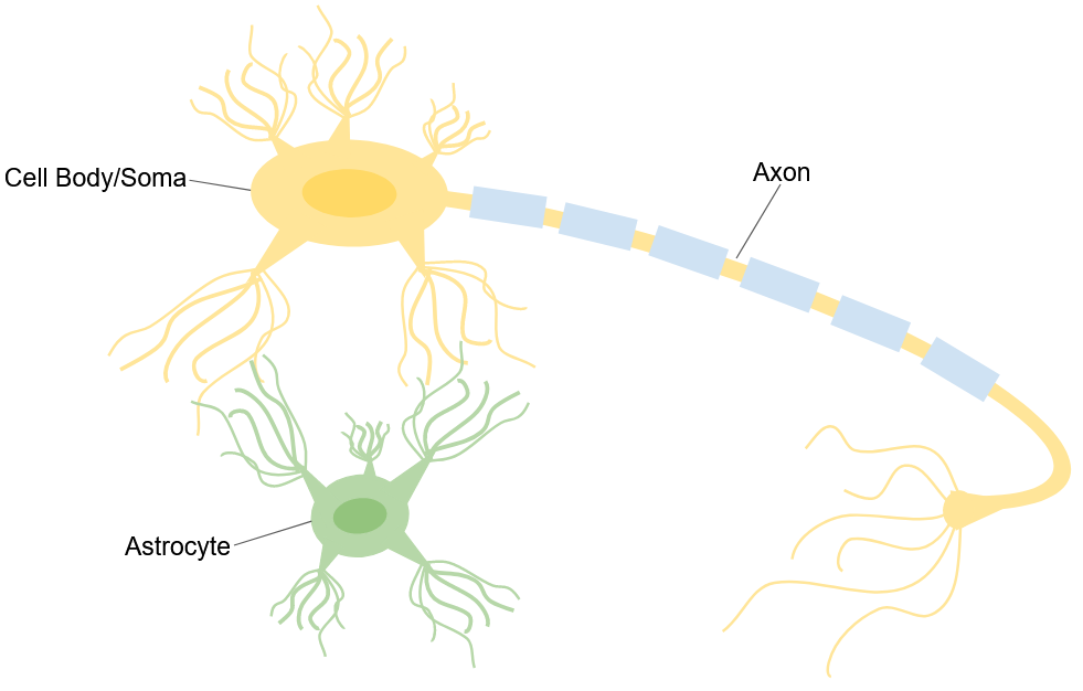
Though estimates of ECVF in white matter are consistent with known physiologic values, gray matter measurements are higher than expected. This is tentatively attributed to glial cells, which differ across gray and white matter, being smaller than the distinguishable soma and thus having their signal contributions attributed to extracellular spaces in SANDI’s compartment model12. This is represented in Figure 5. Using higher b-values can enable greater diffusion sensitivity and potentially delineate these finer compartments. Further work is needed to update the current predictive modeling tools, which are based on stable adult brains, with hydraulic characterizations of the rapidly changing pediatric brain. Intraprocedural dMRI may be beneficial for tracking tissue expansion during infusion, but lies outside this work’s current scope.Conclusion
In white matter, the proposed application of SANDI can provide quantitative maps of extracellular space consistent with estimates previously determined through invasive physiologic measurements. While previous mapping techniques could only estimate ECVF as a fixed constant of 20%, SANDI can represent spatial variation within white matter. Future work will use altered scan parameters to achieve the same in gray matter and move towards application of predictive infusion flow mapping in animal trials.Acknowledgements
We acknowledge GE-Healthcare and UW-Madison for research support
Data were provided by the Human Connectome Project, WU-Minn Consortium (Principal Investigators: David Van Essen and Kamil Ugurbil; 1U54MH091657) funded by the 16 NIH Institutes and Centers that support the NIH Blueprint for Neuroscience Research; and by the McDonnell Center for Systems Neuroscience at Washington University.
References
[1] Sondhi D, Kaminsky SM, Hackett NR, Pagovich OE, Rosenberg JB, De BP, Chen A, Van de Graaf B, Mezey JG, Mammen GW, Mancenido D, Xu F, Kosofsky B, Yohay K, Worgall S, Kaner RJ, Souwedaine M, Greenwald BM, Kaplitt M, Dyke JP, Ballon DJ, Heier LA, Kiss S, Crystal RG. Slowing late infantile Batten disease by direct brain parenchymal administration of a rh.10 adeno-associated virus expressing CLN2. Sci Transl Med. 2020 Dec 2;12(572):eabb5413. doi: 10.1126/scitranslmed.abb5413. PMID: 33268510; PMCID: PMC8056991.
[2] Raghavan R, Brady ML, Sampson JH. Delivering therapy to target: improving the odds for successful drug development. Ther Deliv. 2016 Jul;7(7):457-81. doi: 10.4155/tde-2016-0016. PMID: 27403630.
[3] Mehta AM, Sonabend AM, Bruce JN. Convection-Enhanced Delivery. Neurotherapeutics. 2017 Apr;14(2):358-371. doi: 10.1007/s13311-017-0520-4. PMID: 28299724; PMCID: PMC5398992.
[4] Sampson JH, Raghavan R, Brady ML, Provenzale JM, Herndon JE 2nd, Croteau D, Friedman AH, Reardon DA, Coleman RE, Wong T, Bigner DD, Pastan I, Rodríguez-Ponce MI, Tanner P, Puri R, Pedain C. Clinical utility of a patient-specific algorithm for simulating intracerebral drug infusions. Neuro Oncol. 2007 Jul;9(3):343-53. doi: 10.1215/15228517-2007-007. Epub 2007 Apr 13. PMID: 17435179; PMCID: PMC1907410.
[5] Brady M, Raghavan R, Sampson J. Determinants of Intraparenchymal Infusion Distributions: Modeling and Analyses of Human Glioblastoma Trials. Pharmaceutics. 2020 Sep 21;12(9):895. doi: 10.3390/pharmaceutics12090895. PMID: 32967184; PMCID: PMC7559135.
[6] Syková E, Nicholson C. Diffusion in brain extracellular space. Physiol Rev. 2008 Oct;88(4):1277-340. doi: 10.1152/physrev.00027.2007. PMID: 18923183; PMCID: PMC2785730.[7] Nicholson C, Syková E. Extracellular space structure revealed by diffusion analysis. Trends Neurosci. 1998 May;21(5):207-15. doi: 10.1016/s0166-2236(98)01261-2. PMID: 9610885.
[8] Bender B, Klose U. Cerebrospinal fluid and interstitial fluid volume measurements in the human brain at 3T with EPI. Magn Reson Med. 2009 Apr;61(4):834-41. doi: 10.1002/mrm.21915. PMID: 19191287.
[9] Palombo M, Ianus A, Guerreri M, Nunes D, Alexander DC, Shemesh N, Zhang H. SANDI: A compartment-based model for non-invasive apparent soma and neurite imaging by diffusion MRI. Neuroimage. 2020 Jul 15;215:116835. doi: 10.1016/j.neuroimage.2020.116835. Epub 2020 Apr 11. Erratum in: Neuroimage. 2021 Feb 1;226:117612. PMID: 32289460; PMCID: PMC8543044.
[10] Zhang H, Schneider T, Wheeler-Kingshott CA, Alexander DC. NODDI: practical in vivo neurite orientation dispersion and density imaging of the human brain. Neuroimage. 2012 Jul 16;61(4):1000-16. doi: 10.1016/j.neuroimage.2012.03.072. Epub 2012 Mar 30. PMID: 22484410.
[11] Van Essen DC, Smith SM, Barch DM, Behrens TE, Yacoub E, Ugurbil K; WU-Minn HCP Consortium. The WU-Minn Human Connectome Project: an overview. Neuroimage. 2013 Oct 15;80:62-79. doi: 10.1016/j.neuroimage.2013.05.041. Epub 2013 May 16. PMID: 23684880; PMCID: PMC3724347.
[12] Köhler, S., Winkler, U. & Hirrlinger, J. Heterogeneity of Astrocytes in Grey and White Matter. Neurochem Res 46, 3–14 (2021). https://doi.org/10.1007/s11064-019-02926-x
Figures