3476
Microstructural Precision: Assessing the Reproducibility and Sensitivity of Multidimensional Diffusion Metrics1Forensic and Neurodevelopmental Sciences, Institute of Psychiatry, Psychology and Neuroscience, King's College London, London, United Kingdom, 2Centre for Neuroimaging Sciences, Institute of Psychiatry, Psychology and Neuroscience, King's College London, London, United Kingdom
Synopsis
Keywords: Microstructure, Diffusion/other diffusion imaging techniques, Multidimensional Diffusion
Motivation: The reproducibility and sensitivity of Multidimensional Diffusion (MDD) measures have not been extensively investigated.
Goal(s): Our goal was to assess reproducibility and sensitivity of µFA in comparison with conventional FA metrics for multiple white and grey matter regions.
Approach: Test-retest data was acquired to compute ICC scores (reproducibility) and power calculations (sensitivity) to predict the number of subjects required to detect 1%, 2%, 4% and 5% changes for each metric across multiple regions.
Results: While FA shows the highest reproducibility for both WM and GM, µFA demonstrates enhanced sensitivity to detect microstructural changes for the same percentage difference.
Impact: Our test-retest study demonstrates that Multidimensional Diffusion (MDD) metrics, like µFA, show good reproducibility scores and increased sensitivity in the detection of microstructural changes when compared to equivalent FA changes in both white and grey matter.
Introduction
Compared with conventional diffusion magnetic resonance imaging (dMRI), multidimensional diffusion (MDD) acquisitions offer a clearer representation of microstructural anisotropy, as exemplified by metrics like µFA1. However, the reproducibility and sensitivity of these new metrics compared to the more conventional FA has not yet been extensively investigated.In this study, we aim to quantify reproducibility and specificity of µFA, computed using different approaches (Gamma and Covariance models)2,3 and compare against the equivalent FA measure computed from the same dataset (see fig.1). Specifically, using test-retest data we compute intraclass correlation scores and perform a power calculation to assess reproducibility and sensitivity of the metrics.
Methods
Eleven healthy adults (7 male, 4 female, ages 24-41) were recruited. Each subject underwent two separate MRI scans, with an average five-week interval. Scans were performed using a 3T GE MR750 system and Nova 32-channel head coil. Each scanning session included two consecutive acquisitions with a q-space trajectory diffusion encoding sequence (three diffusion shapes: linear (52 volumes at b-values 100, 700, 1400, 2000), planar (21 volumes at b-values 100, 1000, 2000), and spherical (32 volumes at b-values 100, 700, 1400, 2000)). Data was acquired for both 2.0mm and 2.5mm resolutions (TE = 120ms & 113ms respectively, TR = 6000ms, FOV = 256). All scans were preprocessed for denoising, Gibbs unringing, motion and eddy current correction and maps extracted using the multidimensional diffusion framework4 with gamma and covariance models. All maps where normalised to MNI space and multiple regions of interest were used to extract measurements in select white and grey matter regions. Reproducibility was quantified using the ICC3,15 form while sensitivity was established by performing a power calculation assuming 1,2,4 and 5% changes in the metrics with corresponding effect size, Cohen’s D, computed for all regions.Results and Discussion
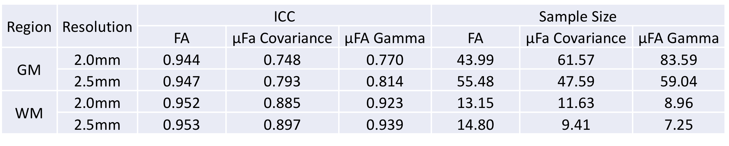
Reproducibility: FA is substantially more reliable in both WM & GM for both resolutions, remaining the most reliable and consistent measure. Both covariance and gamma µFA measures perform relatively well in GM with fair consistency (covariance µFA ICC: 0.793± 0.102, gamma µFA ICC: 0.814 ± 0.087). While similarly consistent in GM, gamma µFA outperforms covariance µFA across all WM ROIs with consistent margin (covariance µFA: 0.897 ± 0.047, gamma µFA: 0.939 ± 0.025). Increasing resolution to 2.00mm resulted in minimal changes in consistency for the majority of WM ROIs (covariance µFA: 0.885 ± 0.067, gamma µFA: 0.923 ± 0.050). However, most GM ROIs exhibited a moderate decrease in consistency for both µFA measures (see table 1).Deep GM regions, such as the Pallidum, tend to have lower ICC values across all measures (FA: 0.782, covariance µFA: 0.360, gamma µFA: 0.366), indicating lower reliability in comparison to cortical GM, such as the frontal and parietal lobes (Left frontal lobe, FA: 0.960, covariance µFA: 0.828, gamma µFA: 0.888). This could be due to the more complex anatomical structures of deep GM or decreased SNR, which tend to make measures less reliable owing to variations in delineation or feature extraction. In comparison, ICC values for WM regions are generally high, indicating good reliability in quantifying WM characteristics. Significant variability is observed between gamma µFA and covariance µFA in certain GM ROIs, particularly within the frontal and parietal lobes (Left parietal lobe, covariance µFA: 0.812, gamma µFA: 0.874). This variance may be attributed to the unique characteristics of these different acquisition methods and their efficacy in quantifying features within regions characterized by low signal intensity.
Sensitivity: Based on the power analysis, it is evident that all white matter ROIs exhibit a substantially lower participant requirement than grey matter to detect a 4% change (average GM: 63.05, WM:11.25). µFA Gamma is the most effective measure in white matter for most ROIs at both 2mm and 2.5mm resolutions (2mm: 8.96, 2.5mm 7.25), however all measures perform well. In contrast, both µFA measures lack power in GM at 2.0mm, however see significant improvement at the lower 2.5mm resolution (see fig.2). Deep grey matter regions consistently show low statistical power across both resolutions.
Conclusions
In summary, FA and µFA both exhibit high reliability in quantifying features of white matter and cortical grey matter, with FA remaining the most reproducible measure, as reflected in the highest ICC values across all measures. However, µFA demonstrates greater sensitivity to microstructural changes, assuming that the same degree of change is comparable between measures. Furthermore, µFA Gamma stands out as the most efficient measure of white matter for the majority of ROIs, irrespective of resolution. These findings show strong support for MDD as a tool for increased sensitivity to microstructural detail for both clinical and research applications.Acknowledgements
No acknowledgement found.References
1. Szczepankiewicz, F., Lasič, S., van Westen, D., Sundgren, P. C., Englund, E., Westin, C. F., ... & Nilsson, M. (2015). Quantification of microscopic diffusion anisotropy disentangles effects of orientation dispersion from microstructure: applications in healthy volunteers and in brain tumors. Neuroimage, 104, 241-252.
2. Westin, C. F., Knutsson, H., Pasternak, O., Szczepankiewicz, F., Özarslan, E., van Westen, D., ... & Nilsson, M. (2016). Q-space trajectory imaging for multidimensional diffusion MRI of the human brain. Neuroimage, 135, 345-362.
3. Topgaard, D. (2017). Multidimensional diffusion MRI. Journal of magnetic resonance, 275, 98-113.
4. Markus Nilsson, Filip Szczepankiewicz, Bjorn Lampinen, Andre Ahlgren, Joao P. de Almeida Martins, Samo Lasic, Carl-Fredrik Westin, and Daniel Topgaard. An open-source framework for analysis of multidimensional diffusion MRI data implemented in MATLAB. Proc. Intl. Soc. Mag. Reson. Med. (26), Paris, France, 2018. http://archive.ismrm.org/2018/5355.html
5. Koo TK, Li MY. A Guideline of Selecting and Reporting Intraclass Correlation Coefficients for Reliability Research. J Chiropr Med. 2016 Jun;15(2):155-63. doi: 10.1016/j.jcm.2016.02.012. Epub 2016 Mar 31. Erratum in: J Chiropr Med. 2017 Dec;16(4):346. PMID: 27330520; PMCID: PMC4913118.
Figures
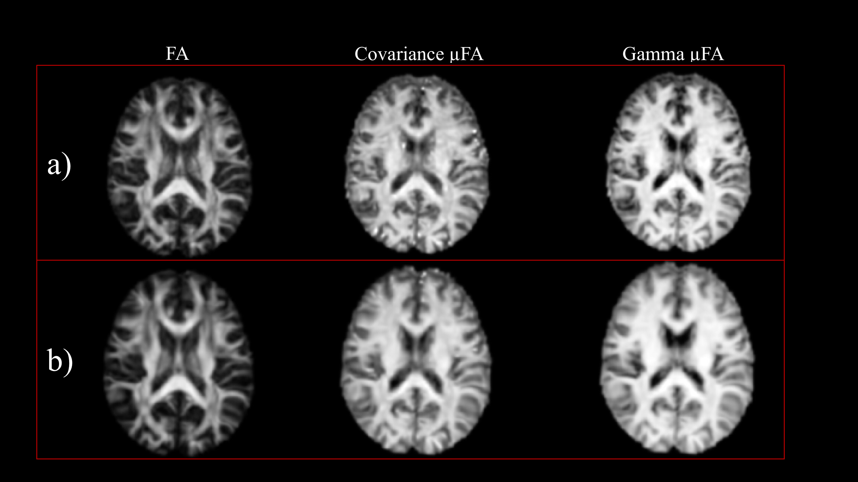
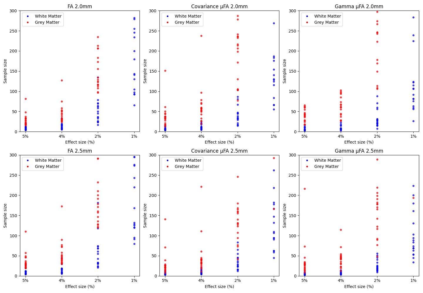
Figure 2: Sample size requirements for detecting 5%, 3%, 2%, and 1% effect size differences in white matter (WM) and grey matter (GM) using conventional FA, covariance µFA, and gamma µFA at 2mm (top row) and 2.5mm (bottom row) resolutions