3475
The variability of diffusion-relaxation multidimensional MRI estimates in the human brain1Multiscale Imaging and Integrative Biophysics Unit, National Institute on Aging, Baltimore, MD, United States, 2Department of Electrical Engineering and Computer Science, Vanderbilt University, Nashville, TN, United States, 3Neuroimaging Research Branch, National Institute on Drug Abuse, Baltimore, MD, United States, 4Department of Chemistry, Lund University, Lund, Sweden
Synopsis
Keywords: Microstructure, Microstructure
Motivation: Multidimensional (MD)-MRI provides valuable subvoxel information. However, its experimental variability has never been investigated.
Goal(s): Assessing the variability of MD-MRI estimates is essential for adaptation to clinical research and widespread use.
Approach: Ten healthy participants were each scanned twice, using a 40-minute 2-mm3 resolution diffusion-relaxation MD-MRI protocol. Agreement, reliability, and repeatability were assessed using Bland-Altman plots, intraclass correlation coefficient, and test-retest variability, respectively.
Results: We demonstrated a good to excellent reliability in the quantification of first- and second-order diffusion parameters. Our findings guide further improvement to the protocol and encourage the use of this MD-MRI framework in clinical research.
Impact: Multidimensional MRI is crucial for investigating tissue microstructure, brain connectivity, and pathology. Here we present an in vivo variability study that shows strong agreement, reliability, and repeatability, especially for diffusion parameters, providing a way forward for clinical research.
Introduction
Diffusion-relaxation multidimensional MRI (MD-MRI) acquisition integrates meso- and microstructural probes1 with chemical composition sensitivity2. MD-MRI data can be analyzed using a model-free approach to obtain intra-voxel distributions of valuable quantitative metrics,3-7 elucidating tissue microstructure,8,9 brain connectivity,10,11 and pathology.12,13 When free gradient waveforms are used in diffusion acquisition,14 it allows for the exploration of frequency-dependent and tensor-related characteristics within the encoding spectrum.15,16An efficient in vivo frequency-dependent MD-MRI acquisition protocol that provides whole brain coverage at 2-mm3 resolution was recently introduced.17 Here, we seek to assess its repeatability by employing a test-retest experimental paradigm with repeated scans of 10 healthy human participants.
Methods
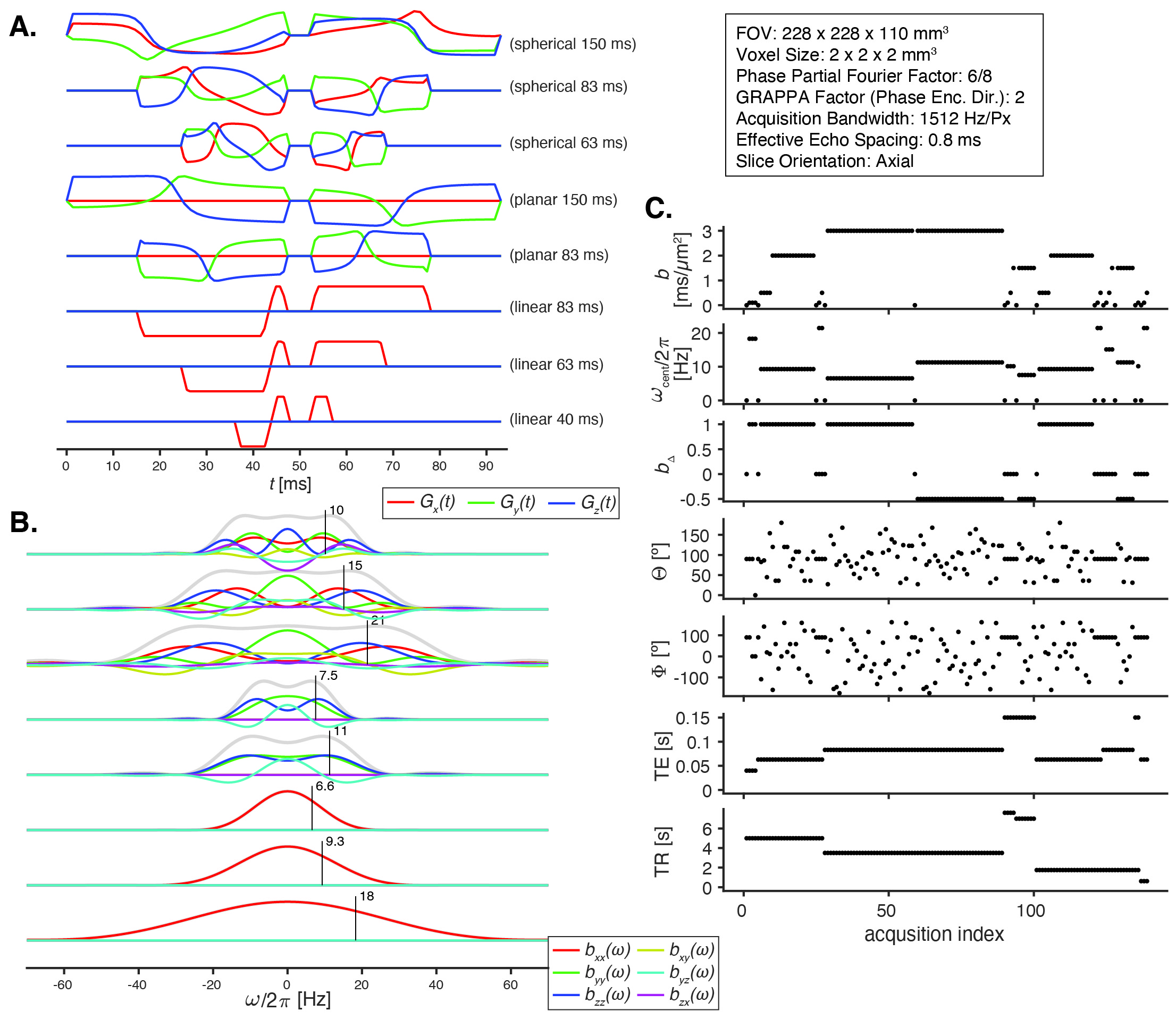
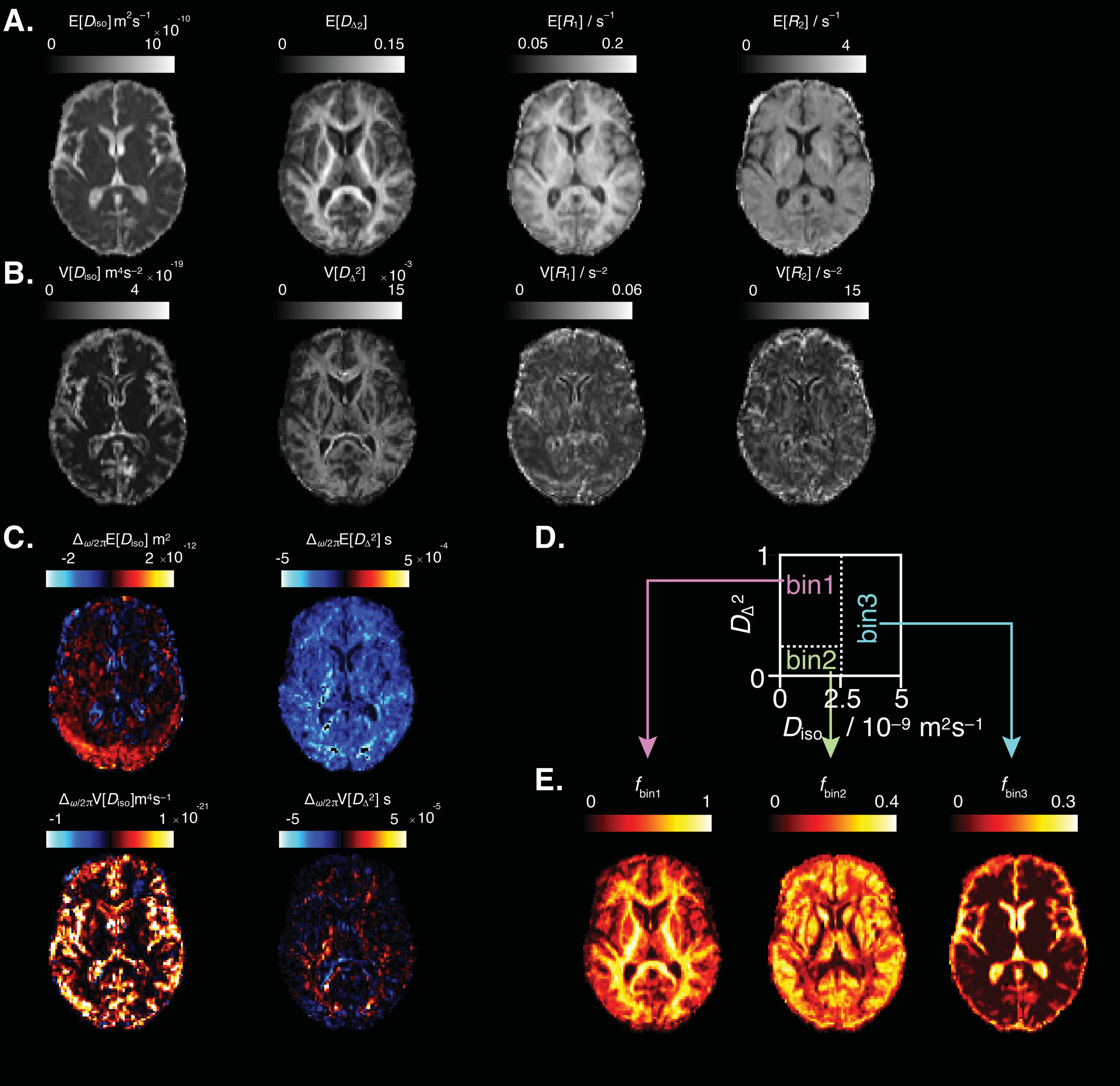
Ten healthy participants were each scanned twice, a few weeks apart, on a 3T Siemens Prisma MRI scanner. Numerically optimized14 linear, planar, and spherical b-tensors were employed with b-values ranging between 0.1 and 3 ms/µm2, in the range of 6.6-21 Hz centroid frequencies (ω/2𝜋), and with different combinations of repetition times, TR=(0.62,1.75,3.5,5,7,7.6) s and echo times, TE=(40,63,83,150) ms, comprising a total of 139 volumes and a 40-minute acquisition time17 (Fig. 1).The data were first preprocessed using the TORTOISE package,18,19 and subsequently processed using the Monte Carlo inversion algorithm16 as implemented in the multidimensional diffusion MRI toolbox.17 Briefly, the b(ω)-TE-TR encoded signal S is modeled as a sum of contributions; the ith component is characterized by its signal weight, fi, tensor-valued diffusion spectrum Di(ω), and longitudinal and transverse relaxation rates R1 and R2 according to a multi-exponential signal model.14 The result is voxelwise D(ω)-R1-R2 distributions, which in this work are expressed in terms of means (E[x]) and variances (V[x]) of isotropic diffusivity, Diso, squared normalized anisotropy DΔ2, relaxation, and relative signal fractions over sub-divisions of the distribution space, fbin1, fbin2, and fbin3, roughly corresponding to white matter (WM), gray matter (GM), and CSF (Fig. 2).9
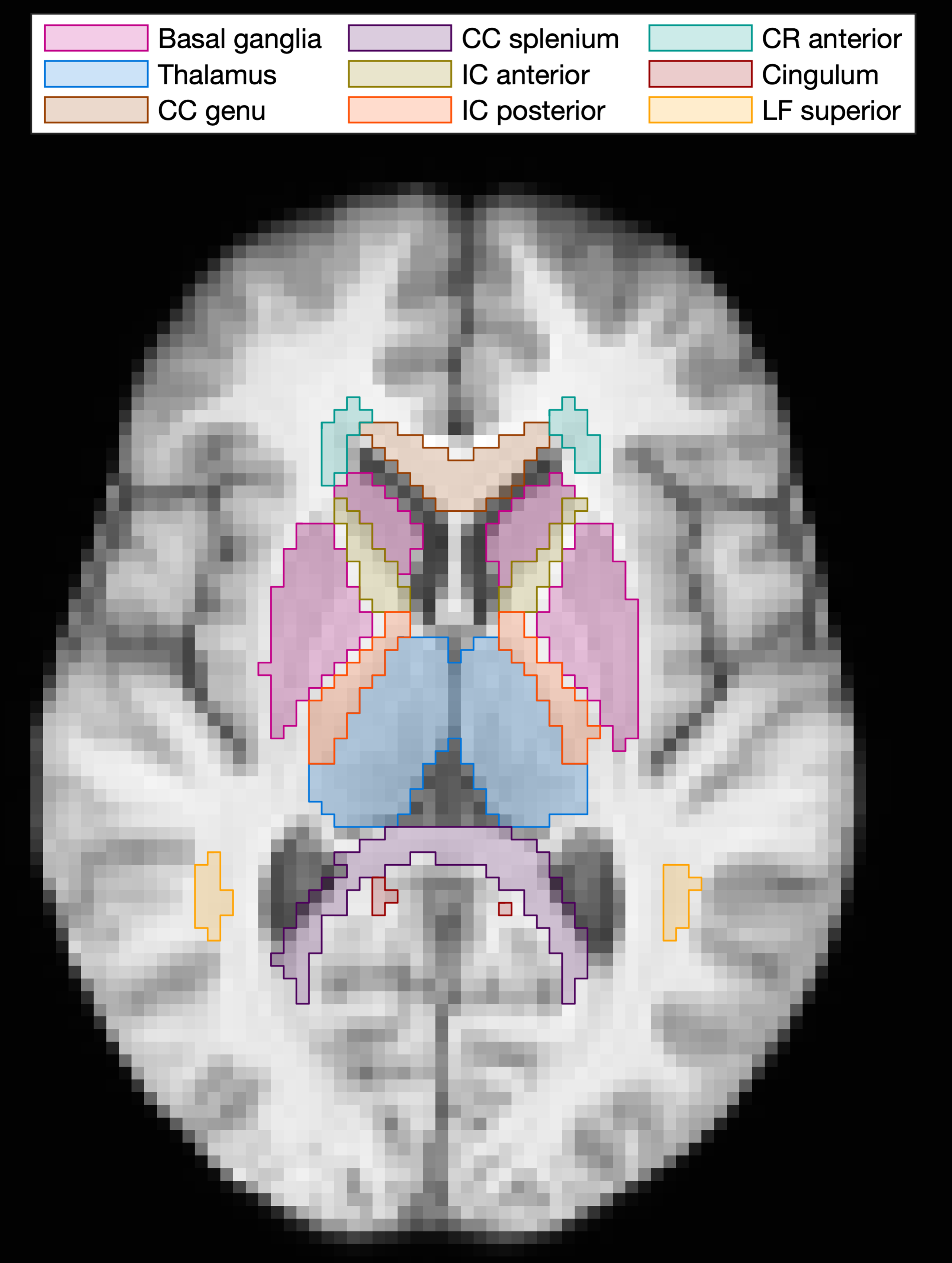
We examined 14 regions of interest (ROIs) that were identified using SLANT21 (Fig. 3), and all analysis was done in native subject space. For each MD-MRI parameter, the test-retest variability (TRV) and intra-class correlation coefficient (ICC) were computed from the two repetition scans22 in each ROI.
Results
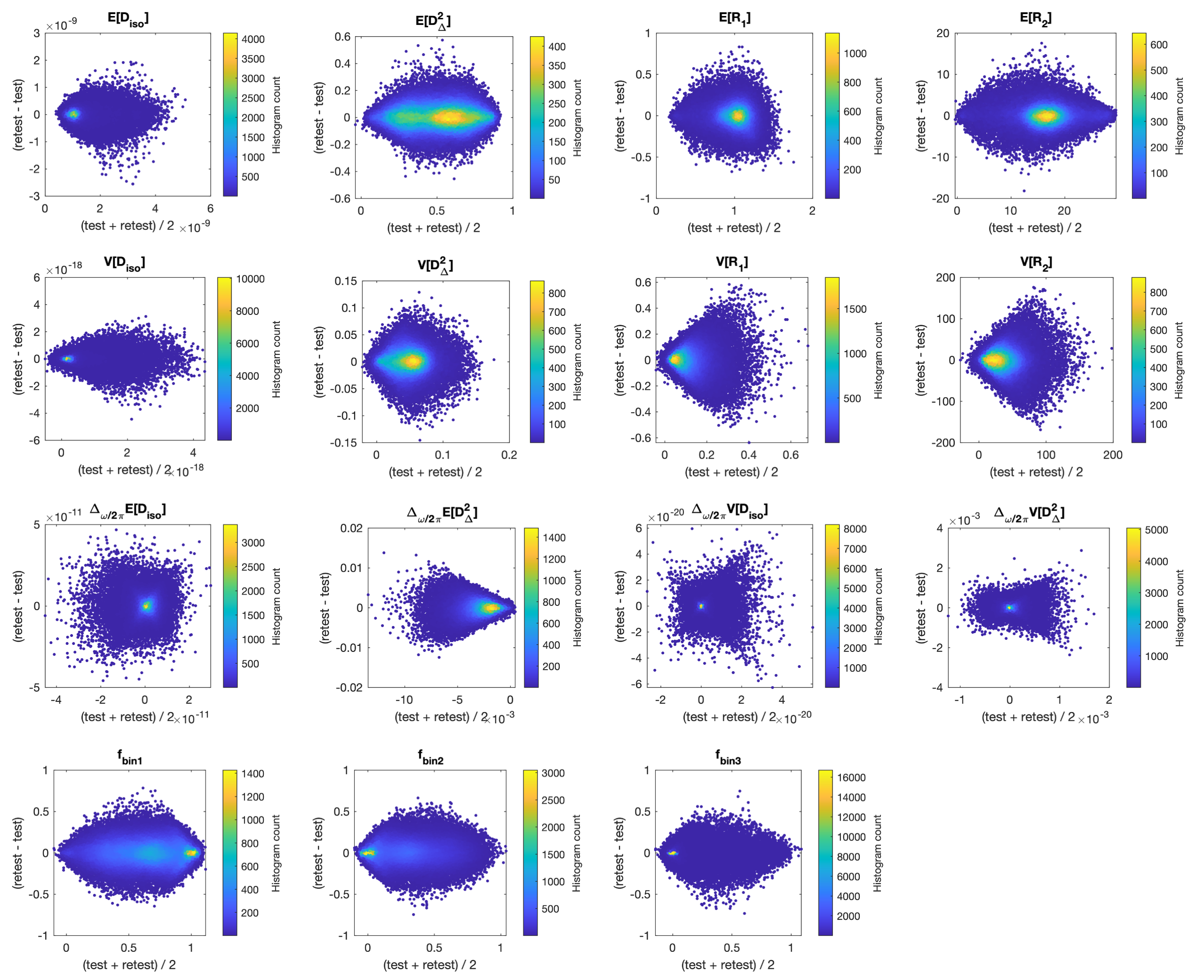
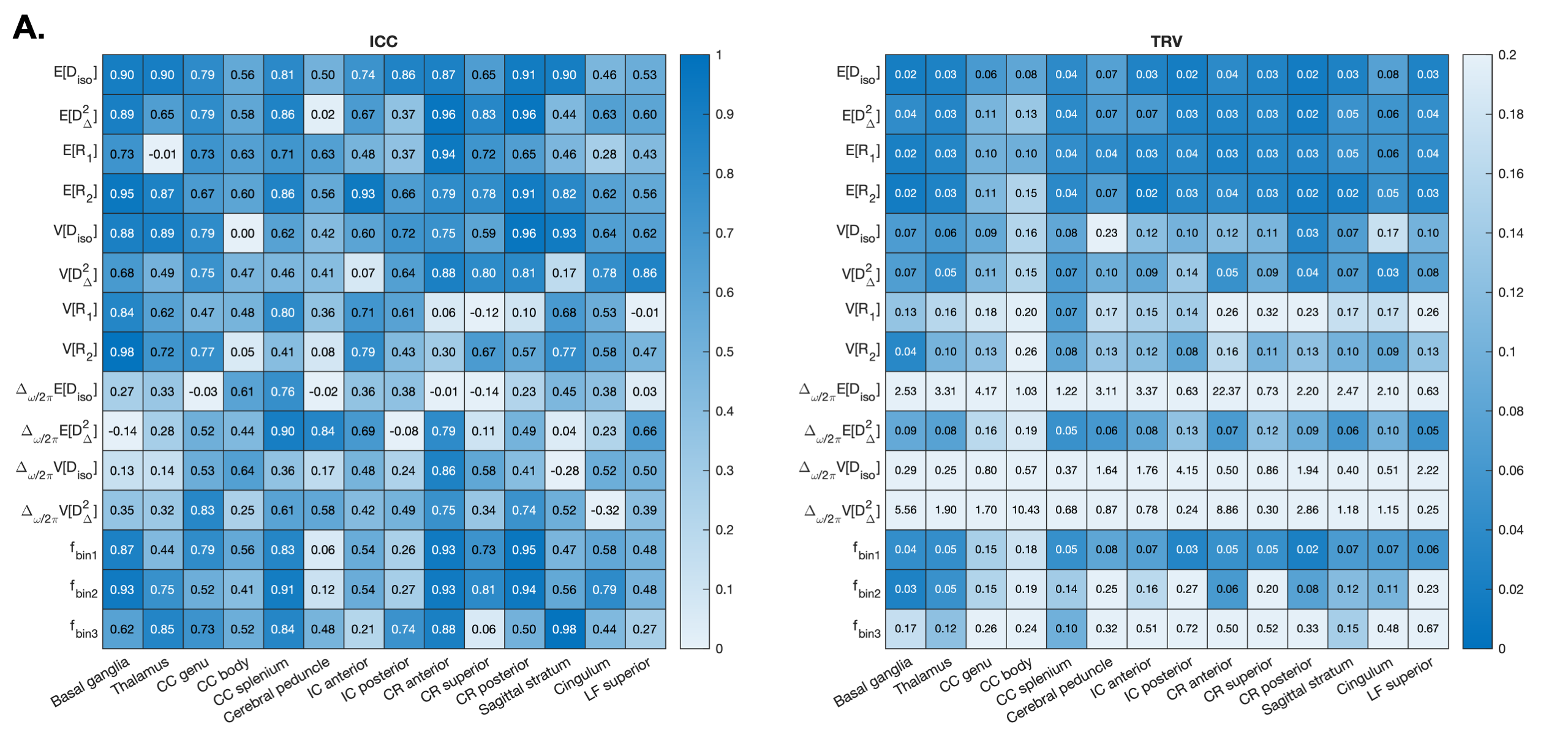
We investigated agreement, reliability, and repeatability between MD-MRI measurements using Bland-Altman plots, ICC, and TRV, respectively.23 Fig. 4 shows Bland-Altman plots that express the agreement between repeated MD-MRI parameters. The plots are symmetric about the y-axis (difference between test and retest), suggesting that there was no bias between the test and the retest measurements. Fig. 5A shows the ICC values for each ROI-averaged MD-MRI parameter, and Fig. 5B shows the corresponding TRV. Generally, the expectation parameters were more repeatable than the variance parameters; diffusion parameters Diso and DΔ2 were more repeatable than relaxation rates R1 and R2; bin fractions f1 and f2 were more repeatable than f3; and the rates of change as a function of diffusion encoding frequency were the least repeatable parameters.Discussion
Our test-retest analyses revealed strong agreement, and varying reliability and repeatability measures for the investigated MD-MRI parameters. Bland-Altman analysis showed lack of systematic errors in all variables. Reliability as expressed by ICC was found to be excellent (ICC>0.75) or good (0.6<ICC<0.74) in most ROIs for all first-order statistical measures, E[X], and for diffusion second-order statistical variables, V[Diso] and V[DΔ2]. The relaxation variance parameters exhibited lower reliability, perhaps due to limited experimental encoding of TE and TR. The rates of change of diffusion parameters as a function of diffusion encoding frequency exhibited low reliability in most ROIs. The experimental diffusion encoding frequency range is expected to be particularly sensitive to length scale of approximately 13µm,24 which is too large for the examined brain regions and could therefore be driving these results. The bin volume fractions are estimated from the Diso-DΔ2 distributions and exhibit moderate-to-good reliability in most ROIs. Volume fraction 3 had lower repeatability than fractions 1 and 2, which could result from it being more susceptible to partial volume effects due to its more binary-like nature as can be seen in Fig. 2E. Repeatability expressed by TRV showed similar trends as the ICC. Reliability and repeatability of the MD-MRI parameters were ROI-dependent, with three ROIs in particular (Cerebral peduncle, IC posterior, LF superior) showing ICC<0.5 for most of the MD-MRI parameters.Conclusion
We present the first in vivo MD-MRI agreement, reliability, and repeatability study. The variability of a range of MD-derived parameters from a 40-minute 2-mm3 resolution protocol was quantified. We demonstrated a good to excellent reliability in the quantification of first- and second-order diffusion parameters. Our findings guide further improvement to the protocol and encourage the use of this MD-MRI framework in clinical research.Acknowledgements
The authors would like to thank Mr. Phil Cholak for facilitating the MRI scans. This work was supported by the Intramural Research Programs of the National Institute on Aging and the National Institute on Drug Abuse of the National Institutes of Health.References
- Callaghan, Paul T., Translational Dynamics and Magnetic Resonance: Principles of Pulsed Gradient Spin Echo NMR (Oxford, 2011; online edn, Oxford Academic, 17 Dec. 2013).
- Tofts P. Quantitative MRI of the brain: Measuring changes caused by disease: John Wiley; 2003.
- Galvosas, P. and P.T. Callaghan, Multi-dimensional inverse Laplace spectroscopy in the NMR of porous media. Comptes Rendus Physique, 2010. 11(2): p. 172-180
- Bernin, D. and D. Topgaard, NMR diffusion and relaxation correlation methods: New insights in heterogeneous materials. Current Opinion in Colloid & Interface Science, 2013. 18(3): p. 166-172
- Song, Y.-Q., et al., Chapter 4: Two-dimensional NMR of Diffusion and Relaxation, in Diffusion NMR of Confined Systems: Fluid Transport in Porous Solids and Heterogeneous Materials. 2017, The Royal Society of Chemistry. p. 111-155
- Benjamini, D. and P.J. Basser, Multidimensional correlation MRI. NMR in Biomedicine, 2020. 33(12): p. e4226
- Zhang, Y. and B. Blümich, Spatially resolved D–T2 correlation NMR of porous media. Journal of Magnetic Resonance, 2014. 242: p. 41-48
- Benjamini, D. and P.J. Basser, Magnetic resonance microdynamic imaging reveals distinct tissue microenvironments. NeuroImage, 2017. 163: p. 183-196
- Martin, J., et al. Nonparametric D-R1-R2 distribution MRI of the living human brain. NeuroImage, 2021. 245: 118753.
- de Almeida Martins, JP, et al. Computing and visualising intra-voxel orientation-specific relaxation–diffusion features in the human brain. Hum Brain Mapp. 2021. 42: 310–328.
- Reymbaut, A, et al. Toward nonparametric diffusion characterization of crossing fibers in the human brain. Magn Reson Med. 2021. 85: 2815–2827.
- Kim, D., et al., Diffusion-relaxation correlation spectroscopic imaging: A multidimensional approach for probing microstructure. Magnetic Resonance in Medicine, 2017. 78(6): p. 2236-2249
- Benjamini, D., Priemer, D.S., Perl, D.P., Brody, D.L., and Basser, P.J. Mapping astrogliosis in the individual human brain using multidimensional MRI. Brain, 2023. 146(3):1212–1226.
- Sjölund, J., et al., Constrained optimization of gradient waveforms for generalized diffusion encoding. Journal of Magnetic Resonance, 2015. 261: p. 157-168
- Lundell, H., Nilsson, M., Dyrby, T.B. et al. Multidimensional diffusion MRI with spectrally modulated gradients reveals unprecedented microstructural detail. Sci Rep, 2019. 9, 9026.
- Narvaez, O., et al. Massively Multidimensional Diffusion-Relaxation Correlation MRI. Frontiers in Physics, 2022. 9.
- Johnson, J.T.E., et al. In vivo disentanglement of diffusion frequency-dependence, tensor shape, and relaxation using multidimensional MRI. bioRxiv 2023.10.10.561702.
- Pierpaoli C, Walker L, Irfanoglu MO, Barnett AS, Chang LC, Koay CG et al. TORTOISE: An integrated software package for processing of diffusion MRI data. In Proceedings of International Society of Magnetic Resonance in Medicine; 2010, p.1597.
- Irfanoglu M.O., Nayak A, Jenkins J, Pierpaoli C. TORTOISEV3: Improvements and New Features of the NIH Diffusion MRI Processing Pipeline, In Proceedings of International Society of Magnetic Resonance in Medicine; 2017, p.3540.
- Nilsson, M., et al. An open-source framework for analysis of multidimensional diffusion MRI data implemented in MATLAB. in Proc Intl Soc Mag Reson Med. 2018
- Yuankai Huo, et al. 3D whole brain segmentation using spatially localized atlas network tiles. NeuroImage, 2019. 194:105-119.
- McGraw, K. O., & Wong, S. P. Forming inferences about some intraclass correlation coefficients. Psychological Methods, 1996. 1:30–46.
- Bartlett, J.W. and Frost, C. Reliability, repeatability and reproducibility: analysis of measurement errors in continuous variables. Ultrasound Obstet Gynecol, 2008.31: 466-475.
- Stepišnik, J. Time-dependent self-diffusion by NMR spin-echo. Physica B: Condensed Matter, 1993. 183(4):343-350.
Figures