3468
TENEXI: Echo time-dependent neurite exchange imaging for in vivo evaluation of exchange time and relaxation time on the Connectome 2.0 scanner1Radiology, Massachusetts General Hospital, Charlestown, MA, United States
Synopsis
Keywords: Microstructure, Gray Matter, Exchange, relaxation, diffusion, time dependence
Motivation: It is challenging to evaluate both T2 relaxation time and exchange time in intra-neurite and extra-cellular spaces of in vivo gray matter.
Goal(s): To evaluate neurite exchange time and compartmental T2 relaxation times using a relaxation-diffusion-exchange protocol on Connectome 2.0.
Approach: We acquired in vivo time-dependent diffusion-relaxation MRI data with multiple diffusion times (13-30ms) and echo times (31-120ms) using the Connectome 2.0 scanner with high-performance gradient system.
Results: The exchange time and intra-neurite/extra-cellular relaxation times are about 10 ms and 120/80 ms across the cortical ribbon, respectively.
Impact: In vivo mapping of water exchange and compartmental relaxation times is achievable in the brain gray matter using the high gradient performance Connectome 2.0 scanner. Multi-contrast MR protocol helps to improve the accuracy and precision in model fitting.
Introduction
Recently, diffusion-relaxation MR protocol has been applied in the human brain to identify the compartmental T2 relaxation time without the consideration of water exchange [1-4]. However, diffusion signals in gray matter (GM) are sensitive to water exchange between compartments [5-9], such as intra-neurite space and extra-cellular space, manifested by the diffusion time(t)-dependence of signals at high b-value [8-9]. Here, we combine the T2 relaxation-weighting into biophysical modeling of anisotropic diffusion and water exchange [10], and perform noise propagation and in vivo scan to demonstrate the feasibility of estimating water exchange time and compartmental relaxation time altogether using a joint time-dependent diffusion-relaxation protocol.Theory
In relaxation-anisotropic Kärger model [10], MR signals in GM is assumed to be contributed by two exchanging Gaussian compartments [5,6,8-10], including an anisotropic "stick"-like neurite compartment and an isotropic extra-cellular space, each with different values of diffusivity and T2 relaxation time (Fig.1A). This model has 6 tissue parameters: neurite volume fraction $$$f$$$, intra-neurite diffusivity $$$D_a$$$, extra-cellular diffusivity $$$D_e$$$, exchange rate $$$r_a$$$ (exchange time=$$$(1-f)/r_a$$$, intra-neurite relaxation time $$$T_{2a}$$$, and extra-cellular relaxation time $$$T_{2e}$$$. The Bloch-Torrey equation for the intra- and extra-neurite magnetization $$$M_a$$$ and $$$M_e$$$ in this system at diffusion wave vector $$$q$$$ and diffusion time $$$t$$$ is given by (Fig.1A) [10]$$\partial_t\left(\begin{array}{c}M_a\\ M_e\end{array}\right)=-\left[{\cal D}q^2+{\cal R}_{ex}+{\cal R}_2\right]\left(\begin{array}{c}M_a\\ M_e\end{array}\right)\,,\quad\quad\quad(1)$$
where
$${\cal D}=\left(\begin{array}{c}D_a\cos^2\theta&0\\0&D_e\end{array}\right)\,,\quad{\cal R}_{ex}=\left(\begin{array}{c}r_a&-r_e\\-r_a&r_e\end{array}\right)\,,\quad{\cal R}_2=\left(\begin{array}{c}T_{2a}^{-1}&0\\0&T_{2b}^{-1}\end{array}\right)\,,$$
$$$r_e=fr_a/(1-f)$$$, and $$$\theta$$$ is the angle between the fiber direction n and gradient direction g.
Given that the T2 relaxation occurs not just in-between but also outside the diffusion pulse pair at $$$t\leq TE$$$, it is required to estimate the exact timing of diffusion gradient applications (Fig.1B) [10]. Under the narrow pulse approximation, we specify the timing of applying the first and second diffusion gradient pulses as $$$t_{g1}$$$ and $$$t_{g2}$$$, such that $$$(t_{g2}-t_{g1})=(\Delta-\delta/3)$$$. The evolution of the transverse magnetization of each compartment is divided into three parts in time, such that [10]
$$q(t)=\begin{cases}\gamma G\delta&t\in[t_{g1},t_{g2}]\\0&{\rm otherwise}\end{cases}$$
with gyromagnetic ratio $$$\gamma$$$ and gradient strength $$$G$$$. The direction-dependent signal is $$$M_a({\rm TE})+M_e({\rm TE})$$$ and its direction-averaged signal is the signal model.
This model is an extension the neurite exchange imaging (NEXI/SMEX) [8-9] with additional T2 variations between compartments, i.e., TE-dependent NEXI (TENEXI).
Methods
In vivo MRI was performed in a subject (43 year-old female) on the Connectome 2.0 scanner (MAGNETOM Connectom.X, Siemens Healthineers, Gmax=500 mT/m, (dG/dt)max=600T/m/s) using a 72-channel head coil [11]. We acquired DWIs using pulse-gradient spin-echo sequence with Gmax≤498 mT/m:1. TE=54ms, Δ/δ=30/6ms, b=1000, 2300, 3500, 4800, 6500,11000,17500 s/mm2
2. TE=54ms, Δ/δ=13/6ms, b=1000, 2300, 3500, 4800, 6500 s/mm2
3. TE=37ms, Δ/δ=13/6ms, b=1000, 2300, 3500, 4800, 6500 s/mm2
For each b-shell, we obtained 32 DWIs and 3 interspersed b=0 images. We acquired additional b=0 images in TE=[31, 70, 85, 100, 120]ms. Other parameters: resolution=2mm isotropic, FOV=220x220 mm2, TR=5100ms, PF=6/8, GRAPPA=2, SMS=2, total scan time=1 hour. We acquired a T1-MPRAGE in 0.9mm isotropic resolution for GM segmentation.
Imaging processing of DWIs and b=0 images was based on the DESIGNER pipeline [12,13]. Spherical mean signals were calculated for each b-shell and time point, and normalized by using the b=0 image of TE=31ms. TENEXI was fitted to normalized signals to estimate tissue parameters in each GM region-of-interest, segmented using FreeSurfer [14].
‘askAdam’ framework is a model fitting tool [15-16], leveraging the efficiency and suitability of the Adam optimizer [15]. We compared the performance of ‘askAdam’ for the ordinary NEXI and TENEXI in the noise propagation of Rician or Gaussian noise at SNR=50. Further, we chose a single parameter set as the ground truth and tested the parameter degeneracy [17-18] of the ordinary NEXI and TENEXI using random or dictionary(prior)-based initialization.
Results
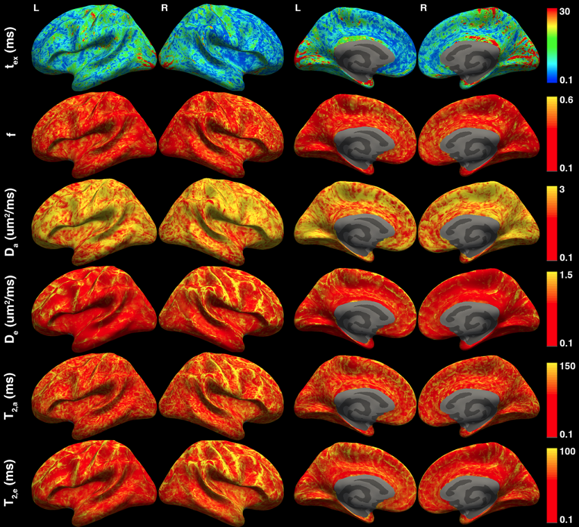
Noise propagation shows that TENEXI has slightly better accuracy of parameter estimation than NEXI (Fig.2). The parameter degeneracy problem occurs both in TENEXI and NEXI for random initialization, whereas dictionary(prior)-based initialization alleviates the problem (Fig.3).Across the cortical ribbon, the estimated exchange time is 9.4±14.1 ms (median ± IQR), intra-neurite T2avalues is 116±60 ms, and the extra-neurite T2e value is 74±41 ms (Fig.4). In general, we observed T2a>T2e in the cortical GM.
Discussion and conclusions
State-of-the-art high gradient diffusion MRI using Gmax=500 mT/m on the Connectome 2.0 scanner enables in vivo evaluation of water exchange effect and compartmental T2 relaxation time in the GM. Parameter degeneracy problem in exchange model can be alleviated by using the dictionary(prior)-based initialization for model fitting.Acknowledgements
This study is support by NIH under the award number: DP5OD031854, R01NS118187, P41EB015896, P41EB030006, U01EB026996, S10RR023401, S10RR019307.References
1. Veraart, J., Novikov, D. S., & Fieremans, E. (2018). TE dependent Diffusion Imaging (TEdDI) distinguishes between compartmental T2 relaxation times. NeuroImage, 182, 360-369.
2. Lampinen, B., Szczepankiewicz, F., Mårtensson, J., van Westen, D., Hansson, O., Westin, C. F., & Nilsson, M. (2020). Towards unconstrained compartment modeling in white matter using diffusion‐relaxation MRI with tensor‐valued diffusion encoding. Magnetic resonance in medicine, 84(3), 1605-1623.
3. Gong, T., Tong, Q., He, H., Sun, Y., Zhong, J., & Zhang, H. (2020). MTE-NODDI: Multi-TE NODDI for disentangling non-T2-weighted signal fractions from compartment-specific T2 relaxation times. Neuroimage, 217, 116906.
4. Gong, T., Tax, CMW., Mancini, M., Jones, DK., Zhang, H., & Palombo, M. (2023) Multi-TE SANDI: Quantifying compartmental T2 relaxation times in the grey matter. Proceedings of ISMRM, 0766.
5. Fieremans, E., Novikov, D. S., Jensen, J. H., & Helpern, J. A. (2010). Monte Carlo study of a two‐compartment exchange model of diffusion. NMR in Biomedicine, 23(7), 711-724.
6. Nilsson, M., Alerstam, E., Wirestam, R., Sta, F., Brockstedt, S., & Lätt, J. (2010). Evaluating the accuracy and precision of a two-compartment Kärger model using Monte Carlo simulations. Journal of Magnetic Resonance, 206(1), 59-67.
7. Lee, H. H., Papaioannou, A., Novikov, D. S., & Fieremans, E. (2020). In vivo observation and biophysical interpretation of time-dependent diffusion in human cortical gray matter. Neuroimage, 222, 117054.
8. Olesen, J. L., Østergaard, L., Shemesh, N., & Jespersen, S. N. (2022). Diffusion time dependence, power-law scaling, and exchange in gray matter. NeuroImage, 251, 118976.
9. Jelescu, I. O., de Skowronski, A., Geffroy, F., Palombo, M., & Novikov, D. S. (2022). Neurite Exchange Imaging (NEXI): A minimal model of diffusion in gray matter with inter-compartment water exchange. NeuroImage, 256, 119277.
10. Stanisz, G. J., Li, J. G., Wright, G. A., & Henkelman, R. M. (1998). Water dynamics in human blood via combined measurements of T2 relaxation and diffusion in the presence of gadolinium. Magnetic resonance in medicine, 39(2), 223-233.
11. Mahmutovic M, Shrestha M, Ramos-Llordén G, et al. A 72-channel Head Coil with an Integrated 16-Channel Field Camera for the Connectome 2.0 Scanner. Submitted to ISMRM 2024.
12. Ades-Aron, B., Veraart, J., Kochunov, P., McGuire, S., Sherman, P., Kellner, E., ... & Fieremans, E. (2018). Evaluation of the accuracy and precision of the diffusion parameter EStImation with Gibbs and NoisE removal pipeline. Neuroimage, 183, 532-543.
13. Tournier, JD., Jeurissen, B., Christiaens, D. (2023). Iterative model-based Rician bias correction and its application to denoising in diffusion MRI. Proceedings of ISMRM, 31, 3795.
14. Fischl, B. (2012). FreeSurfer. Neuroimage, 62(2), 774-781.
15. Chan, KS., Kim, TH., Bilgic, B., Marques, JP. Semi-supervised learning for fast multi-compartment relaxometry myelin water imaging (MCR-MWI). Proceedings of ISMRM, 30, 1639.
16. Marques, JP., Hollander, DD., Chan, KS. Improved R2* and QSM mapping for dummies - ask Adam. Proceedings of ISMRM, 31, 1089.
17. Novikov, D. S., Veraart, J., Jelescu, I. O., & Fieremans, E. (2018). Rotationally-invariant mapping of scalar and orientational metrics of neuronal microstructure with diffusion MRI. NeuroImage, 174, 518-538.
18. Jelescu, I. O., Veraart, J., Fieremans, E., & Novikov, D. S. (2016). Degeneracy in model parameter estimation for multi‐compartmental diffusion in neuronal tissue. NMR in Biomedicine, 29(1), 33-47.
Figures
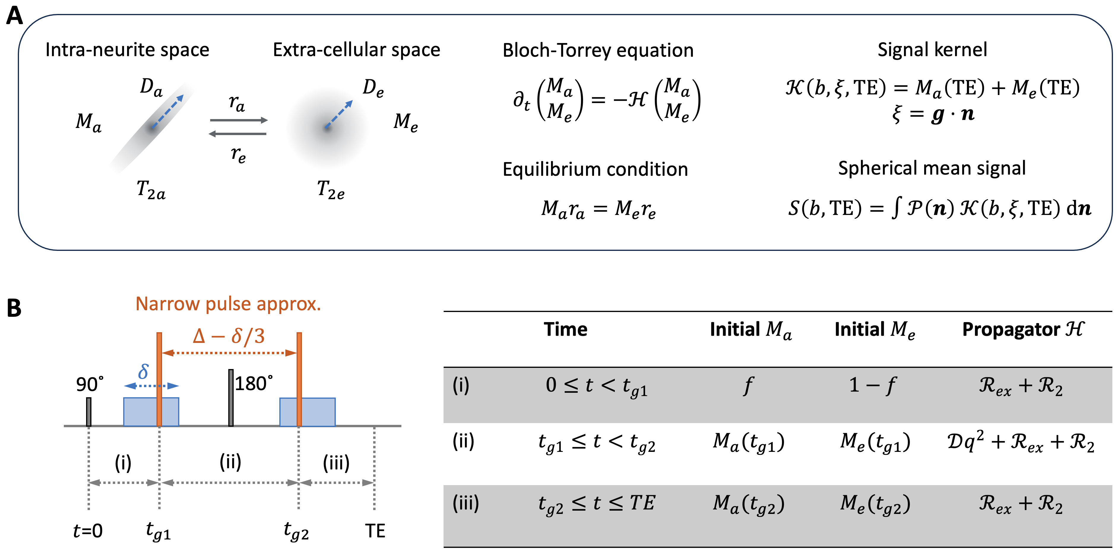
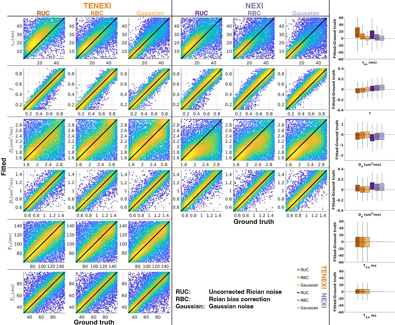
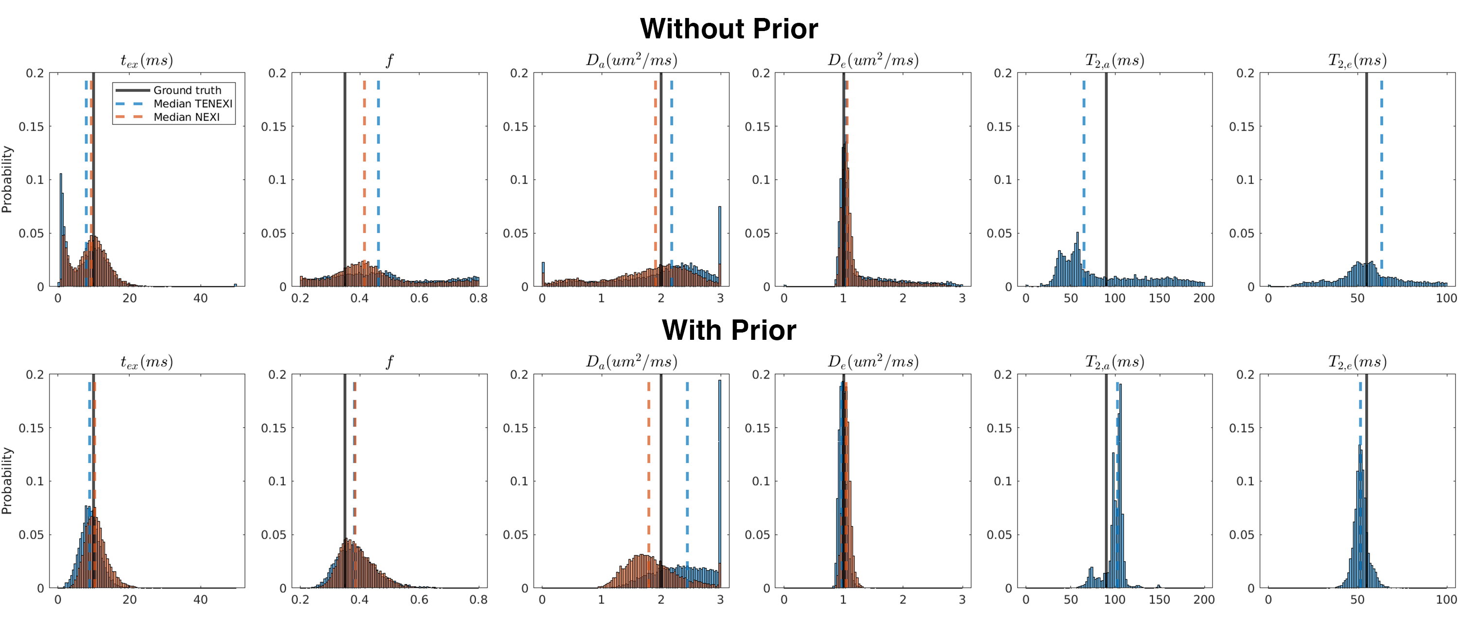
Fig.3 The parameter degeneracy of TENEXI and the ordinary NEXI. The ground truth values of tissue parameters are f=0.35, Da=2 µm2/ms, De=1 µm2/ms, tex=10 ms, T2a=90 ms, T2e=55 ms, p2=0.1. Using the dictionary(prior)-based initialization, we successfully alleviate the parameter degeneracy for the exchange time estimation.