3467
Investigating the impact of magnetisation transfer and water exchange via permeability on diffusion MRI measurements1Wellcome Centre for Integrative Neuroimaging, FMRIB, Nuffield Department of Clinical Neurosciences, University of Oxford, Oxford, United Kingdom
Synopsis
Keywords: Diffusion Modeling, Microstructure
Motivation: Magnetisation transfer (MT) and water exchange across permeable membranes operate on a similar length scale to diffusion and may impact the measured diffusion-weighted MRI (dMRI) signal. This could bias resulting parameter estimates when performing microstructural modelling.
Goal(s): To investigate how magnetisation transfer and water exchange affect dMRI measurements, particularly pore size estimations.
Approach: Monte-Carlo simulations were used to model the dMRI signal affected by MT and cross-membrane exchange in a parallel-plate geometry. Errors in plate separation estimations represent their impact on dMRI estimates.
Results: MT had a limited effect on dMRI pore size estimation, while cross-membrane water exchange can cause large overestimations.
Impact: Pore size estimates from diffusion-weighted MRI have been found to be minimally affected by magnetisation transfer but might be significantly biased by large membrane/structure permeability. New signal models incorporating diffusion and complementary phenomena can now be explored using Monte-Carlo simulations.
Introduction
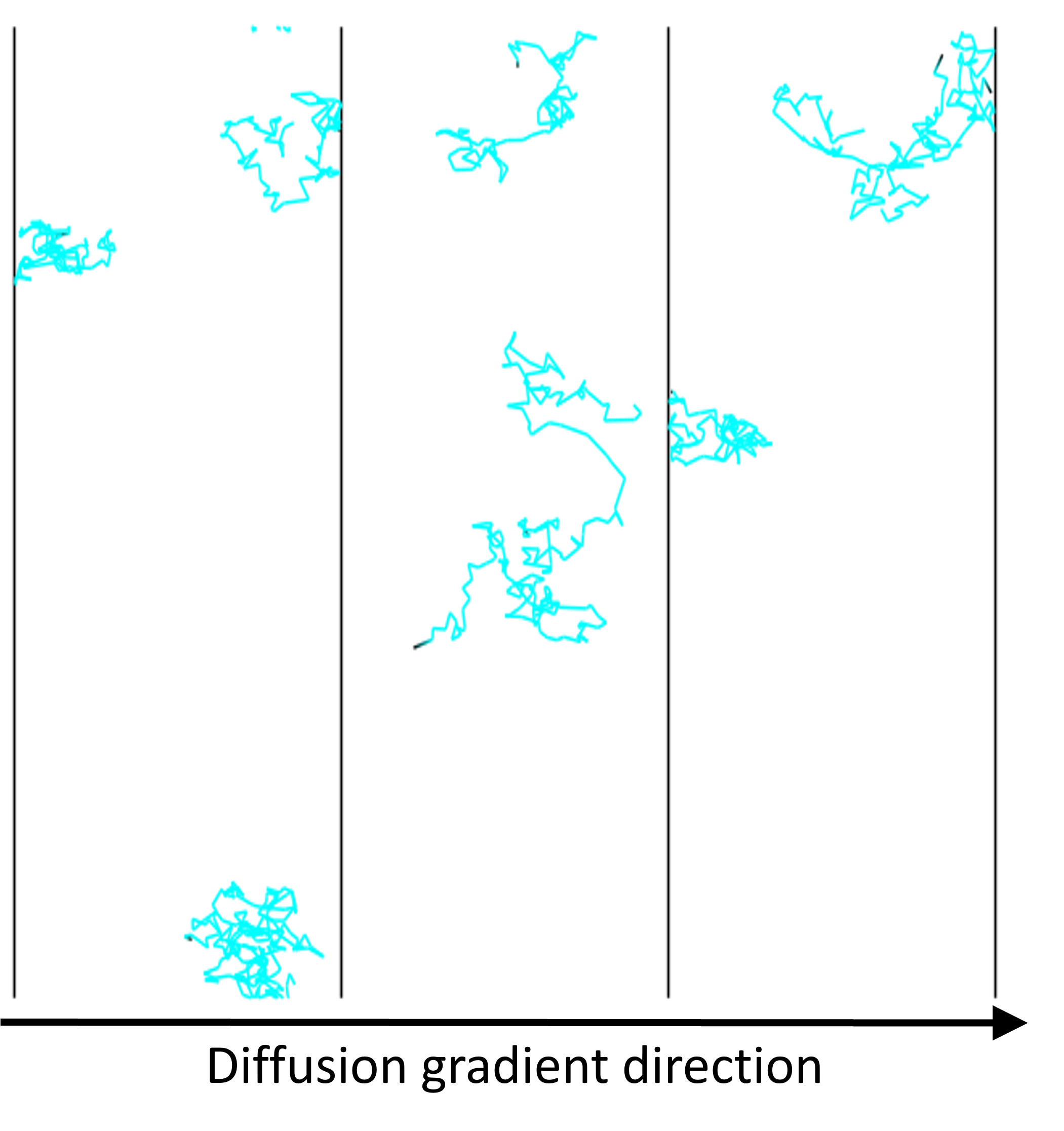
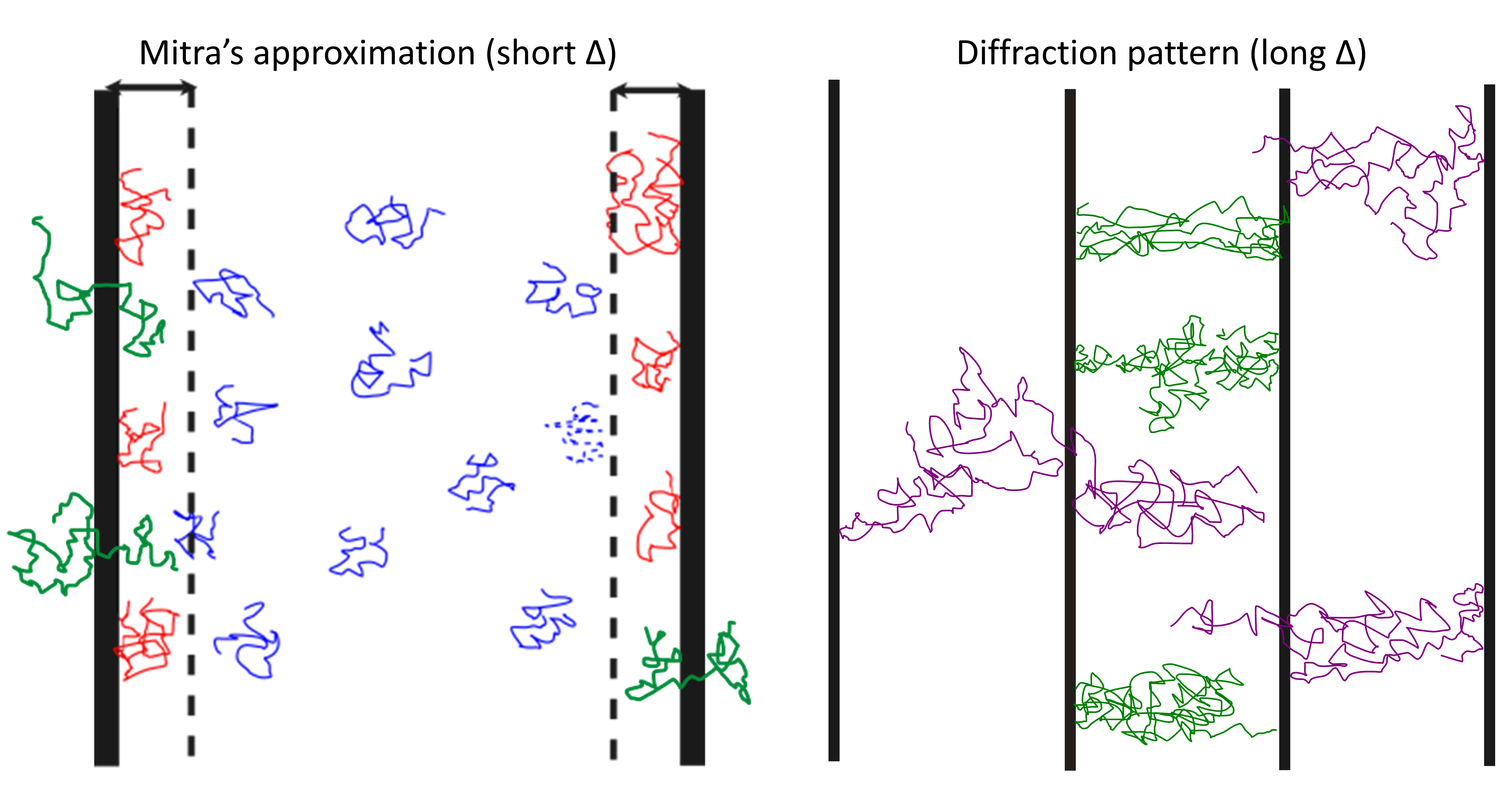
When performing diffusion-weighted MRI (dMRI) experiments, the difference between the diffusion and non-diffusion weighted signal is typically attributed to the impact of diffusion only. However, several other phenomena occur at similar length scales to diffusion, including water exchange across permeable membranes and magnetisation transfer. If these phenomena cause substantial signal deviations, it could bias resulting parameter estimates when performing microstructural modelling, as their signal contributions are often ignored and directly attributed to diffusion effects1,2. We used Monte Carlo simulations to investigate how MT and water exchange impact the measured dMRI signal for a restriction model of repeating equidistant parallel plates (Figure 1).Methods
We performed all simulations using MCMRSimulator (v0.7)3, a novel Monte-Carlo simulator that integrates several microstructural phenomena in a single framework. To simulate MT, Monte-Carlo spins were divided into two exchanging groups: the “bound” pool and the “free” pool. The bound pool is localised at user-generated obstructions and has a very short relaxation time (<1ms4), while the free pool occupies the remaining space. When a spin collides with an obstruction it has a probability (user-defined) of transferring to the bound pool. To simulate permeability-induced exchange, spins can pass through an obstruction with a user-defined probability. We chose the probabilistic approach because it acts on individual spins that we track in Monte-Carlo simulation. To translate these probabilities into macroscopic interpretable effect size, we quantify the MT strength by the apparent T2 decay it induces (i.e., effective T2) and we quantify the exchange by the effective exchange rate between the plate-separated compartments.Two analytical dMRI signal models were chosen as references for comparison with simulation results and were used to estimate plate separations: diffraction pattern at long diffusion times2 and Mitra’s approximation at short diffusion times1. All simulations were performed using diffusion-weighted spin-echo sequences, with an intrinsic diffusivity of 2µm2/ms to mimic tissue fluid.
For the long-diffusion-time regime simulation, 105 spins were simulated between regularly spaced parallel plates 1µm apart with an instantaneous diffusion-weighted gradient applied orthogonally to the plate. Simulations were performed at different MT or exchange rates, with the diffusion time (Δ) set to 40ms to ensure long diffusion time relative to the plate separation. Signal attenuations at q-values 0-13rad/mm were obtained and compared to the analytical solution $$$E(q)=\left(\frac{sin(πqL)}{πqL}\right)^2$$$, where L is the plate separation.
For the short-diffusion-time regime, 106 spins were used to reduce noise floor. The plate separation was changed to 10µm and Δ = 0.1 and 0.4ms which were short relative to the plate separation. The q-value was fixed at 1rad/µm. The plate separation (L) and intrinsic diffusivity (D0) were estimated using the apparent diffusivity D(Δ) at two Δ values (0.1ms, 0.4ms) via $$$D(Δ)≈D_0 \left[1-\frac{4}{9\sqrt{\pi}} \frac{1}{L} (D_0 Δ)^{1/2} \right]$$$.
Results
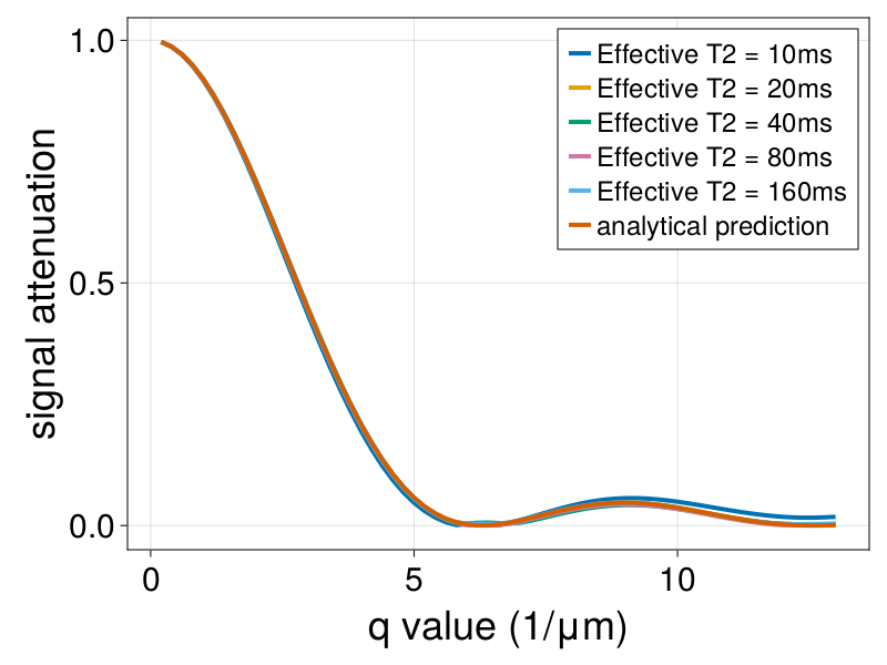
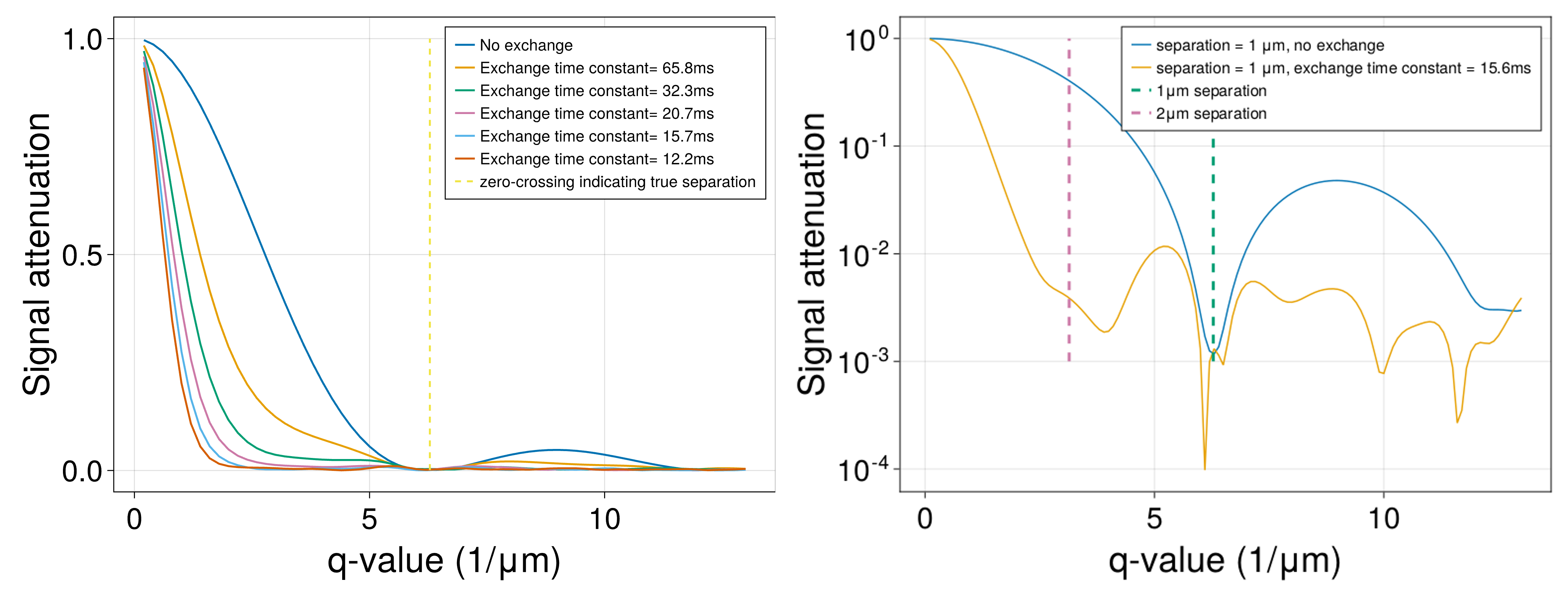
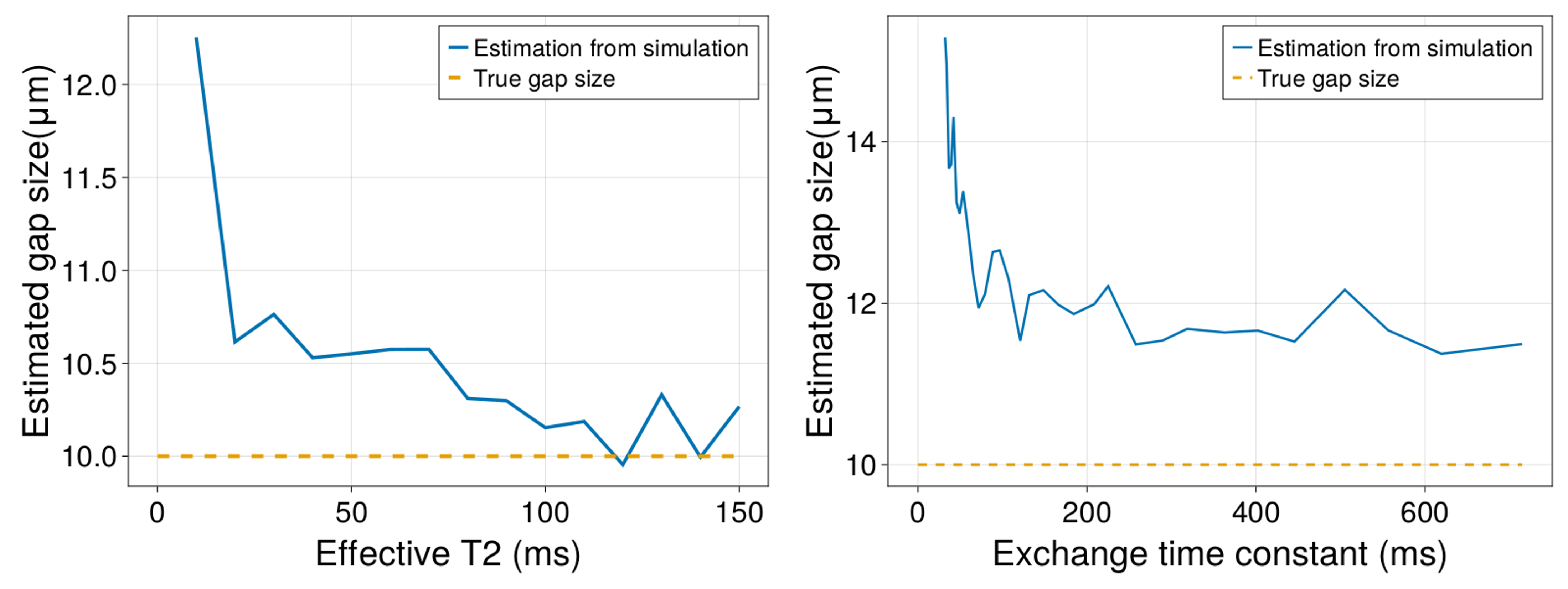
In the long-diffusion-time regime, as shown in Figure 2, realistic MT strengths has no observable effect on the diffraction pattern. Figure 3 shows that exchange reduces the signal amplitude and introduces additional extrema. For the short-diffusion-time regime, Figure 4 shows that both MT and exchange cause Mitra’s approximation to overestimate plate spacing.Discussion
To explain our MT simulation result, we note that some spins will by chance interact more with the obstructions. These spins will both experience more restrictions and have a shorter effective T2 due to MT. This effect is particularly pronounced at short diffusion times in the Mitra’s approximation (Figure 4), where MT causes reduced signal contributions from spins interacting with obstructions, which causes overestimation of compartment sizes. At longer diffusion times, this effect is less pronounced, because spins are more thoroughly mixed and have roughly equal probability to interact with obstructions and experience MT interactions (Figure 2).Permeability-induced exchange reduces the signal amplitude for the diffraction pattern because it allows spins to displace beyond the plates, which causes further dephasing under diffusion encoding. As shown in Figure 5, in Mitra’s approximation, exchange reduces the fraction of spins reflected by obstructions, which leads to an overestimation of gap sizes. At long diffusion times, spins will get reflected at plates that are beyond the two plates enclosing the spin’s initial position. This introduces additional diffraction patterns over the primary diffraction pattern and causes large overestimates.
We aim to build on these findings and use the simulator to investigate other phenomena that impact MRI measurements, alongside alternative MRI modalities.
Conclusion
We used a novel Monte-Carlo simulator that combines different microstructural phenomena to simulate the effect of MT and cross-membrane water exchange on dMRI signal. We found that realistic MT strength doesn’t cause significant errors in short or long diffusion time regimes while exchange causes large errors in both regimes.Acknowledgements
ZZ is supported by University of Oxford and China Scholarship Council. KLM is supported by a Wellcome Trust Senior Research Fellowship (224573/Z/21/Z). BCT is supported by a Wellcome Trust Sir Henry Wellcome Fellowship (222829/Z/21/Z). MC is supported by a Wellcome Trust Collaborative Award (215573/Z/19/Z). The Wellcome Centre for Integrative Neuroimaging is supported by core funding from the Wellcome Trust (203139/Z/16/Z).References
1. Mitra, P. P., Sen, P. N. & Schwartz, L. M. Short-time behavior of the diffusion coefficient as a geometrical probe of porous media. Phys. Rev. B 47, 8565–8574 (1993).
2. Özarslan, E. & Basser, P. J. MR diffusion–“diffraction” phenomenon in multi-pulse-field-gradient experiments. J. Magn. Reson. 188, 285–294 (2007).
3. Cottaar, M. MCMRSimulator.jl. (2022) doi:10.5281/zenodo.7318656.
4. Boucneau, T. et al. In vivo characterization of brain ultrashort-T2 components. Magn. Reson. Med. 80, 726–735 (2018).
Figures