3460
Increasing the realism of in silico cortex substrates for diffusion MRI simulation1BAOBAB, NeuroSpin, Paris-Saclay University, CNRS, CEA, Gif-sur-Yvette, France
Synopsis
Keywords: Simulation/Validation, Microstructure, gray matter
Motivation: Virtual microstructure has been one of the promising approaches to study and validate diffusion MRI microstructure models. Previous works in generating synthetic substrates have only focused on specific cell types, i.e. axons, neurons.
Goal(s): The main goal of this work is to advance modeling of virtual gray matter microstructure and to close the real to simulation gap in brain microstructure geometries.
Approach: We extend a framework of microstructure modeling to generate hybrid substrates that combine purely synthetic cells with virtual cells reconstructed from histology.
Results: Different virtual substrates have been generated with axons and neurons with similar volume fractions in human cortex.
Impact: More realistic virtual microstructure can allow the development of new computational models to map microstructure from diffusion MRI data. It can also be used to validate a whole set of analytical models and evaluate their accuracy.
Introduction
Numerical simulations have provided an interesting approach to studying and validating brain microstructure models1. For this purpose, many methods to generate virtual brain cells have been proposed. However, the proposed methods target only the generation of substrates with specific brain cells of a given type. For example, generating a virtual scene populated only by axons2,3 or neurons of multiple types4. To the best of our knowledge, no method proposed to synthesize virtual microstructure scenes combining neurons and axons. This work aims to close the modeling gap of virtual brain microstructure by extending the MEDUSA framework5 to allow the generation of hybrid virtual substrates. The presented method allows the synthesis of volumes of interest (VOIs), combining neurons with axons with similar volume fractions to values reported on human brain gray matter microstructure6.Methods
Dataset: The NEUROMORPHO dataset7,8 has been used to reconstruct virtual neurons of the human brain in the “.SWC” format.Cell Reconstruction: The reconstruction method creates spheres from the provided cell “.SWC” file. The reconstructed spheres are interpolated to represent synapses and to fill the connections between different connected parts within the neurons. Thus providing the same sphere-atom representation used in MEDUSA5. The reconstruction code is available at the repository9.
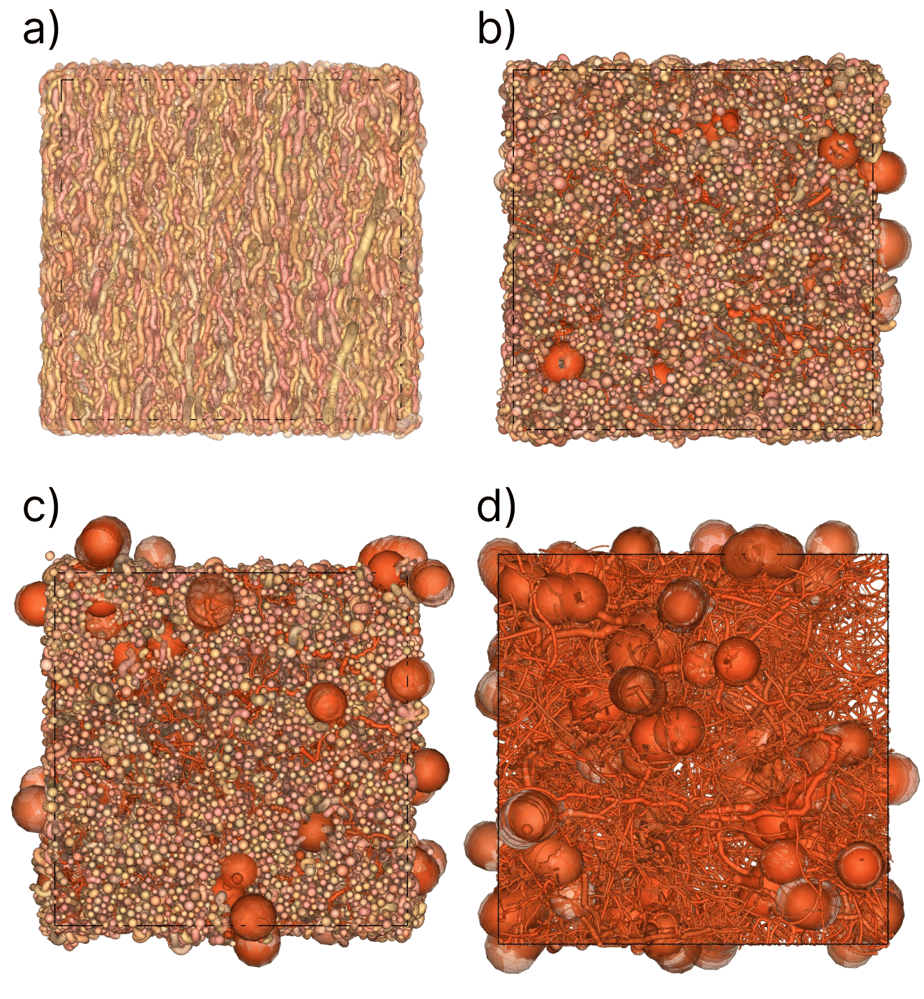
Generation of neuron populations: Four different substrate types were considered with ten generated samples for each.
- a) comprises only axons with the following properties: 85% volume fraction (VF), a diameter distribution following a gamma distribution of mean diameter of 2μm and with a standard deviation of 1μm. The axon population is oriented along the z-axis with 1.49 degrees of angular dispersion and small amount of tortuosity with 0.05μm of magnitude and a 10μm wavelength.
- b) is hybrid, it contains pyramidal neurons from the NEUROMORPHO dataset7,8 with 12% VF and mean soma diameter of 17μm and an axon population with similar properties as a) but with different VF of 42%.
- c) is similar to b) with a less dense pyramidal neuron population with a VF of 6%.
- d) is made of pyramidal neurons only with similar soma diameter distribution as b) and different VF of 15%. The VF values of substrate types b) and c) are comparable to those reported from human brain samples scanned with electron microscopy6. The MEDUSA framework was used for generation and cell de-overlapping to minimize intersections between different cells.
Results
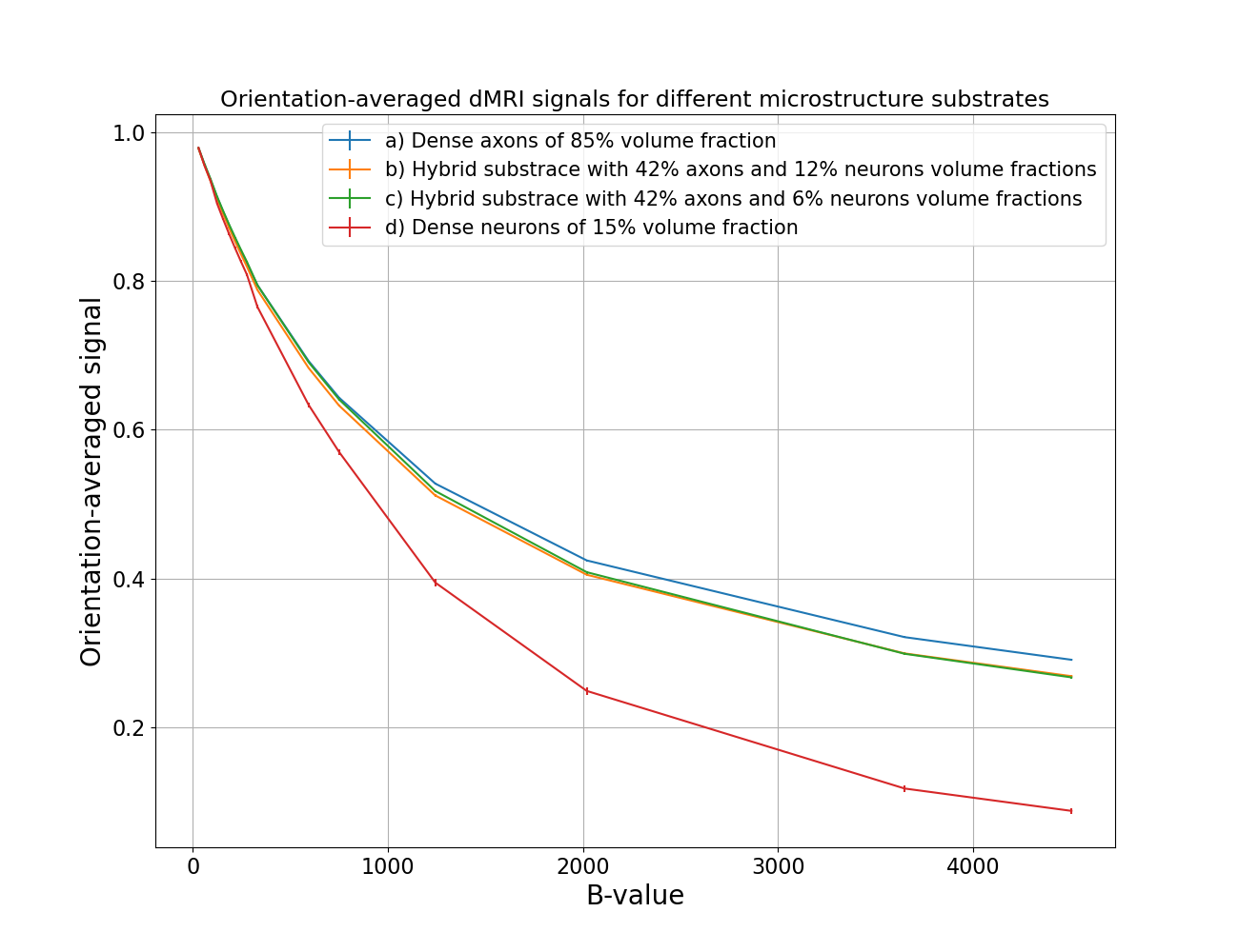
Figure 1 illustrates a reconstructed pyramidal neuron cell from the NEUROMORPHO dataset7,8. The cell is made of a set of overlapping spheres in the same fashion as used in MEDUSA. Figure 2 shows the set of parameters for samples b) and c) as well as VF reference values obtained from real human cortex sample6. Figure 3 shows four samples representing each substrate type generated in a VOI of 100x100x100μm3. The sparse representation of these substrates with sphere atoms allows to keep the memory required for each sample below 60 Mbytes. Finally, the simulated diffusion MRI signals in Figure 4 show a clear distinction between signal attenuations for substrates with axons only compared to substrates with neurons only.Conclusion
This work demonstrates a proof of concept for synthesizing virtual substrates with hybrid cells, thus, increasing the modeling realism of synthetic microstructure. Future work directions include large-scale simulation of microstructure with intra and extra-cellular diffusion, also, exploring the ability to add more components in the synthetic microstructure as blood vessels and glial cells which were not included in this study.Acknowledgements
This project has received funding from the European Union’s Horizon 2020 research and innovation program under grant agreement No 800945 -- NUMERICS -- H2020-MSCA-COFUND-2017References
- Novikov, Dmitry S., et al. "Quantifying brain microstructure with diffusion MRI: Theory and parameter estimation." NMR in Biomedicine 32.4 (2019).
- Callaghan, R., Alexander, D. C., Palombo, M., & Zhang, H. (2020). ConFiG: Contextual Fibre Growth to generate realistic axonal packing for diffusion MRI simulation. NeuroImage, 220, 117107. https://doi.org/10.1016/J.NEUROIMAGE.2020.117107
- Villarreal-Haro, J. L., Gardier, R., Canales-Rodríguez, E. J., Fischi-Gomez, E., Girard, G., Thiran, J. P., & Rafael-Patiño, J. (2023). CACTUS: a computational framework for generating realistic white matter microstructure substrates. Frontiers in Neuroinformatics, 17. https://doi.org/10.3389/fninf.2023.1208073
- Palombo, M., Alexander, D. C., & Zhang, H. (2019). A generative model of realistic brain cells with application to numerical simulation of the diffusion-weighted MR signal. NeuroImage, 188, 391–402. https://doi.org/10.1016/J.NEUROIMAGE.2018.12.025.
- Ginsburger, K., Matuschke, F., Poupon, F., Mangin, J. F., Axer, M., & Poupon, C. (2019). MEDUSA: A GPU-based tool to create realistic phantoms of the brain microstructure using tiny spheres. NeuroImage, 193, 10-24.
- Shapson-Coe, A., Januszewski, M., Berger, D. R., Pope, A., Wu, Y., Blakely, T., Schalek, R. L., Li, P., Wang, S., Maitin-Shepard, J., Karlupia, N., Dorkenwald, S., Sjostedt, E., Leavitt, L., Lee, D., Bailey, L., Fitzmaurice, A., Kar, R., Field, B., … Lichtman, J. W. (2021). A connectomic study of a petascale fragment of human cerebral cortex. BioRxiv, 2021.05.29.446289. https://doi.org/10.1101/2021.05.29.446289.
- Ascoli GA. Mobilizing the base of neuroscience data: the case of neuronal morphologies. Nat Rev Neurosci 2006, 7:318–24.
- https://neuromorpho.org/ (November 2023).
- https://framagit.org/cpoupon/gkg/-/tree/master/python/simulation?ref_type=heads (November 2023)
Figures
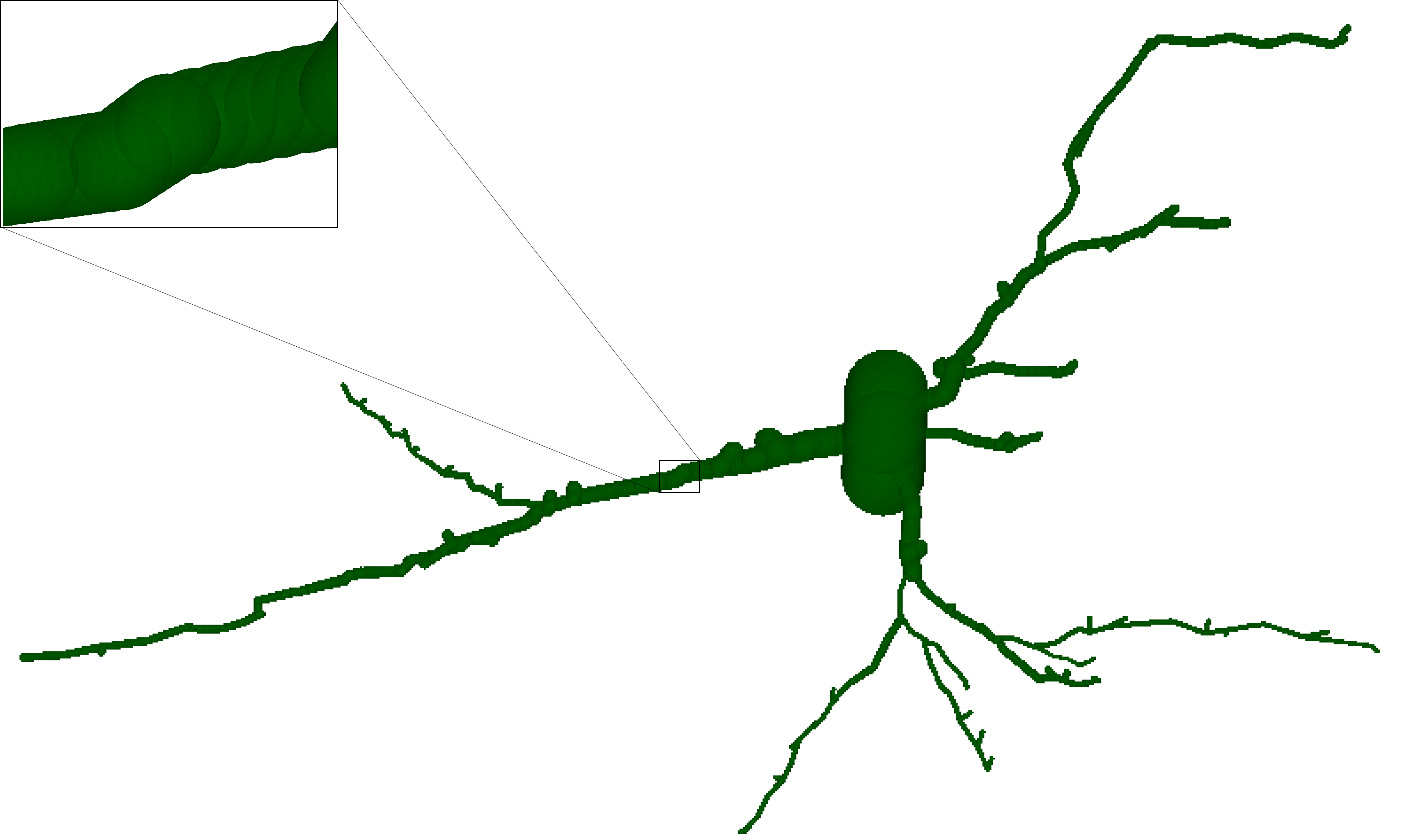
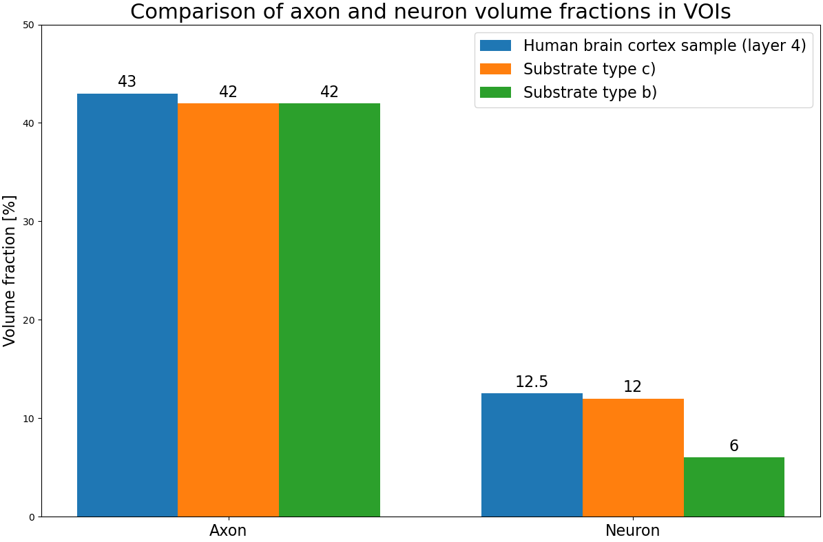
Figure 2: Comparison of the used volume fractions in samples b) and c) with values from human brain cortex sample6.