3457
Shining Light on Degeneracies and Uncertainties in the NEXI and SANDIX Models with µGUIDE1CUBRIC - Cardiff University, Cardiff, United Kingdom, 2Department of Radiology, CHUV, Lausanne, Switzerland, 3UNIL, Lausanne, Switzerland
Synopsis
Keywords: Microstructure, Microstructure
Motivation: New biophysical models of gray matter microstructure have been introduced, with a particular focus on exchange time and soma size estimations. However, the fitting quality of these models has not been studied.
Goal(s): Our goal is to study the feasibility of estimating both exchange time and soma size in a clinical setting.
Approach: We applied µGUIDE, a Bayesian inference framework, to quantify the quality of the fitting of two models from the literature, NEXI and SANDIX, using an extensive protocol, and a clinical one.
Results: Estimations of both exchange time and soma size in clinical setting shows high uncertainty.
Impact: For the first time, we are using µGUiDE on two microstructure models that take into account the exchange between neurites and the extra-cellular space. We applied it to both synthetic and clinical data.
Background
NEXI1, or SMEX2, and SANDIX2 are recent gray matter (GM) microstructure models that focus on either exchange time, soma radius or both estimations. While these models try to better represent the underlying (GM) microstructure, their increased complexity makes the estimation of the model parameters more challenging.For a given model, the choice of acquisition protocol is crucial, as it allows to tune the sensitivity to some tissue parameters. However, rich protocol acquisitions are unsuited for patients due to very long scan time, as opposed to preclinical acquisitions. In clinical settings, where the protocol is less extensive, it becomes necessary to characterize its suitability for estimating microstructure parameters of a given model.
Bayesian inference methods, such as µGUIDE3, aim to estimate full posterior distributions, which allow to quantify the quality of the fitting and highlight degeneracies.
This work aims at studying the fitting quality of NEXI and SANDIX, given two acquisition protocols used in literature, using µGUIDE. We investigate the impact on the fitting of both the acquisition protocol and the noise.
Methods
Model definitions: NEXI1 is a two-compartment model with four microstructure parameters: exchange time between neurites and extra-cellular space (ECS) tex, intra- and extra-neurite diffusivities Di/De, and neurite signal fraction f. SANDIX2 adds a third compartment of impermeable spheres to model soma, adding additional soma signal fraction fs and radius rs, making it more challenging to fit.Protocols: We considered two PGSE protocols. First one is an extensive ex-vivo acquisition protocol, similar to the one used to validate SANDIX2, while the second one is a protocol feasible for in-vivo human acquisitions, the NEXI3T Connectom protocol4.
Simulated data: Synthetic signals were generated following NEXI and SANDIX , using random parameter combinations sampled on biological feasible ranges, for each protocol. Rician noise levels sampled from the clinical data were added to the signals, with a median SNR of 50, to mimic real data acquisitions. 106 synthetic signals were used for training µGUIDE.
Clinical data: Four healthy volunteers were scanned (2 of whom rescanned on a different day). An MPRAGE was acquired for anatomical reference (1-mm isotropic resolution). Diffusion-weighted images were acquired on a 3T Siemens Connectom system using a PGSE EPI sequence with combinations of b-values of 1 (13 directions), 2.5 (25 dir.), 4 (25 dir.), 6 (32 dir.) and 7.5ms/µm² (65 dir.) and Δ=20, 29, 39 and 49ms; δ=9ms, and 15 b=0 images per Δ at 1.8-mm isotropic resolution, TE/TR=76/ 3700 ms, for a total scan time of 45 minutes.
Processing: Multi-shell multi-diffusion time data was preprocessed jointly; steps included MP-PCA magnitude denoising5, Gibbs ringing correction6, distortion and eddy current correction7. The cortical ribbon was segmented on the MPRAGE image using FastSurfer8 and projected onto the diffusion native space using linear registration9. Parametric maps of both models were estimated using non-linear least squares and µGUIDE. We extract three measures from the estimated posterior distributions: maximum-a-posteriori, an ambiguity and an uncertainty measure.
Results and discussion
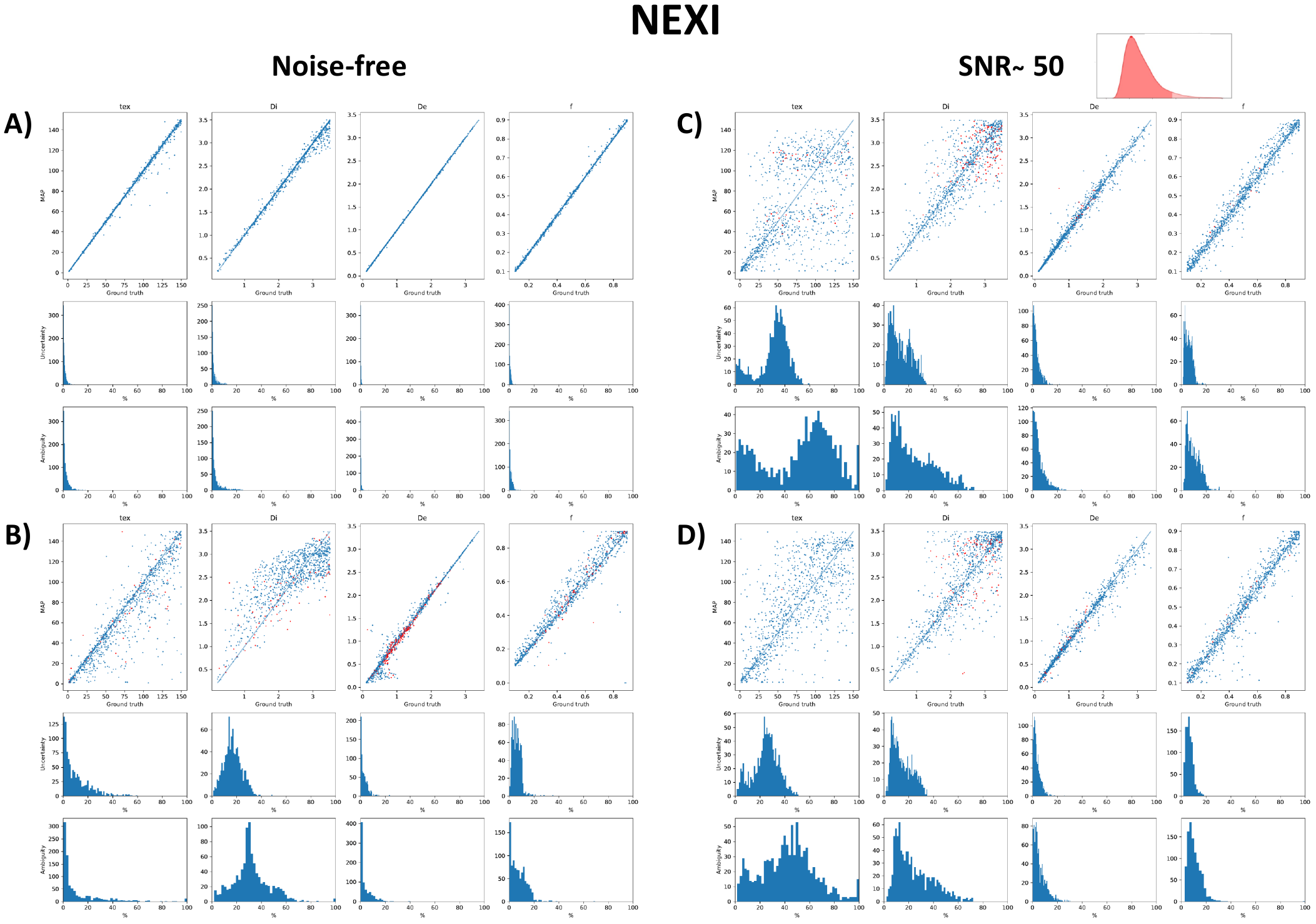
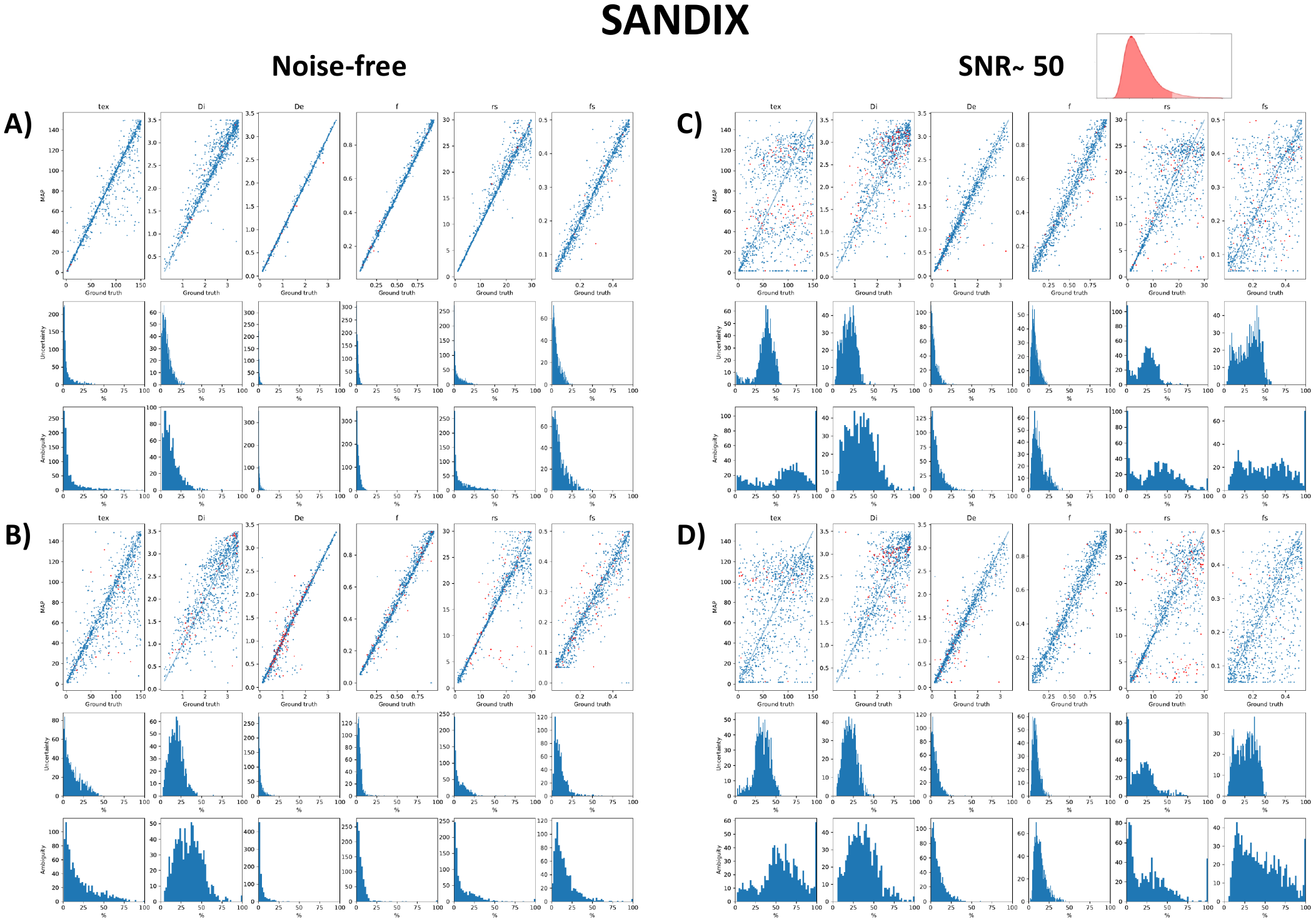
Fig.1&2: The extensive protocol acquisition allows to remove almost all degeneracies and substantially reduce the uncertainty of the estimates. However, this protocol is heavily impacted by noise presence , as the signal for high b-values is mostly lost. The fitting quality of the Connectom protocol is less affected by the presence of noise, as it relies on smaller b-values. However, even when considering a noise-free scenario, this protocol shows bias and variance in parameter estimates.Estimates using NEXI with the Connectom protocol and a realistic noise level (Fig.1.D) demonstrates that tex estimates are mostly unreliable, while the model shows good capacity to estimate De and f. In the presence of noise, it seems impossible to jointly estimate tex and rs accurately (Fig.2.C&D).
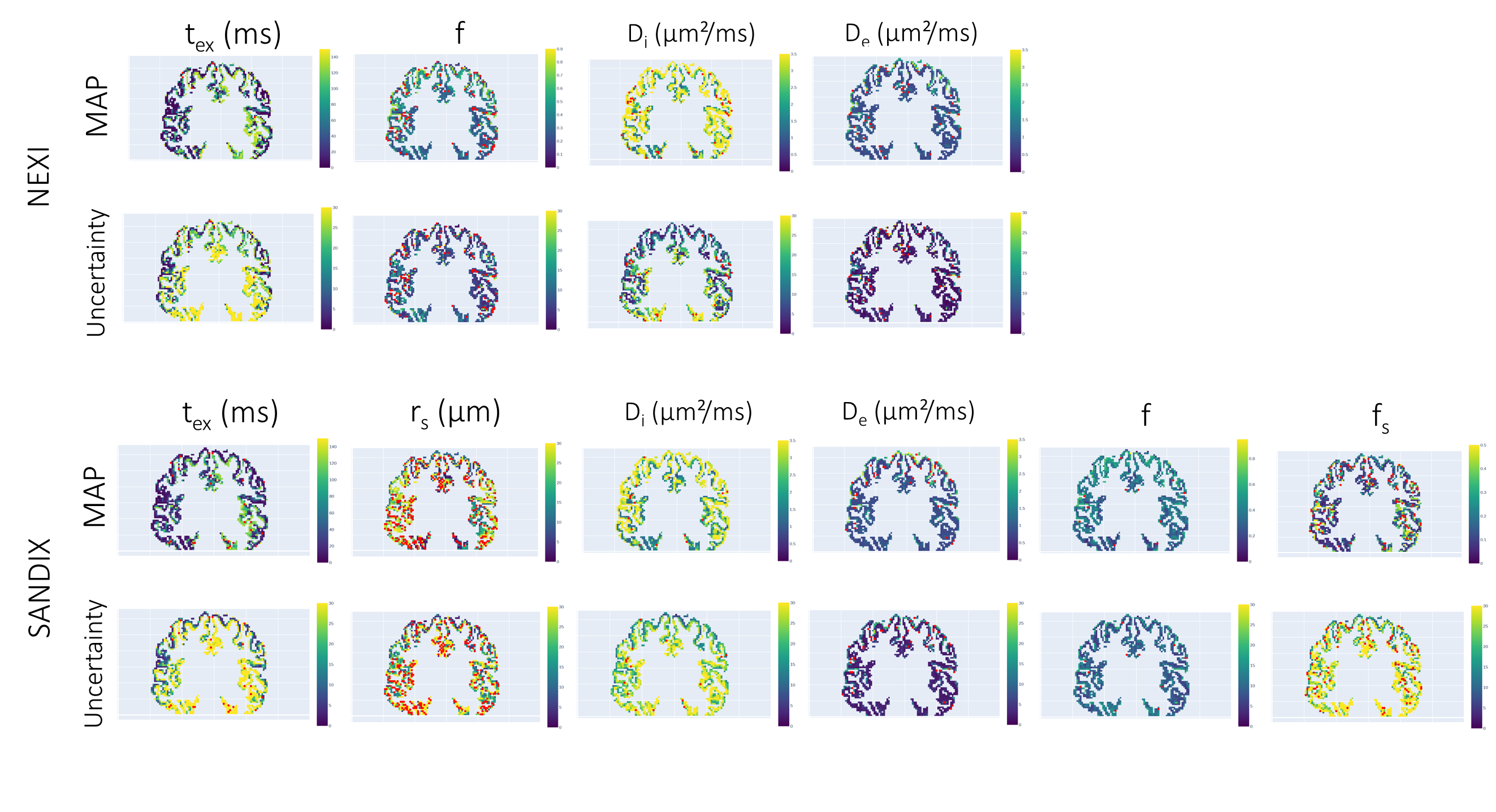
Fig.3: High uncertainties, indicating low confidence, are obtained for tex in the model NEXI. De and f estimates using SANDIX show a low uncertainty, as expected from the simulations.
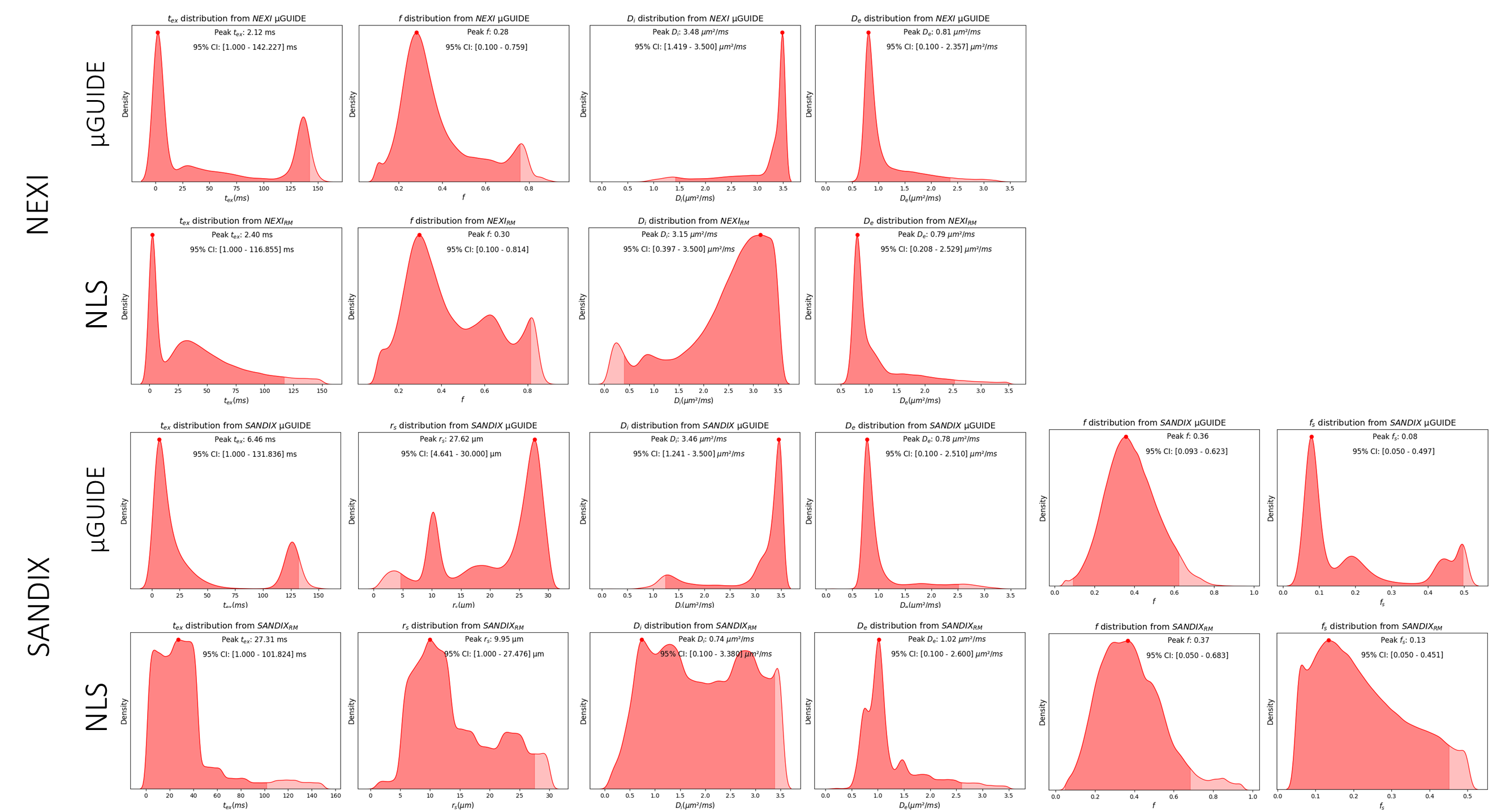
Fig.4 compares the estimations obtained on the cortical ribbons of all the participants using a non-linear least square fitting method and µGUIDE. Only estimates with uncertainty and ambiguity <50% were kept on µGUIDE’s estimations. Similar results are obtained on NEXI, while SANDIX shows substantial differences for tex, radius and Di. NEXI and SANDIX give the same estimates of extra-neurite diffusivity for all methods.
Conclusion
Our results reveal that each protocol exhibits unique strengths. The extended protocol remains resilient to noise, while the Connectom protocol also demonstrates robustness. The second protocol's limitation is its low coverage of b-values and diffusion times. Our results obtained with µGUiDE align with those obtained using Non-linear least squares, further bolstering our confidence in the former's learning capabilities.Acknowledgements
MJ and MP are supported by UKRI Future Leaders Fellowship (MR/T020296/2).References
[1] Jelescu, NeuroImage 2022; [2] Olesen, NeuroImage 2022; [3] Jallais et al., ISMRM 2023; [4] Uhl et al., arXiv 2023; [5] Veerart, NeuroImage 2016; [6] Kellner, MRM 2016; [7] Andersson, NeuroImage 2016; [8] Henschel, NeuroImage 2020; [9] Avants et al., Insight j, 2009Figures