3455
GEM: a unifying model for Gray Matter microstructure1Department of Radiology, CHUV, Lausanne, Switzerland, 2UNIL, Lausanne, Switzerland
Synopsis
Keywords: Microstructure, Diffusion/other diffusion imaging techniques, Diffusion modeling, Tissue characterization, Soma, Exchange
Motivation: Gray Matter lacks a unified microstructure model, unlike White Matter. This study introduces the Generalized Exchange Model (GEM) to unify Gray Matter models, introducing exchange with simple structures.
Goal(s): Evaluate GEM performance, compare it with other Gray Matter models, and assess its potential for clinical MRI.
Approach: GEM parameters and equations are detailed and validated with simulated data. GEM is applied on clinical MRI data, and compared to other Gray Matter model estimates.
Results: GEM successfully unifies Gray Matter diffusion models, offering plausible estimations in clinical MRI revealing microstructural patterns. Future research will optimize data acquisition and assess accuracy.
Impact: The introduction of the Generalized Exchange Model holds significant implications. For scientists, it provides a unified framework, potentially simplifying and enhancing Gray Matter modeling, promoting consistency in research. Clinicians may benefit from improved specificity in diagnosing neurological conditions.
Background
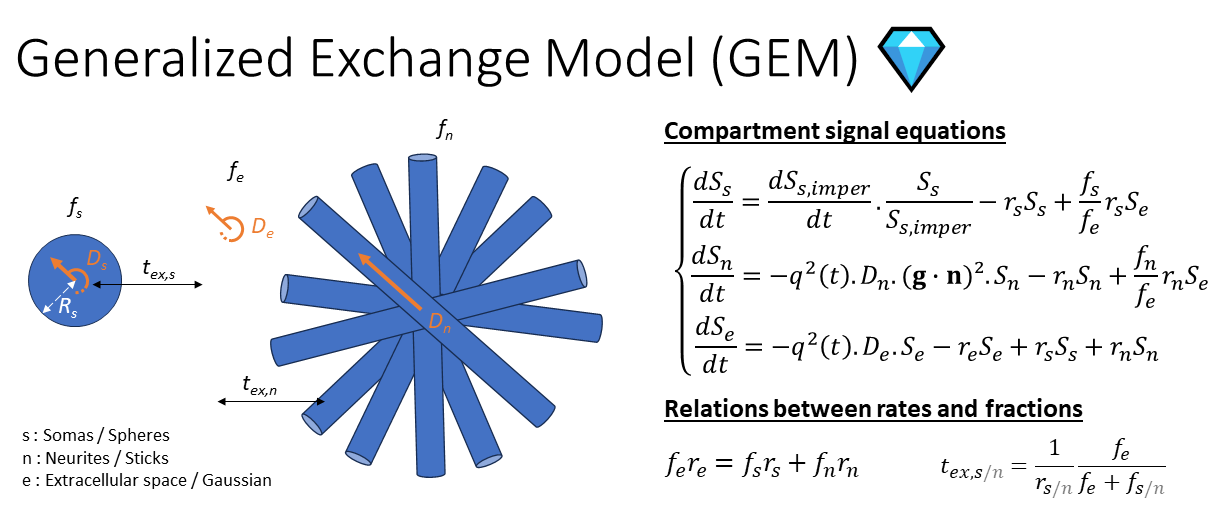
While White Matter models have the Standard Model of Diffusion1, Gray Matter (GM) models lack such a unifying framework. Two of the most extensively studied GM models, NEXI2, proposed in parallel as SMEX3, and SANDI4, correspond to extreme cases where soma and neurite membranes are either considered fully and partially permeable, respectively (NEXI), or impermeable (SANDI). In this work, we propose a means of quantifying exchange in simple structures, such as spheres or cylinders. This enables us to define a new three-compartment model, the Generalized Exchange Model (GEM), with soma and neurites, each exchanging with the extracellular space (ECS). In such a framework, most GM diffusion models can be understood as special cases of GEM. SANDIX3 corresponds to the case where tex,s is infinite, SANDI to the case where both exchange times are infinite, NEXI to the case where the fraction fs is 0, with two implementations: NEXINPA in the case of the Narrow Pulse Approximation2 (δ →0) and NEXIAWP for Actual Wide Pulses3 . We aimed to test the performance of GEM in the human cortex in vivo on a clinical MRI scanner, using an acquisition protocol originally designed for the NEXI model5. We then compared the GEM parameter estimates to the other GM models and to literature values.Methods
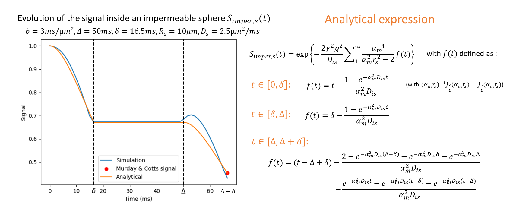
Theory: GEM parameters and equations are laid out in Fig.1. In the first order, the signal from a compartment exchanging with other compartments follows the equation: $$\frac{dS}{dt}=\frac{dS_{imper}}{dt}\frac{S}{S_{imper}}-rS+\sum_{i}{\alpha_i}r_{ext,i}S_{ext,i}$$ where S(t) and Simper(t) correspond to the signal in the permeable and impermeable cases, r to the mean life rate of a particle in the compartment and $$$\sum_{i}{\alpha_i}r_{ext,i}S_{ext,i}$$$ to the external contributions of the other compartments. Obtaining the signal in the presence of exchange is a matter of expressing the signal Simper(t). In the case of a sphere, we can derive it from the spherical mean displacement formula6 (Fig.2). Simulated data: Numerical PGSE diffusion signal was simulated inside a single impermeable sphere of radius Rs=10µm and bulk diffusivity Ds=2.5µm²/ms using the MC/DC simulator7 with 50’000 walkers and parameters b=3ms/µm², Δ=50ms, δ=16.5ms . This result was used to validate the analytical expression (Fig.2). Clinical data: Six healthy volunteers (3M, 26.5±1.1 years old) were scanned on a 3T system (MAGNETOM Prisma, Siemens Healthcare, Erlangen, Germany) and rescanned on a different day. Acquisition: An MPRAGE was acquired for anatomical reference (1-mm isotropic resolution). Diffusion-weighted images were acquired using a PGSE-EPI research sequence with the following parameters: b-values=1.00, 2.00, 3.20, 4.44 and 5.00ms/µm², 20 directions per shell, diffusion times Δ=28.3, 36.0, 45.0, 55.0 and 65.0ms, δ=16.5ms, 4 b=0 images, spatial resolution=2x2x2 mm3, total scan time: 35min. Processing: Multi-shell multi-diffusion time data were preprocessed jointly using a standard pipeline8. The GM region of interests (ROIs) from the Desikan-Killiany-Tourville atlas9 were segmented on the MPRAGE image using FastSurfer10 and projected onto the diffusion native space using linear registration11. Parametric maps of NEXINPA, NEXIAWP, SANDI, SANDIX and GEM, all corrected for Rician Mean12, were estimated from the powder-average signals using non-linear least-squares. The spatial distribution of NEXI features was also examined using inflated brain surfaces obtained using Connectome Workbench13.Results and discussion
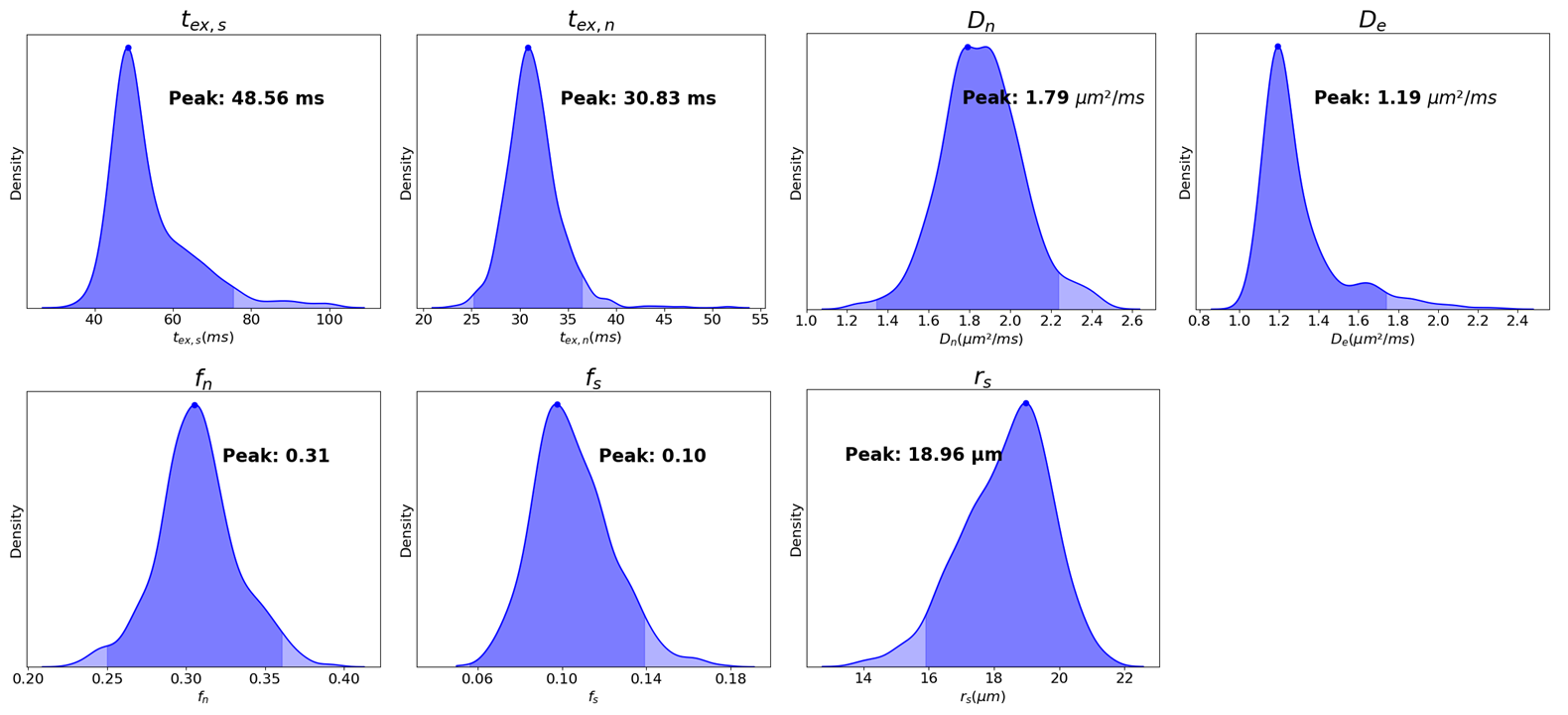
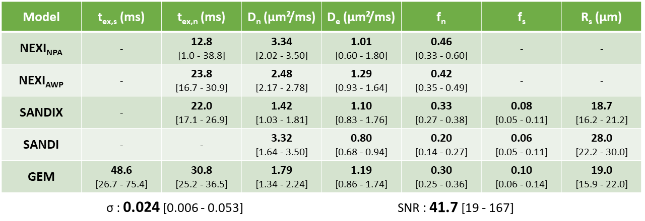
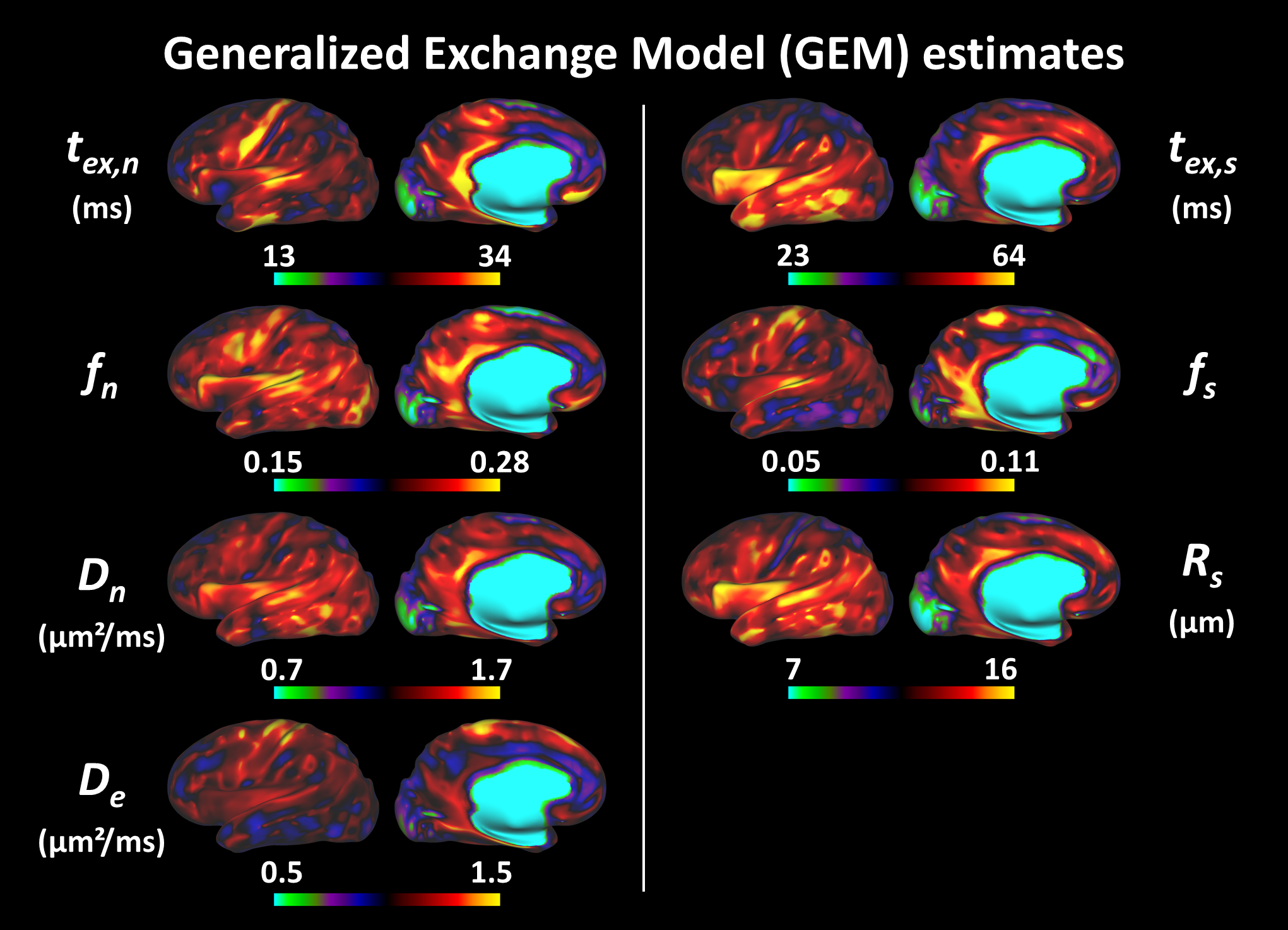
The GEM signal calculation can be accelerated by using the analytical solution in the form of matrix exponential between times δ and Δ. For the other intervals, the ODE solver solve_ivp from scipy.integrate14 was used. Once applied on the clinical data and despite the potential degeneracy of the model, the ROI medians of all subjects were well centered for each parameter (Fig.3). Compared to other GM models, GEM estimates are very close to those of SANDIX, while giving a plausible tex,s, of the same order of magnitude as tex,n (Fig.4). The GEM intra-cellular fractions seem very low, but neurite diffusivities are more biologically plausible15,16 than those obtained with SANDI or NEXINPA. The soma radii appear to be more moderate than those obtained with SANDI, even though they remain high compared to previous studies and histology3,17. The cortical surface maps finally show that this model is sensitive to certain recognizable patterns (Fig.5) such as the longer neurite exchange time on the sensorimotor cortex or on the temporal lobe, matching higher myelination and previously reported in humans using NEXINPA12.Conclusion
GEM was derived and successfully estimated in the human cortex on data acquired on a clinical scanner. Estimated parameters are similar to those obtained with other GM models and literature values. The optimal acquisition for estimating this model has yet to be established. One possible approach would be to vary δ. Future work will also focus on estimating its accuracy and precision using synthetic data.Acknowledgements
The authors thank Thorsten Feiweier for providing access to the PGSE-EPI research sequence. This work was supported by SNSF Eccellenza grant PCEFP2_194260 and ERC Starting Grant ‘FIREPATH’, SERI no. MB22.00032.References
[1] Novikov, NMR in Biomedicine 2018 [2] Jelescu, NeuroImage 2022 [3] Olesen, NeuroImage 2022 [4] Palombo, NeuroImage 2020 [5] Uhl, Proceedings of the ISMRM Diffusion Workshop 2022 [6] Neuman, Journal of Chemical Physics 1974 [7] Rafael-Patino, Frontiers in Neuroinformatics 2020 [8] Ades-Aron, NeuroImage 2018 [9] Klein and Tourville, Frontiers in Neuroscience 2012 [10] Henschel, NeuroImage 2020 [11] Avants et al., Insight j, 2009 [12] Uhl, Arxiv 2023 [13] Marcus, Frontiers in Neuroinformatics 2011 [14] Virtanen, Nature Methods 2020 [15] Dhital, NeuroImage 2019 [16] Howard, NeuroImage 2022 [17] Cardona, PLoS biology 2010Figures