3454
Multiscale sample entropy analysis of resting-state fMRI over the lifespan1USC Stevens Neuroimaging and Informatics Institute, Keck School of Medicine at USC, Los Angeles, CA, United States
Synopsis
Keywords: fMRI Analysis, Data Analysis, Multiscale Sample Entropy, Complexity
Motivation: Multiscale sample entropy (MSE) is a common complexity metric used in functional Magnetic Resonance Imaging (fMRI), yet it has not been applied to analyze the evolution across age groups.
Goal(s): The goal of this study is to analyze the evolution of fMRI complexity over the lifespan using MSE.
Approach: Resting-state fMRI data of 526 subjects age 6 – 85 years were analyzed with a linear mixed-effect model (LME) of MSE in different brain regions.
Results: Overall, a decrease in mean gray matter complexity was observed after puberty. LME model showed significant decrease in complexity with age in middle frontal and superior frontal gyrus.
Impact: The findings of this study shed light on the evolution of brain complexity with development and aging and may provide benchmark for detecting aberrant complexity in brain disorders.
Introduction
Exploring and understanding of the development, maturation and aging process becomes important as the aging population increase. Over the years, aging of human brain was studied using different modalities of medical imaging data. In recent years, complexity of resting state Functional Magnetic Resonance Imaging (rs-fMRI) data has shown significant differences between young and elderly populations1 and faster decline in patients with Alzheimer’s disease2. There are multiple methods to measure complexity in time series data such as entropy-based methods and fractal dimension-based methods. Various studies have evaluated the test-retest reliability of these methods and their applications in exploring different characteristics of physiological data3. Using complexity metrics on physiological data is an evolving branch of time series analysis and novel complexity metrics have been introduced over the recent years. A recent study assessed Hurst Exponent over the lifespan and observed significant decrease of complexity with age in frontal and partial lobes and increase of complexity in insula, limbic lobe, occipital lobe, and temporal lobe4. Multiscale Sample Entropy (MSE), which is a modified version of the sample entropy and a prevalent complexity metric in fMRI time series data analysis, has so far not yet been used to study development and healthy aging over the lifespan. In this study we investigate the changes in MSE over the lifespan.Methods
The study included Nathan Klein Institute (NKI) – Rockland Sample rs-fMRI data of 526 subjects age ranging from 6 to 85 years, acquired using the following parameters (TR = 1400 ms; 2 mm isotropic; duration = 10 mins; slices = 64). The enhanced NKI project was aimed at creating a large scale (number of participants > 1000) community sample of participants across the lifespan5,6. Data pre-processing was performed in CONN-toolbox and included motion realignment, regression of motion parameters and derivatives, regression of cerebrospinal fluid and white matter signal fluctuations using a-CompCorr, linear drift removal using a high-pass filter, coregistration to individual T1 and normalization to MNI template space. Subjects were divided into 5 groups using hierarchical clustering (Table 1): Age 6-13 (Pre-Puberty, 19 Females, 22 Males), 14-25 (Puberty, 68 Females, 64 Males), 26-38 (Young Adult), 39-63 (Middle Age, 32 Female, 43 Males), 64-85 (Senior, 67 Females, 35 Males). Resting state fMRI-complexity was estimated by means of MSE using an in-house complexity toolbox (LOFT Complexity Toolbox 2.0). MSE was calculated as the area-under-the-curve across entropy at τ = 13 scales, with pattern matching length m = 2 and pattern matching threshold r = 0.3. Average complexity of gray matter (GM) voxels and 14 regions of interest (ROIs) were calculated for each subject. To assess global GM changes across the lifespan, we used a second order exponential function. For regional analyses we applied a linear mixed effect model for age, gender, and their interaction (age × gender) with global mean GM values as a covariate.Results
Mean rsfMRI-complexity of global GM shows an increase from age 6 to 25 and a gradual decrease after age 25 (Figure 1). Maximum rsfMRI-complexity in GM was observed during puberty to adolescence and young adulthood (Age 14 – 25). Linear mixed-effect model when accounting for the overall GM effect indicated reduction of rs-fMRI complexity with age in all cortical areas but did not reach statistical significance (Table 2). However, we observed statistically significant age-by-gender interactions in prefrontal, superior frontal, middle frontal, inferior frontal, lateral parietal, lateral temporal, medial temporal, and precentral regions (p-value < 0.05) (Figure 2). Amongst these areas, the effects were strongest in middle frontal and superior frontal regions (p-value < 0.01).Discussion
Overall, the inverted hockey stick-shape of rsfMRI-complexity in GM replicates the rapid maturation of cortical areas during childhood reaching its peak during adolescence and then gradual but slower decrease with healthy aging. The most interesting findings were observed in superior and middle frontal ROIs; areas that compose the dorsolateral prefrontal cortex (dlPFC) (Figure 2). The dlPFC shows late maturation throughout young adulthood and later in life a decline which is associated with first gain and then loss of executive function7. These findings of lifespan changes in rsfMRI-complexity are in agreement with other MRI measures of cortical development and maturation and thus rsfMRI-complexity presents a new characteristic of brain functionality.Acknowledgements
This study was supported by the National Institutes of Health R01AG066711.References
1. Smith RX, Yan L, Wang DJ. Multiple time scale complexity analysis of resting state FMRI. Brain Imaging Behav. 2014;8(2):284-91.2. Wang B, Niu Y, Miao L, Cao R, Yan P, Guo H, Li D, Guo Y, Yan T, W.u J, Xiang J, Zhang H. Decreased Complexity in Alzheimer's Disease: Resting-State fMRI Evidence of Brain Entropy Mapping. Front Aging Neurosci. 2017;9:378.
3. Guan S, Wan D, Zhao R, Canario E, Meng C, Biswal BB. The complexity of spontaneous brain activity changes in schizophrenia, bipolar disorder, and ADHD was examined using different variations of entropy. Hum Brain Mapp. 2023;44(1):94-118.
4. Dong J, Jing B, Ma X, Liu H, Mo X, Li H. Hurst Exponent Analysis of Resting-State fMRI Signal Complexity across the Adult Lifespan. Front Neurosci. 2018;12:34.
5. Nooner KB, Colcombe SJ, Tobe RH, et al. The NKI-Rockland Sample: A Model for Accelerating the Pace of Discovery Science in Psychiatry. Front Neurosci. 2012; 16;6:152.
6. Tobe RH, MacKay-Brandt A, Lim R, et al. A longitudinal resource for studying connectome development and its psychiatric associations during childhood. Sci Data. 2022; 9, 300.
7. MacPherson SE, Phillips LH, Della Sala S. Age, executive function, and social decision making: a dorsolateral prefrontal theory of cognitive aging. Psychol Aging. 2002;17(4):598-609.
Figures
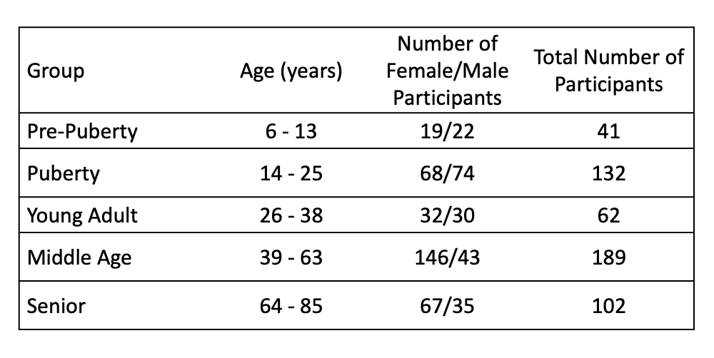
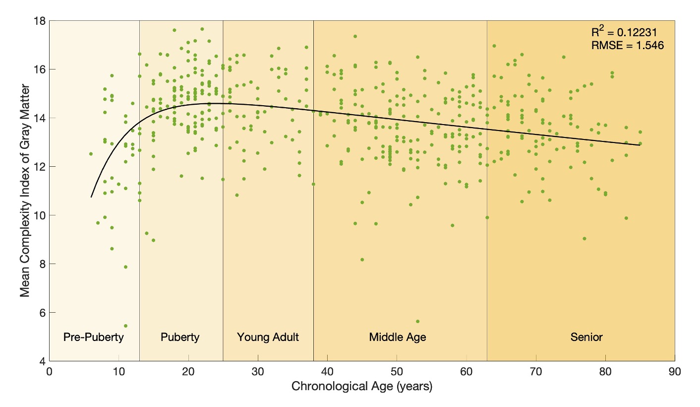
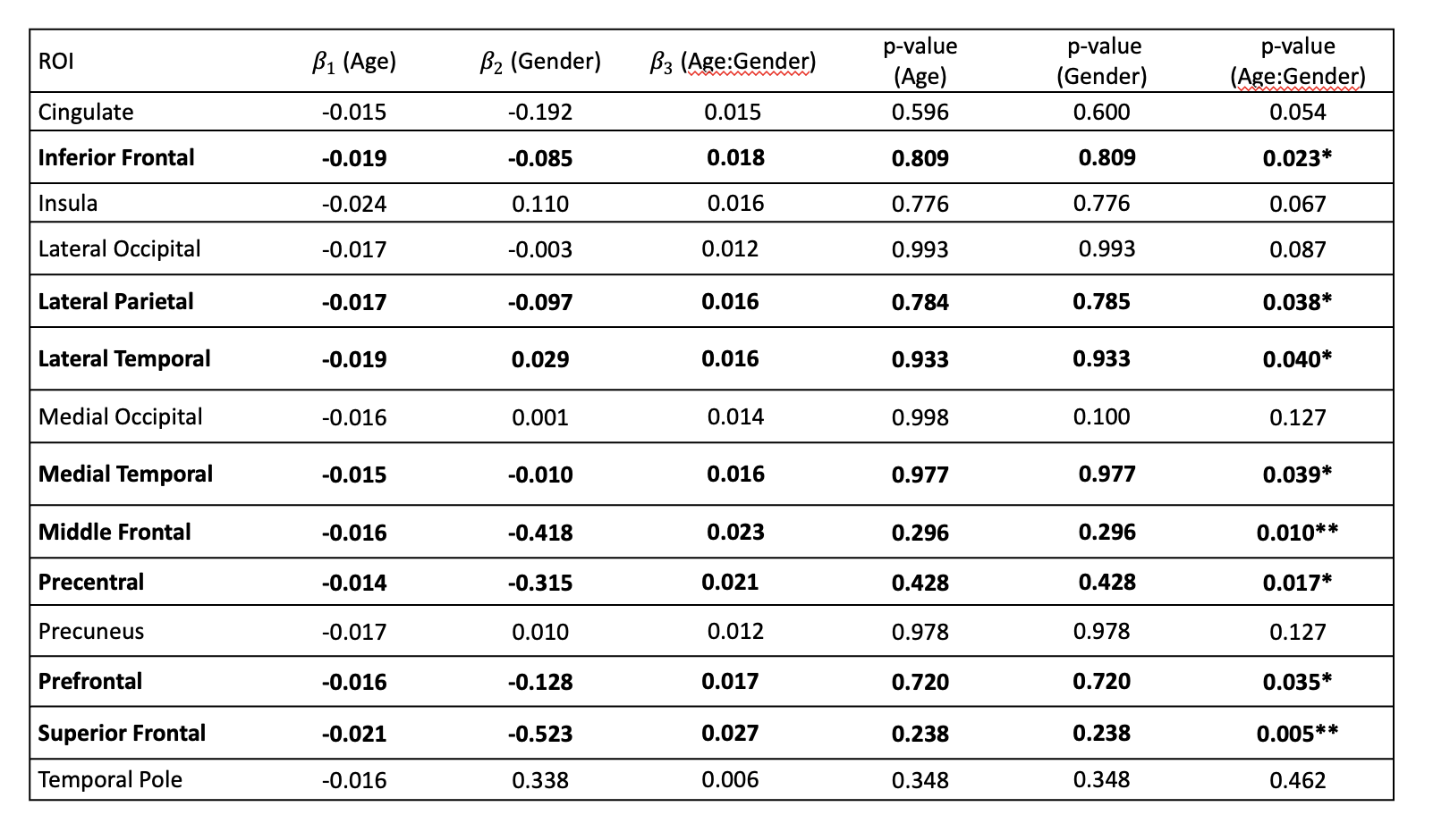

Figure 2: Visualization of the regions of interest showing significant age×gender interaction effects.