3449
Association of blood pressure metrics with amplitudes of physiological brain pulsations1Diagnostic Radiology, University of Oulu, Oulu, Finland
Synopsis
Keywords: fMRI Analysis, Brain, Cardiovascular, fMRI (resting state), Hypertension, Neurofluids
Motivation: Vascular factors, like blood pressure, play a role in brain disorders, but their pathological mechanisms are still unknown.
Goal(s): The study's objective was to examine the impact of blood pressure (systole/diastole) on the physiological pulsations assessed through ultrafast resting-state functional magnetic resonance imaging.
Approach: We quantified brain pulsation amplitudes with the ALFF method and employed multiple linear regression to model the influence of blood pressure metrics on very low-frequency, respiratory, and cardiovascular pulsations.
Results: Systolic blood pressure positively correlated with vasomotor pulsation amplitudes, while both systolic and diastolic pressures were positively correlated with cardiovascular pulsation amplitudes.
Impact: Precise knowledge regarding the influence of blood pressure on brain pulsations plays a pivotal role in future developments of treatments, particularly in conditions like Alzheimer's disease, where blood pressure is a significant risk factor affecting cerebral function.
Introduction
High blood pressure is a significant risk factor for Alzheimer's disease and impacts cerebral hemodynamics [1]. This study probes the relationship between blood pressure and brain pulsatility using magnetic resonance encephalography (MREG), which captures slow (circa 1 Hz) vasomotor waves and respiration pulsations from the venous blood oxygenation level dependent (BOLD) signal, and detects higher frequency CSF space and blood flow dynamics including arterial impulses, this without temporal aliasing [2]. We posit that blood pressure correlates with disruptions in these physiological pulsations. To test this, we assessed physiological fluctuation amplitudes in a diverse healthy adult cohort, examining their association with blood pressure metricss through multiple regression analyses. Our findings aim to clarify how systemic vascular factors could influence cerebral physiology and potentially contribute to neuropathology.Methods
One hundred and twenty-five (125) healthy subjects participated in the study. During image preprocessing, three subjects were excluded because of partial data corruption, and six subjects were excluded because of excessive head movement, leaving 116 subjects to the study. Blood pressure was measured in a supine position just before the scan.Subjects were scanned using a Siemens 3T SKYRA scanner with a 32-channel head coil and a 3D whole brain MREG sequence (TR 100ms, TE 3.6ms, flip angle 25, matrix=643, FOV=192mm) [3]. Data preprocessing involved motion spike removal with AFNI's 3dDespike, standard FSL pipeline with high-pass filtering at 0.008 Hz, motion correction using FSL MCFLIRT, and brain extraction with FSL BET [4]. Anatomical MPRAGE images aided in registering MREG data to MNI152 standard space. Subjects with excessive motion (>0.3 mm mean or >1.5 mm at any timepoint) were excluded.
A modified ALFF method was used to study amplitude of fluctuation (AF) of very low frequency (VLF), respiratory, and cardiovascular pulsations [2,5]. The frequency band for VLF was 0.01-0.1 Hz, while the bands for respiration and cardiovascular were 0.1 Hz wide, centered around the individual bands (i.e., center of the band ± 0.05 Hz). For every subject, the time courses of each voxel from whole brain temporal MREG data were transformed using AFNI 3dPeriodogram to the frequency domain via a fast Fourier transformation, which yielded the voxel-wise power spectrum. The square root of the power spectral density was calculated, and amplitudes calculated over the frequency bands of interest were summed to obtain a corresponding AF map. The AF value in any given voxel represents the total voxel-wise amplitude of a chosen frequency band [5].
Voxel-wise comparisons between different AF maps were performed by a two-sample t-test using a non-parametric threshold-free permutation test (5000 permutations) implemented in vlisa_2ndlevel from LIPSIA [6]. A multiple linear regression model was used to assess the association between blood pressure metrics (systolic and diastolic) and AF metrics, in which each subject’s age, sex, and BMI were included as nuisance variables. In the voxel-based statistical tests we used a whole brain mask including white matter, grey matter, and CSF. The tests were corrected for the family-wise error rate (FWER) at a significance level of p<0.05.
Results
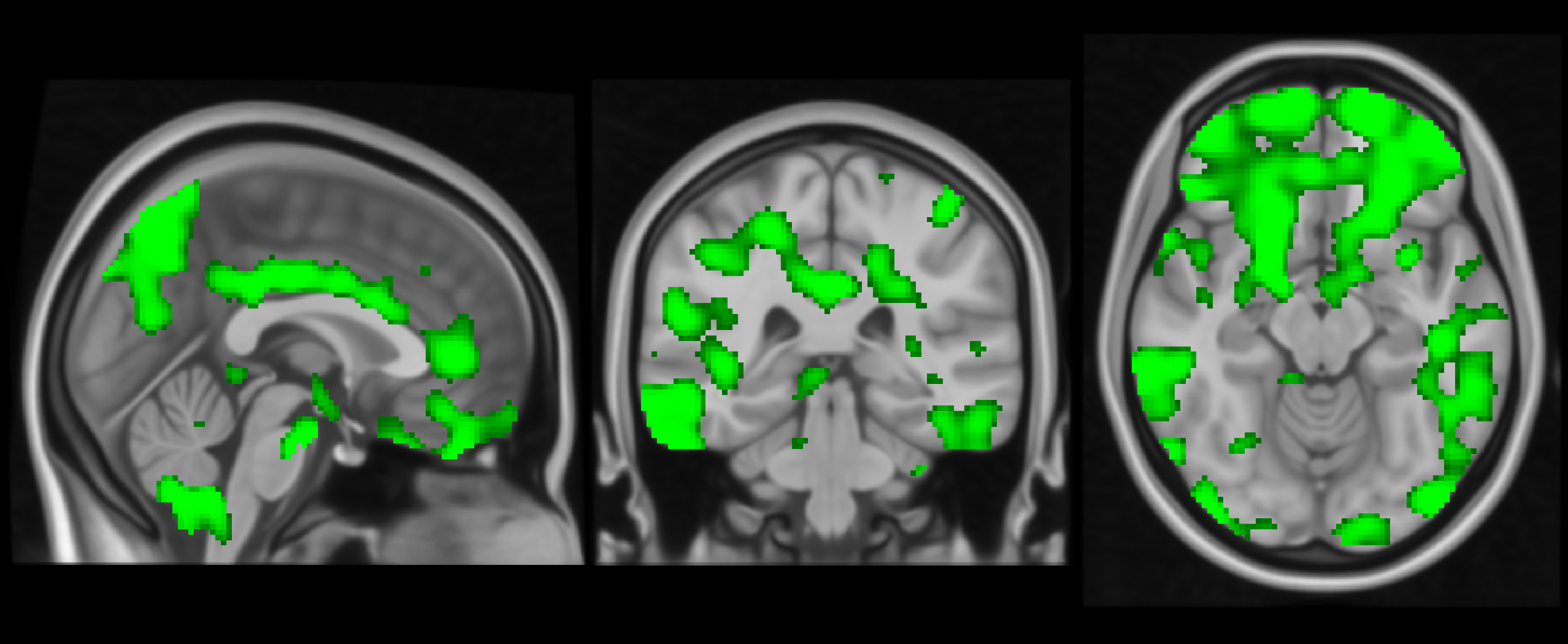
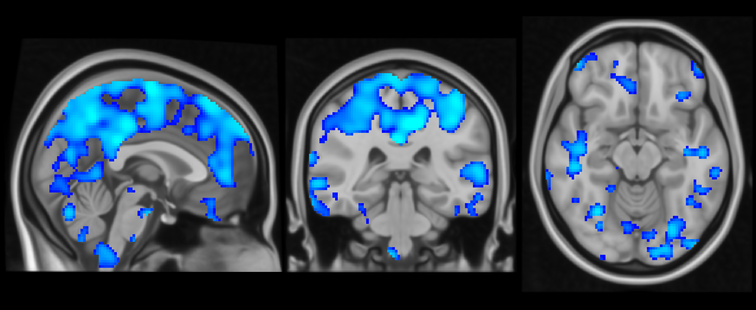
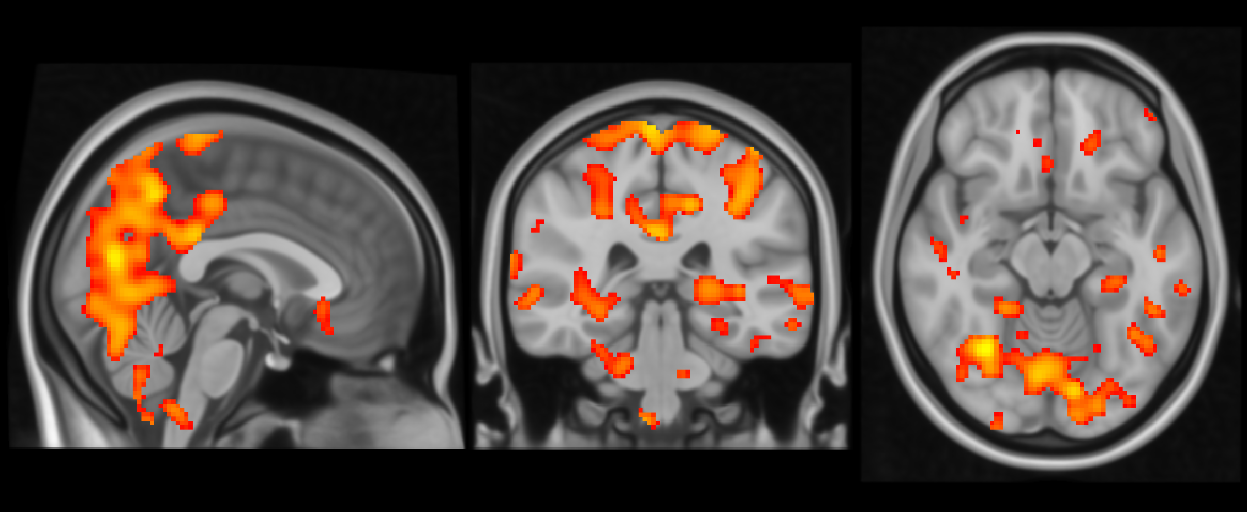
Diastolic pressure reduction diminishes vasomotor and cardiovascular pulsation amplitudes. For vasomotor waves, involved areas are the prefrontal cortex, parietal lobes, cingulate gyrus, occipital lobes, and anterior insular cortex. Cardiovascular pulsations implicate the cingulate cortex, insula, prefrontal cortex, and thalamus. Systolic pressure elevation increases cardiovascular pulsation amplitude, affecting the frontal lobes, temporal lobes, parietal lobes, occipital lobes, and thalamus.Discussion
Reductions in diastolic pressure may lead to diminished vasomotor and cardiovascular pulsation amplitudes, reflecting increased arterial stiffness [7]. This potentially hampers the characteristic arterial pulsatility, resulting in decreased perfusion and heightened resistance that could constrain blood flow and trigger vasoconstriction [8], limiting pulsation amplitude during diastole.Conversely, increased systolic pressure enhances cardiovascular pulsation amplitudes and this could be due to the augmented pulsatile stress during the cardiac cycle's peak, and can possibly improve cerebral perfusion and enhance pulsation transmission into the cerebral microvasculature [9].
Alterations in both vasomotor and cardiovascular pulsations indicate a complex interaction between systemic blood pressure and brain hemodynamics, with potential implications for cerebral circulation and glymphatic function, possibly affecting neurodegenerative progression [10,11].
Conclusion
The findings indicate both vasomotor and cardiovascular pulsations are sensitive to blood pressure changes, indicating a complex interplay between systemic cardiovascular dynamics and cerebral hemodynamics. These changes could influence the progression of neurodegenerative processes through effects on the cerebral circulation and glymphatic system.Acknowledgements
The authors wish to acknowledge CSC – IT Center for Science, Finland, for computational resources.References
1. Lee CJ, Lee JY, Han K, Kim DH, Cho H, Kim KJ, et al. Blood Pressure Levels and Risks of Dementia: a Nationwide Study of 4.5 Million People. Hypertension. 2022 Jan;79(1):218–29.
2. Raitamaa L, Huotari N, Korhonen V, Helakari H, Koivula A, Kananen J, et al. Spectral analysis of physiological brain pulsations affecting the BOLD signal. Human Brain Mapping. 2021;42(13):4298–313.
3. Zahneisen B, Hugger T, Lee KJ, LeVan P, Reisert M, Lee HL, et al. Single shot concentric shells trajectories for ultra fast fMRI. Magn Reson Med. 2012 Aug;68(2):484–94.
4. Jenkinson M, Beckmann CF, Behrens TEJ, Woolrich MW, Smith SM. FSL. NeuroImage. 2012 Aug 15;62(2):782–90.
5. Yu-Feng Z, Yong H, Chao-Zhe Z, Qing-Jiu C, Man-Qiu S, Meng L, et al. Altered baseline brain activity in children with ADHD revealed by resting-state functional MRI. Brain and Development. 2007 Mar;29(2):83–91.
6. Lohmann G, Stelzer J, Lacosse E, Kumar VJ, Mueller K, Kuehn E, et al. LISA improves statistical analysis for fMRI. Nature Communications. 2018 Dec;9(1).
7. Webb AJS. Progression of Arterial Stiffness is Associated With Midlife Diastolic Blood Pressure and Transition to Late‐Life Hypertensive Phenotypes. JAHA. 2020 Jan 7;9(1):e014547.
8. Chen Y, Shen F, Liu J, Yang GY. Arterial stiffness and stroke: de-stiffening strategy, a therapeutic target for stroke. Stroke Vasc Neurol. 2017 Jun;2(2):65–72.
9. De Montgolfier O, Pinçon A, Pouliot P, Gillis MA, Bishop J, Sled JG, et al. High Systolic Blood Pressure Induces Cerebral Microvascular Endothelial Dysfunction, Neurovascular Unit Damage, and Cognitive Decline in Mice. Hypertension. 2019 Jan;73(1):217–28.
10. Iliff JJ, Wang M, Zeppenfeld DM, Venkataraman A, Plog BA, Liao Y, et al. Cerebral Arterial Pulsation Drives Paravascular CSF–Interstitial Fluid Exchange in the Murine Brain. J Neurosci. 2013 Nov 13;33(46):18190–9.
11. Van Veluw SJ, Hou SS, Calvo-Rodriguez M, Arbel-Ornath M, Snyder AC, Frosch MP, et al. Vasomotion as a Driving Force for Paravascular Clearance in the Awake Mouse Brain. Neuron. 2020 Feb;105(3):549-561.e5.
Figures