3448
Autonomic Regression in fMRI during Alertness: Insights from Respiration, Pupil Size, and PPG Amplitude1Institute of Biomedical Engineering, Boğaziçi University, Istanbul, Turkey, 2Department of Radiology, Acibadem University, Istanbul, Turkey, 3Boğaziçi University, Istanbul, Turkey
Synopsis
Keywords: fMRI Analysis, fMRI, physiology, regression, task
Motivation: Understanding autonomic regulation during a cognitive task is vital for unraveling its neural mechanisms. This study delves into autonomic regression in fMRI, examining the roles of respiration, pupil size, and PPG amplitude during alertness.
Goal(s): Our goal is to enhance our understanding of the autonomic nervous system's response to alert conditions by analyzing fMRI data and modeling with various physiological signals.
Approach: Through the examination of respiration, pupil size, and PPG amplitude and fMRI, we aim to reveal patterns of autonomic regression.
Results: The results uncover significant associations between autonomic parameters and fMRI, shedding light on the neuro-physiological correlates of cognitive stress.
Impact: Our findings advance strategies for managing systemic variations during alert conditions, providing valuable insights for fMRI researchers.
Aim
Aim:To assess the impact of autonomic correction on spatiotemporal patterns in the relationship between fMRI and systemic signals, we employed PPG amplitude and pupil size as key indicators. Stress-induced hormonal responses heighten alertness and pulse rate, prompting our investigation into the potential consequences of removing autonomic and behavioral effects from fMRI data. We hypothesized that given the strong co-variation between autonomic signals and fMRI data, the elimination of these components may negatively affect outcomes. This research aims to evaluate the contribution of autonomic processes to spatiotemporal correlations and active brain regions during a cognitive task.Methods
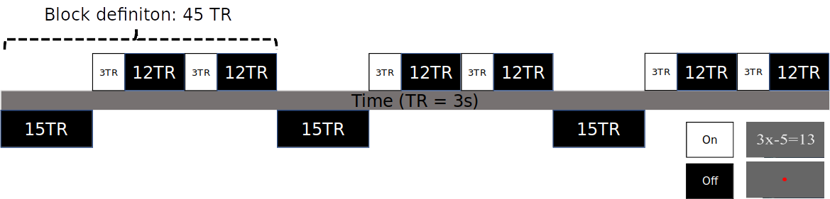
FMRI data were obtained at 3 T with gradient-echo-EPI (FA = 90, TR = 3 s, TE = 36 ms, in-place resolution = 2.5 mm, number of TRs = 135). Cognitive task was elicited by an arithmetic task requiring solving an equation with one unknown. Problems were displayed against a grey background with a fixation dot, repeatedly as shown in Fig. 1. Preprocessing of fMRI data followed the suggested ‘afni_proc’ pipeline (AFNI (1)), including removal of signal drifts, slice-timing correction, realignment of consecutive volumes, registration to MNI template, smoothing (3 mm full width at half maximum), and regression of motion parameters while removing outliers (threshold = 0.2). PPG and respiratory signals were collected with a pulse oximeter attached to the fingertip and respiratory bellows, respectively. PPG amplitude (PPG-AMP), as an index of peripheral vascular volume (2), and respiratory volumes per time (RVT) (3) were calculated. An MRI-compatible camera was used to track a subject’s eye movement. Pupil diameters were recorded automatically as a secondary measure of sympathetic activity. So far we acquired 3 subjects data, and performed the following analysis:Event-locked analysis: We averaged event-locked physiological signals (based on cardiac, respirotry and pupil size variations) and fMRI responses witin grey matter, task (e.g., Visual, IPS) and non-task (e.g., Insula) related regions. We performed voxel-wise correlations of PPG-AMP and pupil diameter with fMRI across subjects. General Linear Modelling (GLM). Each preprocessed fMRI data set will be subjected to General Linear Modelling (GLM). This part will include all timing information regarding mental task according to the experiment. The modeling step will be combined with regression of motion parameters and their derivatives (3dDeconvolve) in ‘afni_proc’. To incorporate further regressors of no interest, as described below, we will include each time-series in ‘afni_proc’, 3dDeconvolve function. Various methods have been developed to reduce the effects of cardiac and respiratory cycles in the fMRI data (4). Among them, we will use one of the most common approaches: RETROICOR (5). RETROICOR models cardiac and respiratory variations using a low-order Fourier series with time-varying cardiac and respiratory phases, which will be included in the GLM as nuisance regressors and removed, as implemented in AFNI's “RetroTS.m”. To evaluate the contribution of better captured autonomic processes on the correlation patterns, we will perform a regression analysis employing RETOICOR + RVT, PPG-AMP, and pupil diameter time-series, adding time-shifted versions. To our knowledge, this will be the first study incorporating PPG-AMP or pupil diameter as regressors during wake conditions in humans.
Results
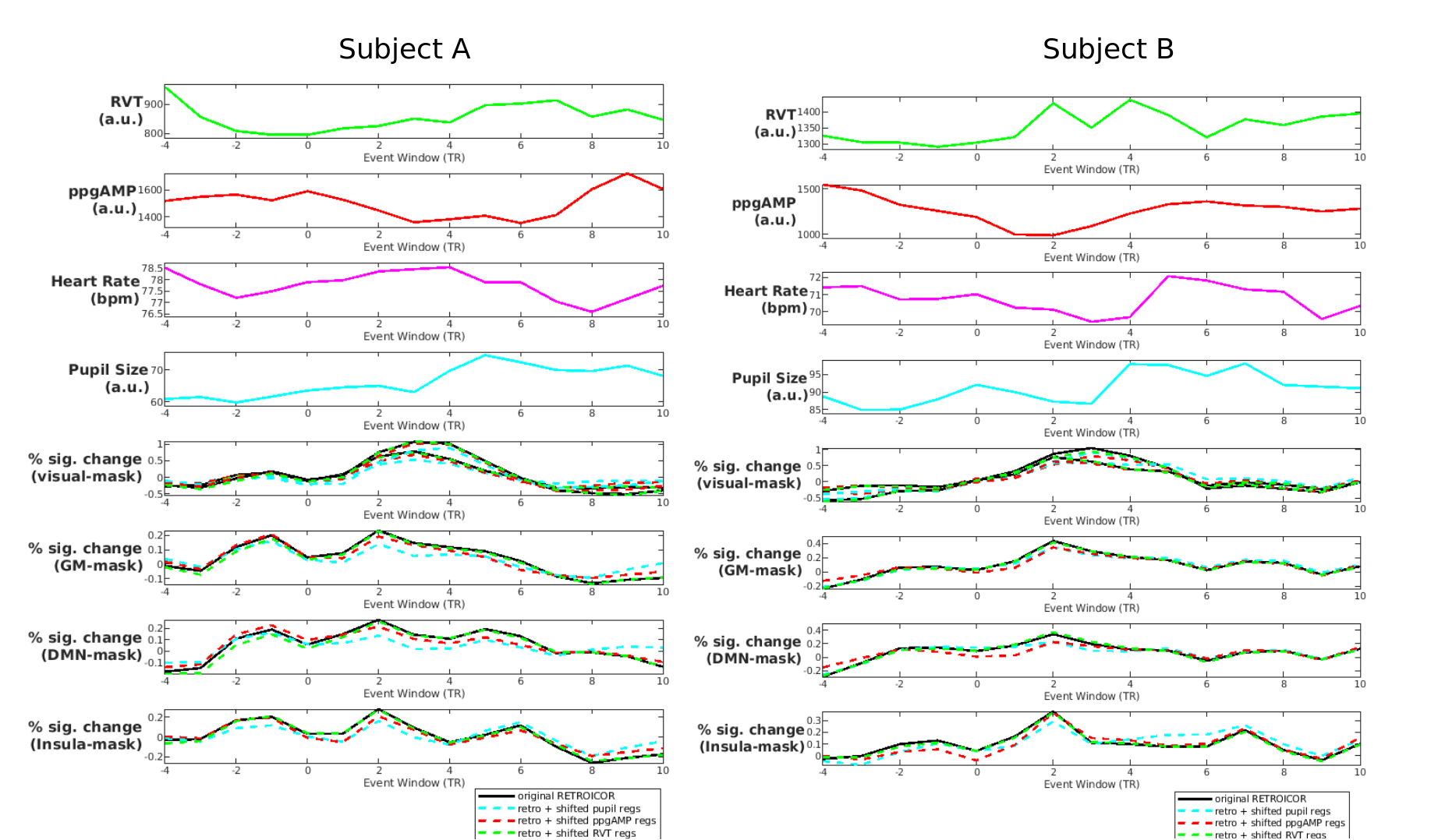
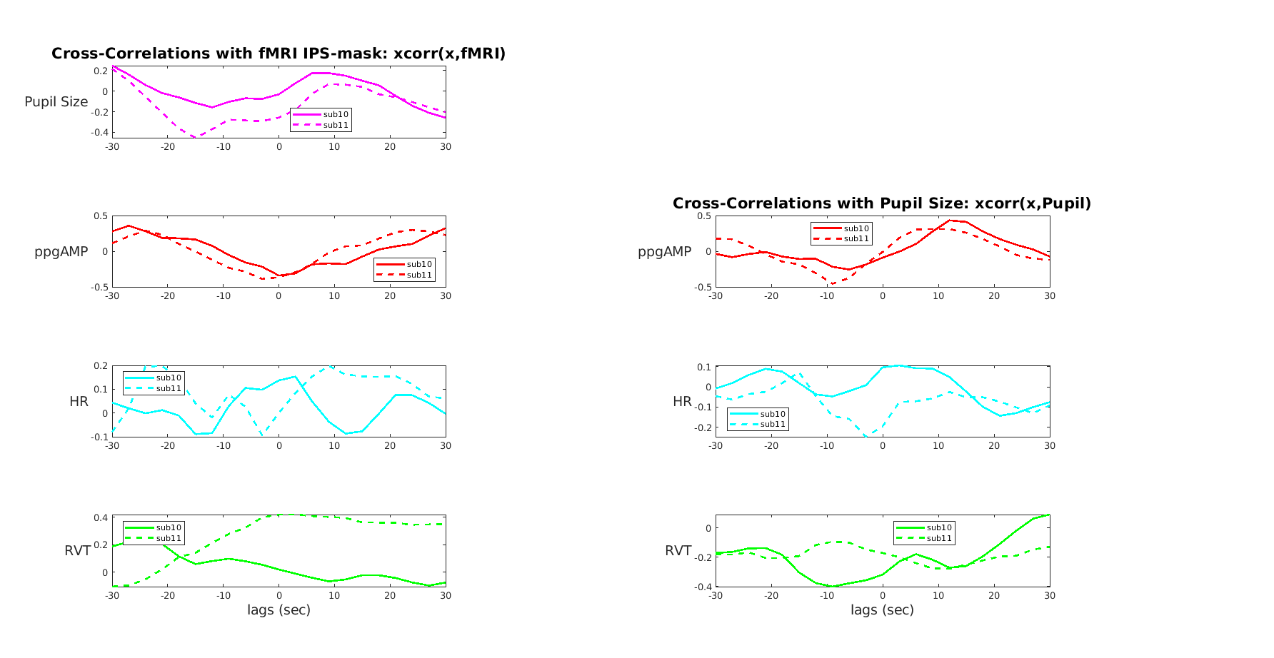
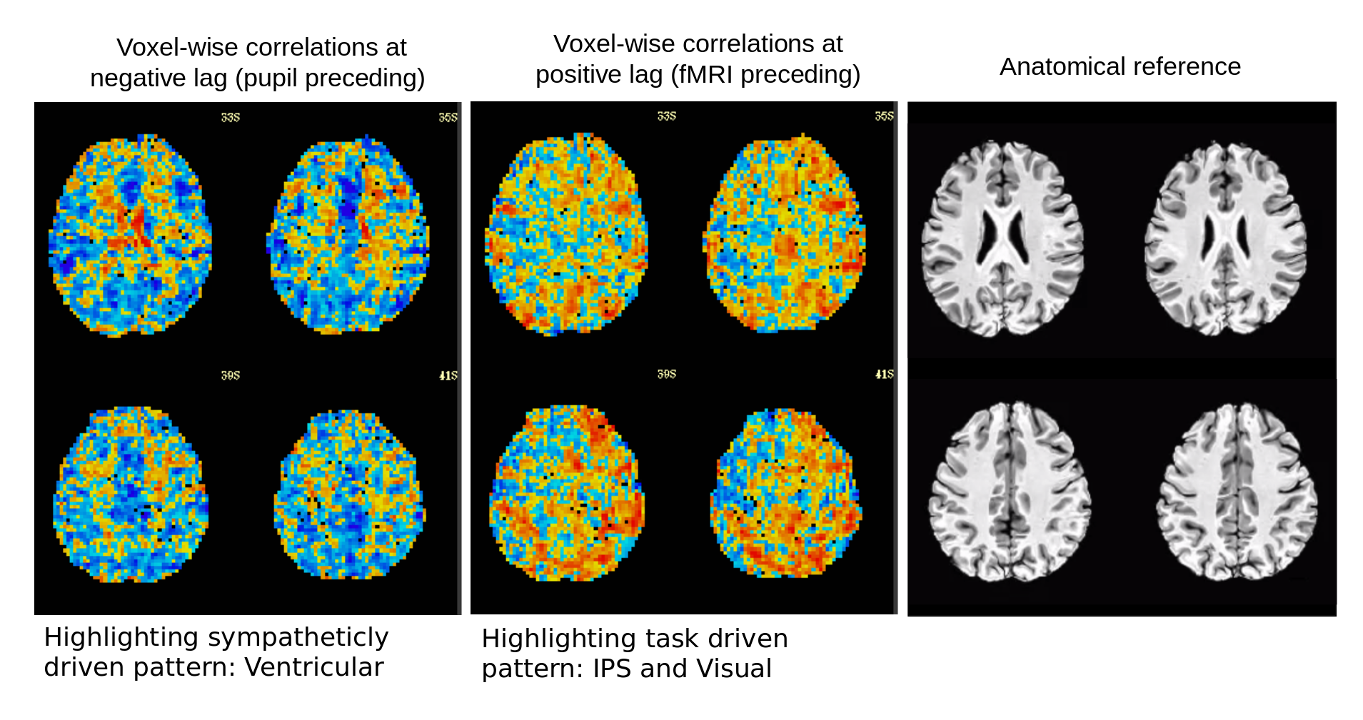
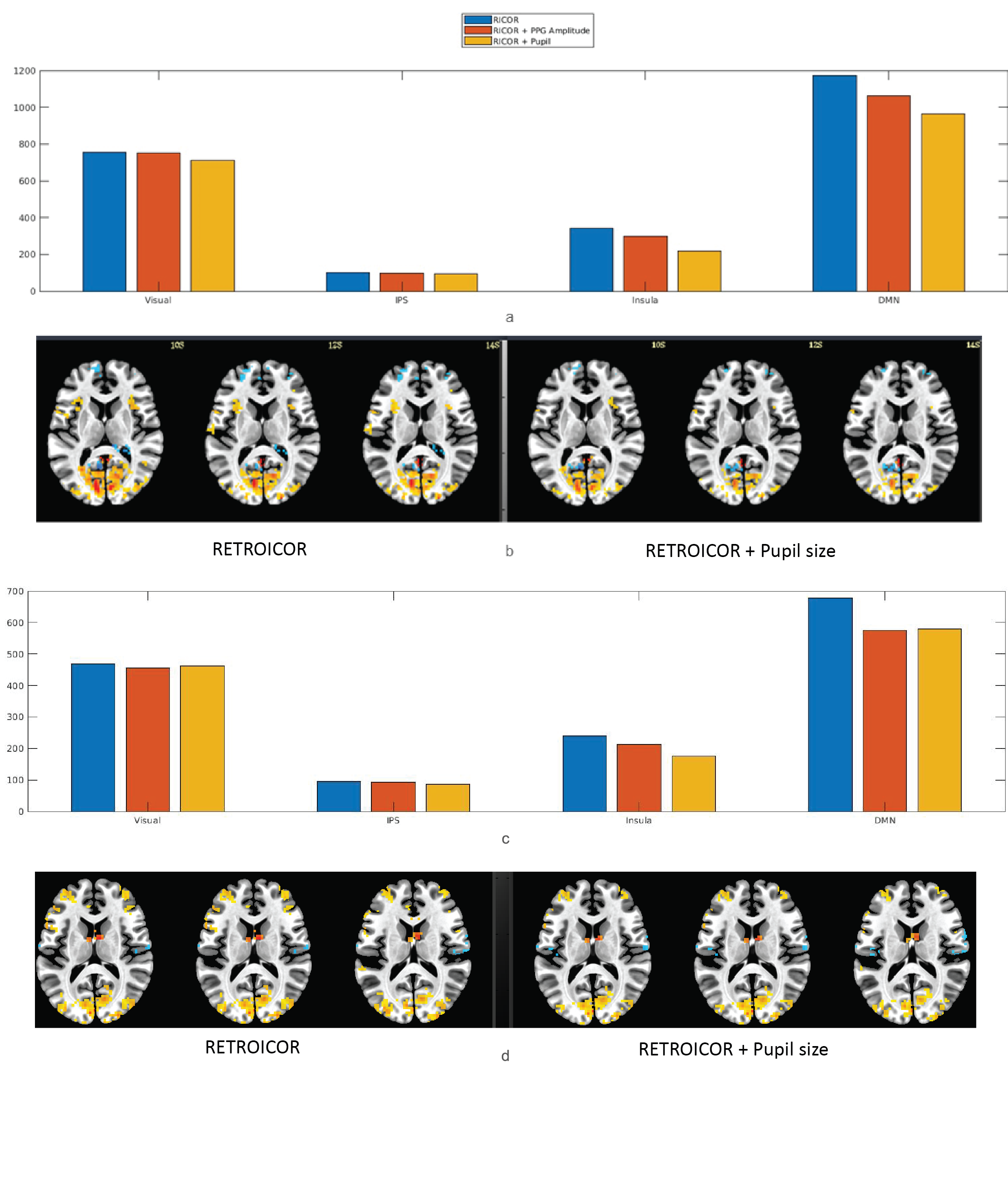
The event-locked plots revealed task-related increases in the fMRI signal, particularly in task-related areas like IPS and visual regions, peaking at 6-9 seconds (2-3TR) in accordance with the hemodynamic response function. Although variations in the contribution of other autonomic signals to the task correlation were observed across two subjects, for both subjects, pupil size increased after a 6- second (2TR) lag peaking around 12 seconds. Conversely, the negative correlation between pupil and fMRI observed in the negative lags of the cross-correlation plot was interpreted as the decrease in the global fMRI signal due to sympathetic activity associated with pupil dilation. This time dependent relationship between pupil size and fMRI signal was also illustrated in the cross-correlation maps revealing sympathetic-driven patterns (around ventricular regions) for negative lags and task-driven patterns around task-related regions (IPS and visual) for positive lags.Furthermore, comparison of activated voxel counts and GLM activity patterns of the fMRI data with and without the removal of time-shifted autonomic regressors revealed that the activity especially in DMN and insular region has reduced after the removal of pupil size and PPG-AMP. Negative activations were also reduced in ventricular regions with autonomic regression.
Conclusion
Our results revealed that contributions of the sympathetic activity to fMRI signal which could not be revealed with RVT, can be revealed with other autonomic signals like PPG-AMP and pupil size. Especially, the pupil size may also have additional advantages over PPG-AMP as well, which needs further demonstration.Acknowledgements
This research is supported by TUBITAK 2232 grant.References
1. Cox RW. AFNI: software for analysis and visualization of functional magnetic resonance neuroimages. Computers and biomedical research, an international journal. 1996;29(3):162-73.2. Özbay PS, Chang C, Picchioni D, Mandelkow H, Moehlman TM, Chappel-Farley MG, et al. Contribution of systemic vascular effects to fMRI activity in white matter. Neuroimage. 2018;176:541-9.3. Birn RM, Diamond JB, Smith MA, Bandettini PA. Separating respiratory-variation-related fluctuations from neuronal-activity-related fluctuations in fMRI. Neuroimage. 2006;31(4):1536-48.4. Caballero-Gaudes C, Reynolds RC. Methods for cleaning the BOLD fMRI signal. Neuroimage. 2017;154:128-49.5. Glover GH, Li TQ, Ress D. Image-based method for retrospective correction of physiological motion effects in fMRI: RETROICOR. Magn Reson Med. 2000;44(1):162-7.Figures