3447
Effects of Caffeine Intake on Brain, CSF, and Autonomic Signals: An EEG-fMRI Study1Institute of Biomedical Engineering, Boğaziçi University, Istanbul, Turkey, 2Department of Radiology, Acibadem University, Istanbul, Turkey
Synopsis
Keywords: fMRI Analysis, fMRI, multimodal imaging, physiology, caffeine
Motivation: Understanding caffeine's influence on the brain, CSF flow, and autonomic signals is essential. Recent research highlights the dynamic nature of these processes, creating a compelling need to investigate how caffeine impacts them.
Goal(s): This study aims to elucidate how caffeine, as a stimulant affecting sympathetic activity, affects brain activity, CSF flow, and autonomic signals.
Approach: Using an EEG-fMRI setup, we monitor individuals following caffeine intake to assess its effects on neural, CSF, and autonomic dynamics.
Results: The study's results transform our understanding of caffeine's impact, guiding scientists, clinicians, and patients to make right choices about caffeine consumption's effects on autonomic function, and health.
Impact: The results of this study can improve our understanding of caffeine's influence on neural and physiological processes, towards novel experiment design and analysis strategies.
Aim
This study seeks to explore the connections between caffeine intake, brain activity, cerebrospinal fluid (CSF) flow, autonomic signals during rest. We will analyze the correlations among specific brain regions, CSF flow patterns, autonomic signals, and EEG power in individuals post-caffeine consumption, emphasizing caffeine's distinct impact on these neural, physiological, and autonomic processes, providing insights into its influence on the "fight or flight" response. Through these objectives, we aim to enhance our understanding of the complex relationship between caffeine, brain function, and physiological dynamics.Methods
Resting state fMRI data were obtained at 3 T Siemens scanner with gradient-echo-EPI (FA = 90, TR = 3 s, TE = 36 ms, in-place resolution = 2.5 mm, number of TRs = 135). EEG data was collected simultaneously with 32 channel head coil (Brain Products). 4 subjects were included in the analyses, 2 of which underwent simultaneous EEG-fMRI acquisition. Repeated scans were performed following immediate intake of caffeine pills, first scan 10 minutes second scan 30 minutes following intake. The subjects were not informed if the pill was caffeine or placebo. Preprocessing of fMRI data followed the suggested ‘afni_proc’ pipeline (AFNI (1)), including removal of signal drifts, slice-timing correction, realignment of consecutive volumes, registration to MNI template, smoothing (3 mm full width at half maximum), and regression of motion parameters. PPG and respiratory signals were collected with a pulse oximeter attached to the fingertip and respiratory bellows, respectively. Respiratory volumes per time (RVT) (2) were calculated.Brain Products Analyzer's template approach was used to eliminate the MRI gradient artifact in the contaminated EEG data. This method required using average artifact subtraction (AAS) (3) before downsampling the data to 250 Hz. We semi-automatically identified R-peaks in QRS events in the ECG channel and then did an average template subtraction to remove cardioballistic artifacts. To further remove artifacts caused by eye blinks and muscular movement, independent component analysis (ICA) was applied across the channels. After initial cleaning, the EEG data was subjected to a band-pass filter with a frequency range of 0.2-35 Hz, and Fz was used as a reference channel. To investigate lag dependent correlations, we used various regions of interest, e.g., Insula, DMN, Visual, Motor, and calculated cross-correlations 1) among themselves, 2) with CSF signal (4th ventricle), and 3) EEG alpha power. Additionally we also perfomed heat maps based on min and max correlations.
Results
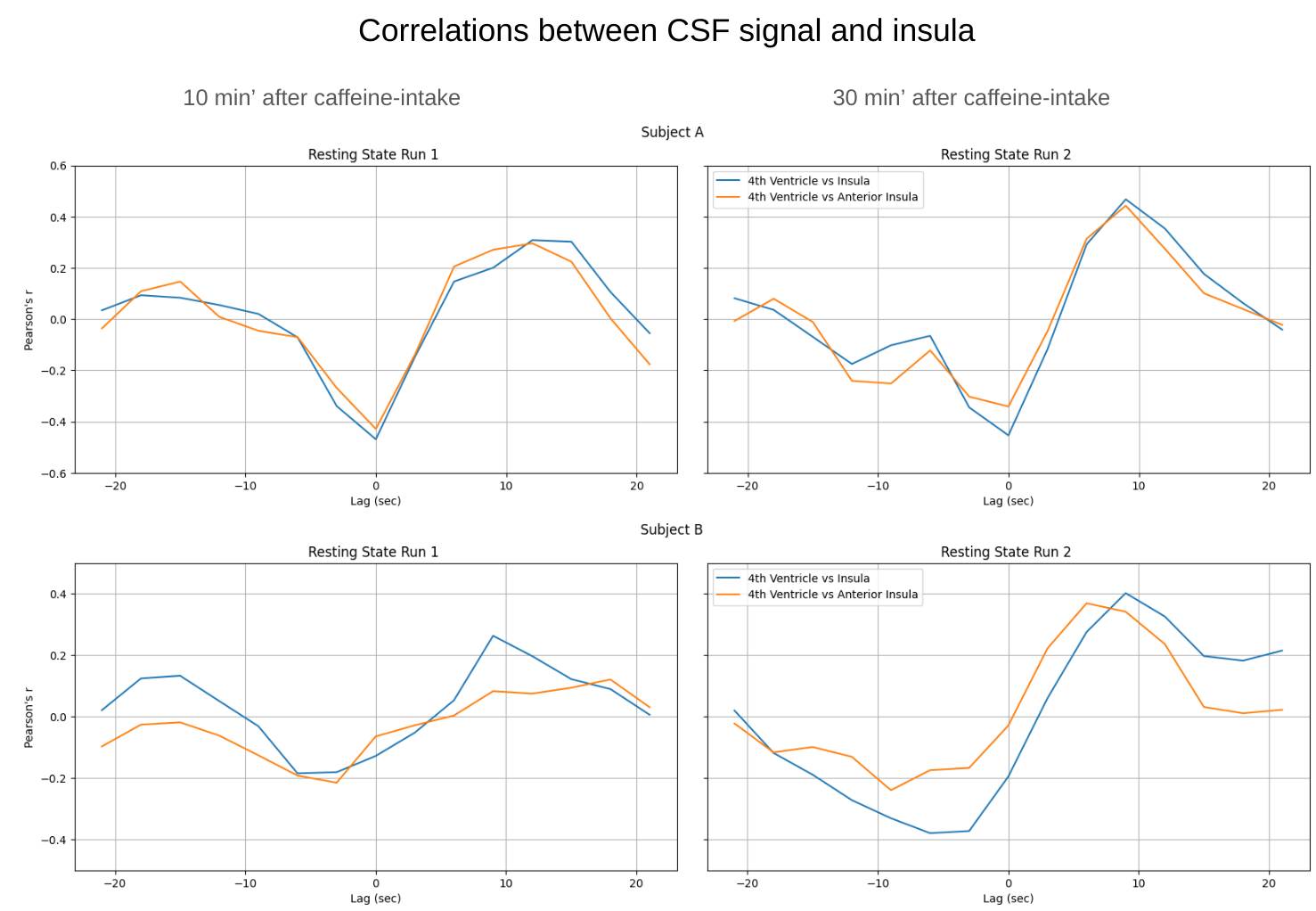
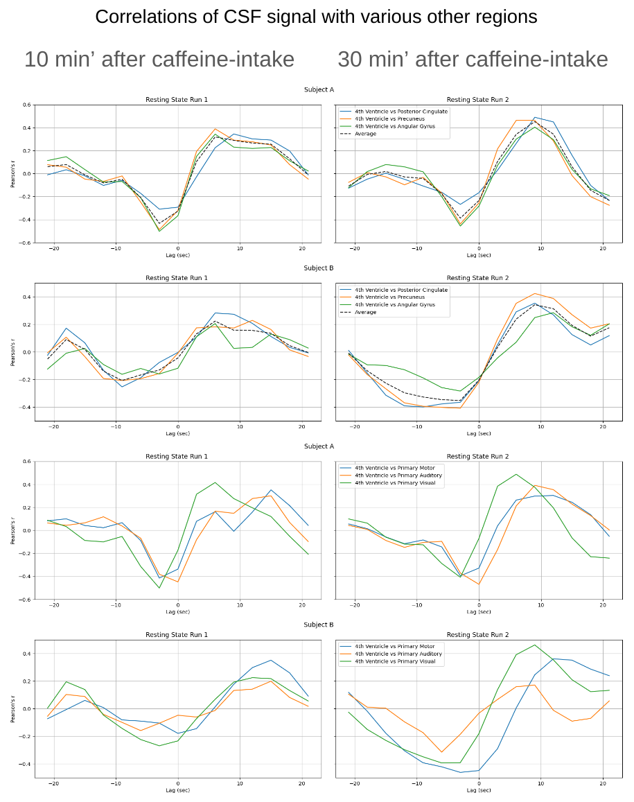
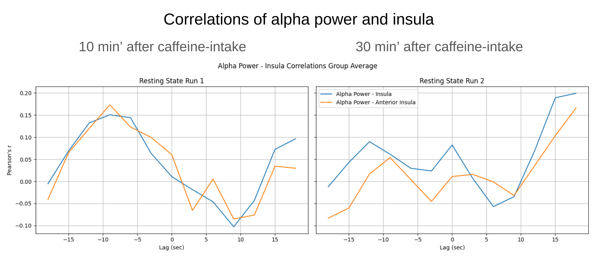
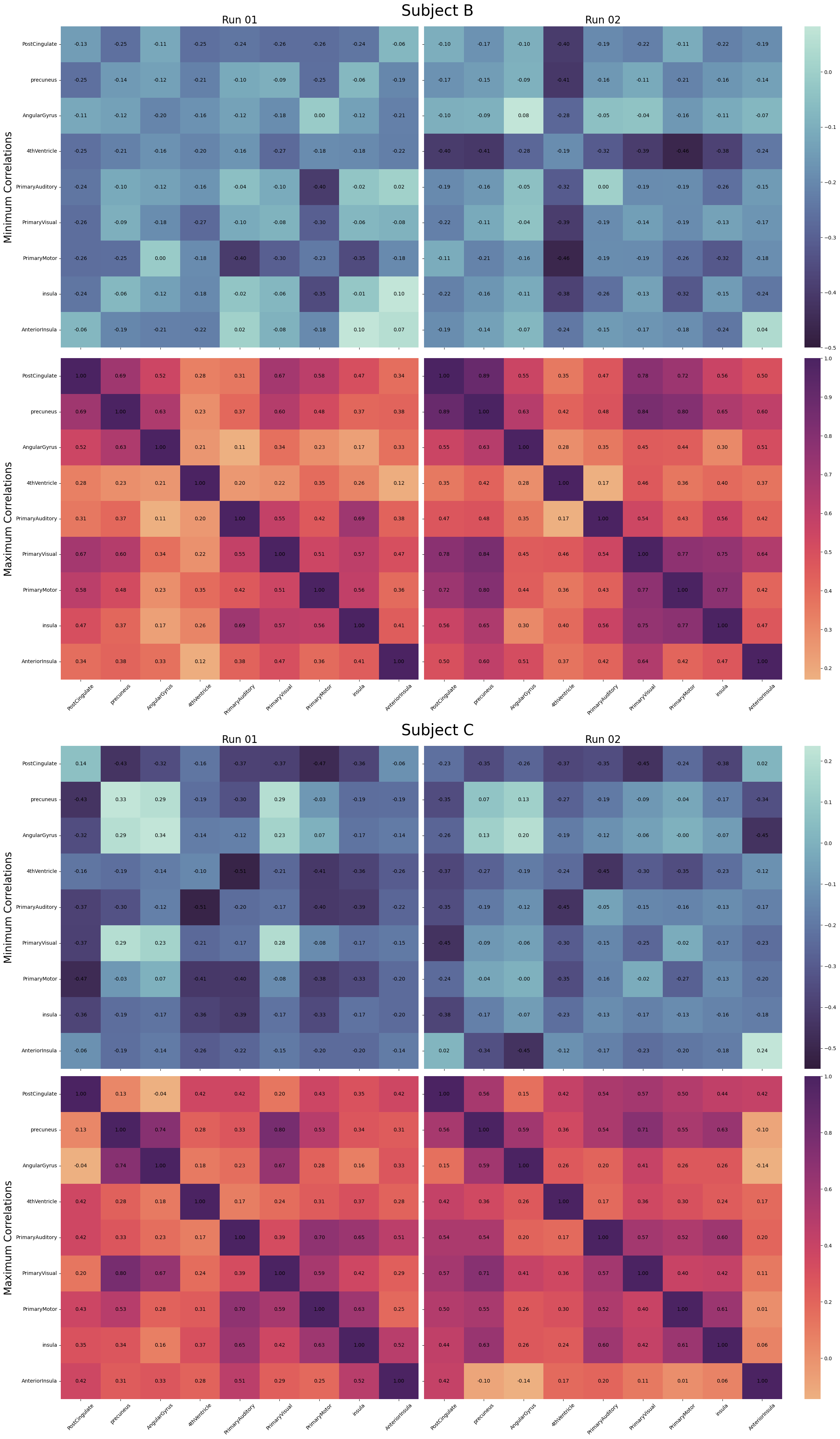
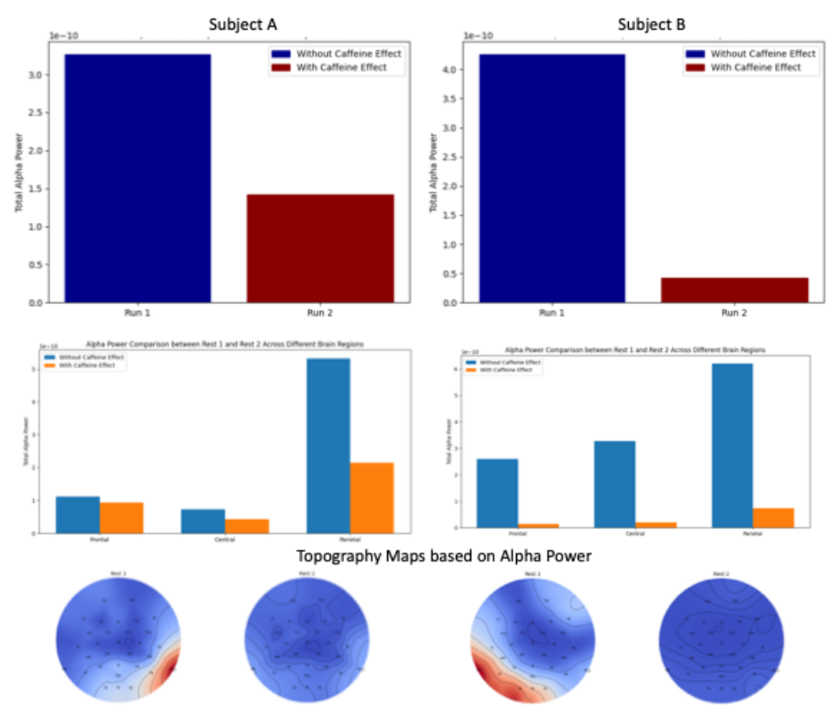
Following caffeine intake, our study observed a notable increase in the positive lag correlation between the 4th ventricle and the insula. Fig. 1 visually illustrates this effect with two subjects, where a positive lag indicates that the 4th ventricle signal lags behind the insula signal.The insula, known for its involvement in alertness, particularly the anterior insula as a component of the ventral attention network, displayed an increasing positive peak in our study, whether considering the entire insula or just the anterior portion. Expanding our analysis to include various brain regions, such as the default mode network (DMN) regions (posterior cingulate, precuneus, and angular gyrus) and primary visual, motor, and auditory cortices, we observed a general increase in positive peaks for all correlations between consecutive resting state scans (run 1-2). Notably, the positive peak of the primary visual cortex appeared to be the most influenced by caffeine, likely attributed to the subjects' eyes remaining open during the resting state scans. In some cases, we noted a negative correlation between CSF signal and brain regions, possibly linked to varying levels of drowsiness. To explore this further, we intend to expand our investigation with a larger subject sample. Correlation heatmaps in figure 4 shows for two subjects, the change of minimum and maximum values of the mentioned peaks for all regions of interest. A decrease is observed for CSF correlations of subject B while both subject B and C show an increase in positive peak in correlations of CSF with DMN and primary visual region as well as insula. Furthermore, in regions of the insula, correlations with the 4th ventricle were reduced. We observed an alpha power reduction following caffeine intake based on our EEG analysis (Fig. 3). Subjects A-B both showed decreased alpha band brainwave activity after caffeine intake, indicating heightened alertness and cognitive activation (Fig. 5). The central region was demonstrating the most significant change. Topographical maps confirmed a clear decrease in alpha wave activity in the central region.Conclusion
In conclusion, our findings suggest a multifaceted interplay between caffeine's stimulant effects on cerebrospinal fluid flow, increased sympathetic tone, and the resulting alterations in brain signals (Ozbay et al. 2019, Picchioni et al., 2020).Acknowledgements
This research is funded by TUBITAK 2232 grant.References
1. Cox RW, Computers and biomedical research, 1996.
2. Birn RM et al. Neuroimage, 2006
3. Allen, P. J. et al., NeuroImage 2000
4. Ozbay P et al., Neuroimage, 2019
4. Picchioni D et al., Neuroimage, 2020
Figures