3445
Sex Differences and Age-Related Decline in Absolute Cerebral Metabolic Rate of Oxygen (CMRO2) Consumption1Faculty of Health, Charles Darwin University, Darwin, Australia, 2Departments of Radiology and Clinical Neurosciences, University of Calgary, Calgary, AB, Canada, 3Department of Radiology, Medical College Wisconsin, Milwaukee, WI, United States, 4GE Healthcare, Calgary, AB, Canada, 5Hotchkiss Brain Institute, University of Calgary, Calgary, AB, Canada, 6Departments of Biomedical Engineering and Electrical & Software Engineering, University of Calgary, Calgary, AB, Canada
Synopsis
Keywords: fMRI Analysis, fMRI, cerebral metabolism, calibrated fMRI, aging, sex
Motivation: Resting CMRO2 is a marker of brain health that may inform typical and pathological brain aging. However, there is conflicting literature describing how CMRO2 changes across the lifespan, which may be influenced by extraneous variables such as sex and end-tidal partial pressure of carbon dioxide (PETCO2).
Goal(s): This study aimed to evaluate CMRO2 changes across the lifespan, after considering these possible confounding variables.
Approach: Dual-calibrated BOLD fMRI quantified grey matter absolute CMRO2 in 83 participants.
Results: Sex and age significantly predicted CMRO2. Females had higher CMRO2 than males, and CMRO2 decreased with increasing age for females.
Impact: Grey matter CMRO2 decreases in normal healthy aging. Considering both sexes, the CMRO2 decline rate was -0.88 per year, after accounting for PETCO2 and sex. When males and females were analysed separately, females only showed a significant decline with age.
Introduction
Understanding the typical aging process is essential for the early detection of age-related diseases such as dementia. Resting cerebral metabolic rate the oxygen (CMRO2) is a marker of energy utilisation and therefore an index of brain health. However, whether resting CMRO2 changes in typical aging remains a gap in the literature1. Numerous prior studies have investigated how the typical aging process affects resting CMRO2, with conflicting results reported. Increases in global CMRO2 have been reported with age using TRUST MRI2, although other work using this technique demonstrated no differences between older and younger adults3. This finding of no change is consistent with PET data indicating that CMRO2 remains stable with increasing age4, 5. Conflicting findings from PET and dual-calibrated BOLD fMRI however have suggested that CMRO2 decreases with increasing age6-8. Possible causes of these conflicting findings include differences due to biological sex and baseline PETCO2. Sex differences have been observed, with women having higher CMRO2 than men9. Differences in PETCO2 have also been described as significantly contributing to variations in oxygen extraction fraction (OEF), which is inherently related to CMRO210. This cross-sectional study aimed to characterise CMRO2 changes across the lifespan, accounting for sex and PETCO2. This was achieved using dual-calibrated BOLD for whole-brain quantification of absolute CMRO2 using hyperoxic and hypercapnic respiratory manipulations11-14, and a novel multiband multi-echo pseudo-continuous ASL (pCASL) fMRI sequence for simultaneous ASL and BOLD acquisition15, 16.Methods
Participants: 83 healthy adults (M = 39.9±14.6 years, range = 18-75 years, 47 F) participated in this study, which was approved by the Conjoint Health Research Ethics Board at the University of Calgary.MRI data acquisition: Data were collected on a 3T GE MRI (Discovery MR750) with a 32-channel head coil from Nova Medical. A 20-minute multiband multi-echo pCASL protocol was implemented with respiratory manipulations for the quantification of absolute CMRO2, TR/TE1/TE2 = 3028/10.5/29.8 ms, flip angle = 85°, multiband factor = 3, labelling duration = 1450 ms, PLD = 1000 ms, voxels = 3.5 x 3.5 x 4 mm with 1-mm slice gap, 400 volumes. A high-resolution structural 3D T1w BRAVO sequence (1mm3) and a 3D pCASL for resting cerebral blood flow (CBF) were also collected.
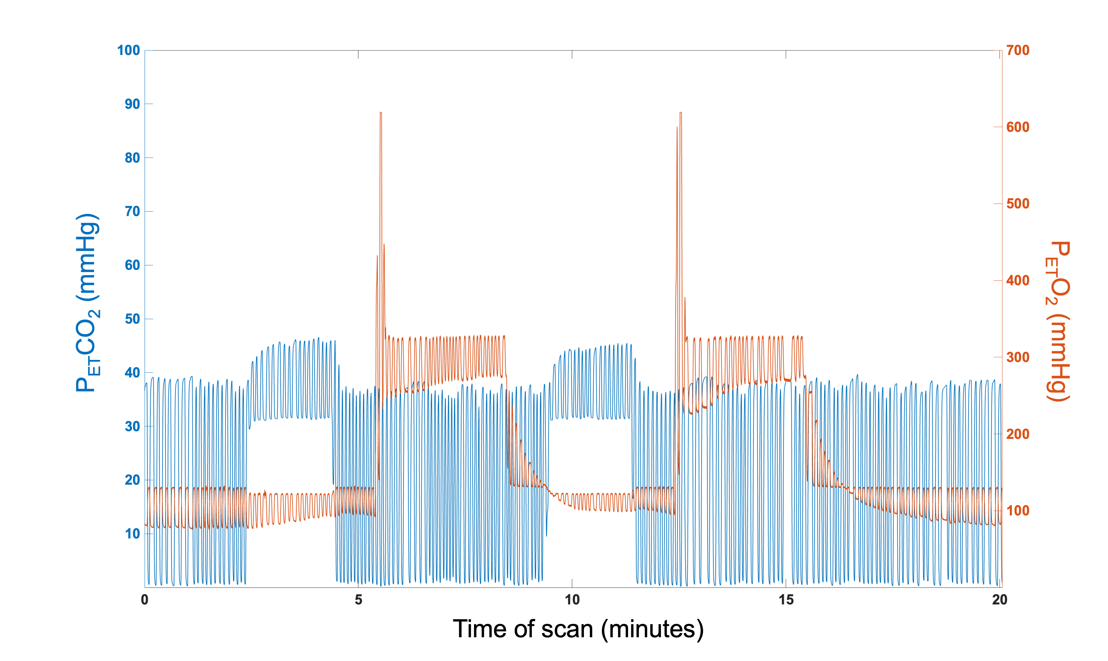
Respiratory manipulations: An MRI-compatible breathing circuit was used to achieve mild hypercapnia (5% CO2, 2x2-minutes) and hyperoxia (50% O2, 2x3-minutes), interleaved with medical air (normocapnia)12, 13, 17. The fixed-concentration automatic gas delivery system consisted of a Digital Flo-Box, Mass Flow Controllers (Sierra Instruments, Monterey, CA) and Gemini O2 and CO2 Monitor and Gas Analyzer (CWE Incorporated). Breathing traces (CO2 and O2) were continuously sampled. Figure 1 demonstrates an example breathing trace.
Data analysis: Motion correction and registration between CBF-weighted (echo 1), BOLD-weighted (echo 2) fMRIs and the T1W image was performed using MATLAB and Advanced Normalization Tools18 followed by spatial smoothing (5mm FWHM). Tissue segmentation of the T1w images was performed in SPM1219 with all subsequent analyses limited to grey matter. First-level fMRI analyses and quantification of CMRO2 were performed in FSL and MATLAB. Absolute CMRO2 was calculated using the calibration model11 which enabled the whole-brain quantification of OEF. CMRO2 was calculated from the product of OEF, CBF and CaO2 (arterial O2 content). CaO2 was inferred from end-tidal partial pressure of O2 measurements. Grey matter CMRO2 was extracted and averaged for each participant. Multiple regression analyses was performed in IBM-SPSS with predictor variables age, sex and PETCO2 and the outcome variable of mean grey matter CMRO2. Further regressions were performed on males and females separately.
Results
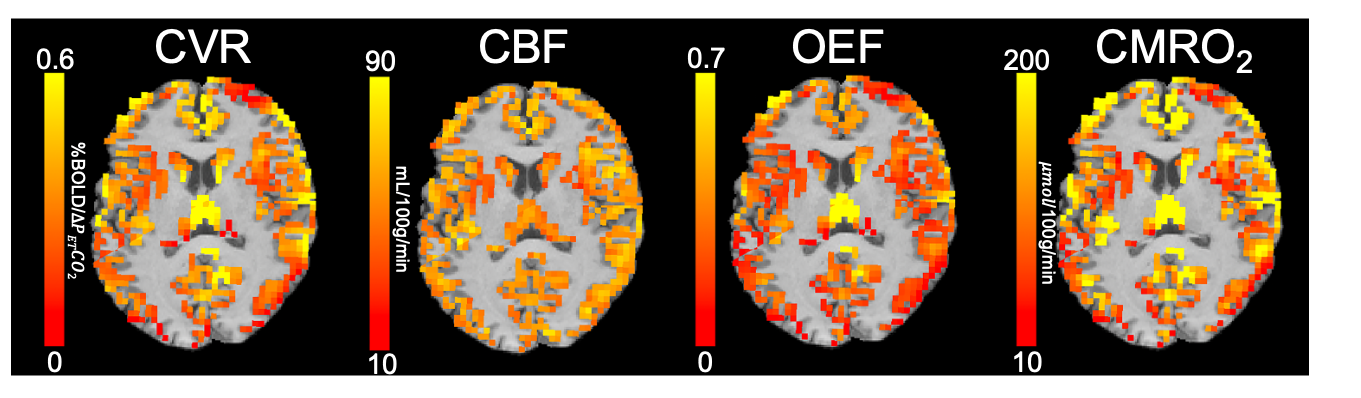
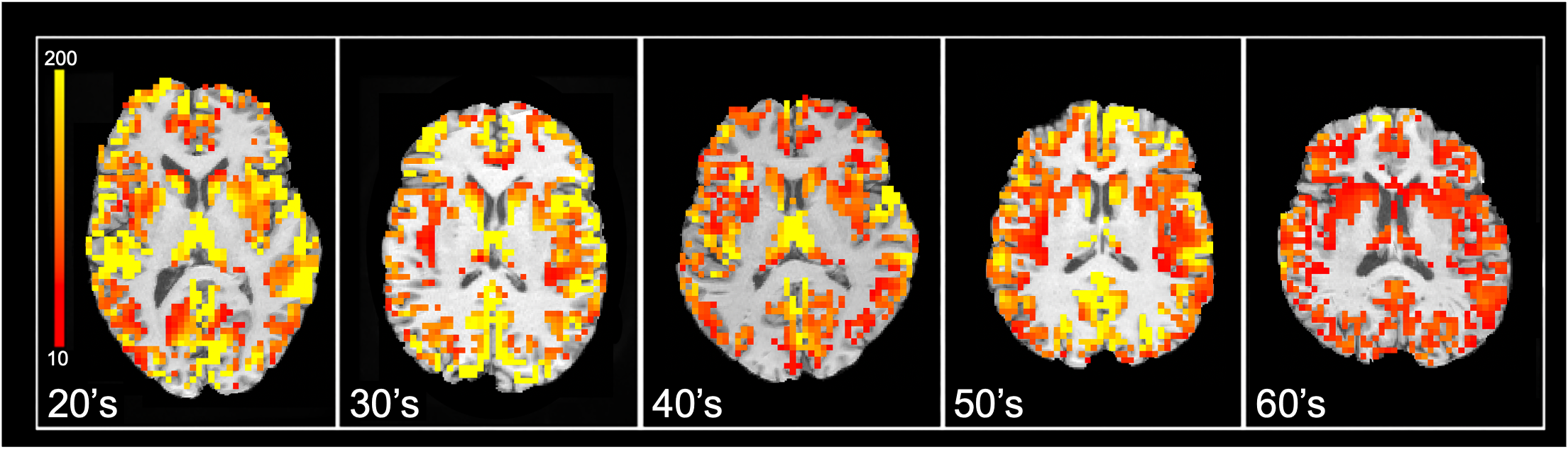
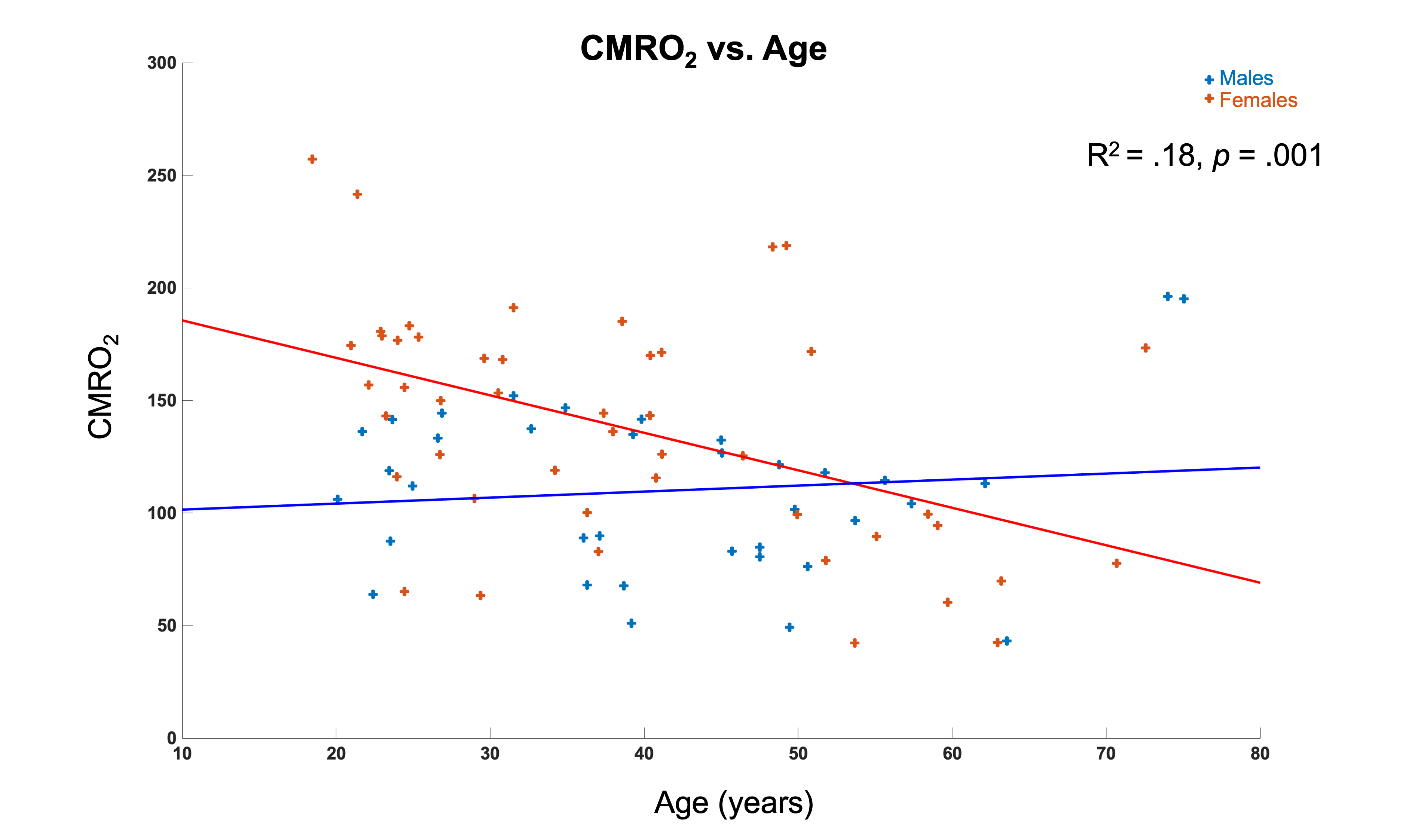
One outlier was identified but retained due to negligible effects. Figure 2 demonstrates maps calculated for CMRO2 estimation. Figure 3 shows exemplar individual CMRO2 images from participants in each decade of life. The multiple regression model was significant for the whole group, F(3, 79) = 5.83, p = .001, R2 = .18, Adjusted R2 = .15. Individual predictors age (p = .01) and sex (p = .005) significantly predicted CMRO2. Mean CMRO2 was lower in males (M = 110.0±36.6 ) than females (M = 138.1±51.5 ). Figure 4 shows the scatterplot of CMRO2 on age for both sexes. Separate regressions showed that CMRO2 decreased with age for females (p < .001) but not males (p = .48).Conclusion
This work supports prior findings from PET and calibrated BOLD5-7 demonstrating that CMRO2 decreases in typical aging. However, further examination of factors associated with dual-calibrated fMRI including optimal approaches for calculating OEF20 and CMRO2 changes during respiratory manipulations is required. Overall, these results suggest that absolute CMRO2 may represent an imaging biomarker associated with healthy and atypical aging.Acknowledgements
GBP acknowledges support from the Campus Alberta Innovates Chair program, the Canadian Institutes for Health Research (FDN-143290), and the Natural Sciences and Engineering Research Council (RGPIN-03880). MEM acknowledges support from Start-up funding at UCalgary and a Natural Sciences and Engineering Research Council Discovery Grant (RGPIN-03552) and Early Career Researcher Supplement (DGECR-00124).References
1. MacDonald ME and Pike GB. MRI of healthy brain aging: A review. NMR Biomed 2021; 34: e4564. 20210606. DOI: 10.1002/nbm.4564.
2. Lu H, Xu F, Rodrigue KM, et al. Alterations in cerebral metabolic rate and blood supply across the adult lifespan. Cerebral cortex (New York, NY: 1991) 2011; 21: 1426-1434. DOI: 10.1093/cercor/bhq224 [doi].
3. Catchlove SJ, Macpherson H, Hughes ME, et al. An investigation of cerebral oxygen utilization, blood flow and cognition in healthy aging. PLoS One 2018; 13: e0197055. 2018/05/22. DOI: 10.1371/journal.pone.0197055.
4. Pantano P, Baron JC, Lebrun-Grandié P, et al. Regional cerebral blood flow and oxygen consumption in human aging. Stroke 1984; 15: 635-641. DOI: 10.1161/01.str.15.4.635.
5. Aanerud J, Borghammer P, Rodell A, et al. Sex differences of human cortical blood flow and energy metabolism. J Cereb Blood Flow Metab 2017; 37: 2433-2440. 20160101. DOI: 10.1177/0271678X16668536.
6. Aanerud J, Borghammer P, Chakravarty MM, et al. Brain energy metabolism and blood flow differences in healthy aging. J Cereb Blood Flow Metab 2012; 32: 1177-1187. 20120229. DOI: 10.1038/jcbfm.2012.18.
7. Leenders KL, Perani D, Lammertsma AA, et al. Cerebral blood flow, blood volume and oxygen utilization. Normal values and effect of age. Brain 1990; 113 ( Pt 1): 27-47. DOI: 10.1093/brain/113.1.27.
8. De Vis JB, Hendrikse J, Bhogal A, et al. Age-related changes in brain hemodynamics; A calibrated MRI study. Hum Brain Mapp 2015; 36: 3973-3987. 2015/07/14. DOI: 10.1002/hbm.22891.
9. Lu H, Xu F, Rodrigue KM, et al. Alterations in cerebral metabolic rate and blood supply across the adult lifespan. Cereb Cortex 2011; 21: 1426-1434. 20101104. DOI: 10.1093/cercor/bhq224.
10. Jiang D, Lin Z, Liu P, et al. Normal variations in brain oxygen extraction fraction are partly attributed to differences in end-tidal CO. J Cereb Blood Flow Metab 2020; 40: 1492-1500. 20190805. DOI: 10.1177/0271678X19867154.
11. Wise RG, Harris AD, Stone AJ, et al. Measurement of OEF and absolute CMRO2: MRI-based methods using interleaved and combined hypercapnia and hyperoxia. Neuroimage 2013; 83: 135-147. 2013/06/13. DOI: 10.1016/j.neuroimage.2013.06.008.
12. Bulte DP, Kelly M, Germuska M, et al. Quantitative measurement of cerebral physiology using respiratory-calibrated MRI. Neuroimage 2012; 60: 582-591. 2011/12/22. DOI: 10.1016/j.neuroimage.2011.12.017.
13. Germuska M and Wise RG. Calibrated fMRI for mapping absolute CMRO. Neuroimage 2019; 187: 145-153. 20180329. DOI: 10.1016/j.neuroimage.2018.03.068.
14. Gauthier CJ, Desjardins-Crépeau L, Madjar C, et al. Absolute quantification of resting oxygen metabolism and metabolic reactivity during functional activation using QUO2 MRI. Neuroimage 2012; 63: 1353-1363. 2012/08/16. DOI: 10.1016/j.neuroimage.2012.07.065.
15. Cohen AD, Nencka AS and Wang Y. Multiband multi-echo simultaneous ASL/BOLD for task-induced functional MRI. PLoS One 2018; 13: e0190427. 20180201. DOI: 10.1371/journal.pone.0190427.
16. Cohen AD, Nencka AS, Lebel RM, et al. Multiband multi-echo imaging of simultaneous oxygenation and flow timeseries for resting state connectivity. PLoS One 2017; 12: e0169253. 20170302. DOI: 10.1371/journal.pone.0169253.
17. Merola A, Germuska MA, Murphy K, et al. Assessing the repeatability of absolute CMRO. Neuroimage 2018; 173: 113-126. 20180214. DOI: 10.1016/j.neuroimage.2018.02.020.
18. Avants BB, Tustison NJ, Song G, et al. A reproducible evaluation of ANTs similarity metric performance in brain image registration. Neuroimage 2011; 54: 2033-2044. 2010/09/17. DOI: 10.1016/j.neuroimage.2010.09.025.
19. Ashburner J and Friston KJ. Unified segmentation. NeuroImage 2005; 26: 839-851. DOI: 10.1016/j.neuroimage.2005.02.018.
20. Germuska M, Merola A, Murphy K, et al. A forward modelling approach for the estimation of oxygen extraction fraction by calibrated fMRI. Neuroimage 2016; 139: 313-323. 20160606. DOI: 10.1016/j.neuroimage.2016.06.004.
Figures