3438
Dual-FOV MB-SWIFT for simultaneous functional imaging of the brain and spinal cord1Center for Magnetic Resonance Research, Department of Radiology, University of Minnesota, Minneapolis, MN, United States, 2A. I. Virtanen Institute for Molecular Sciences, University of Eastern Finland, Kuopio, Finland
Synopsis
Keywords: Functional Connectivity, fMRI, dual FOV
Motivation: Functional neuroimaging target either the brain or the spinal cord, but usually not both because simultaneous acquisitions from distant fields of view are challenging with conventional MRI.
Goal(s): To establish a novel MRI approach for artefact-free, quiet fMRI simultaneously from brain and spinal cord, avoiding the need of dynamic shimming.
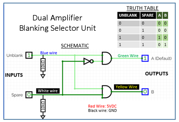
Approach: Our approach is to use MB-SWIFT in a simultaneous dual-FOV configuration with a Dual Amplifier Blanking Selector Unit (DABSU) that employs two transmitters and two receiver channels.
Results: The results established feasibility of the novel approach for task-based fMRI and connectivity analyses between lumbar spinal cord and brain in rat.
Impact: The method could be used for unprecedented investigations of the central nervous system, and could be extended to any two separate parts of the body.
Introduction
Developing novel strategies that enable simultaneous acquisitions of distant sites of the central nervous system (CNS) is critically needed to study CNS networks beyond the brain. Targeting two distant FOVs is challenging with conventional MRI, primarily due to the difficulties of reaching adequate homogeneity of the magnetic field. Towards solving the challenges of CNS studies, we exploited a zero-echo time MRI approach, namely a 3D MRI technique entitled Multi-Band SWeep Imaging with Fourier Transformation (MB-SWIFT) [1, 2]. We previously demonstrated the feasibility of MB-SWIFT for detecting brain activation in human studies [3], and proved its technical benefits for studying the function of the rat brain [4-6] and spinal cord [7] separately. Our hypothesis is that, without the need of dedicated shimming solutions, MB-SWIFT can adequately image two different FOVs simultaneously at distant CNS locations, i.e., brain and lumbar spinal cord.Method
Two coils with 2 cm diameters were used, the first covering the rat brain, and the second positioned on the L1-L2 level of the rat’s spinal cord at 9.4T. A Dual Amplifier Blanking Selector Unit (DABSU) was developed with two transmitter and two receiver channels to excite and collect signal responses from two FOVs (Figure 1). With DABSU, a ‘blanking on’ signal could be given to 2 RF independent amplifiers. For the power calibrations, we developed and tested an automatic power calibration procedure, which operates sequentially for both channels using spin-echo pulse sequence with volume selection.Eight male Sprague–Dawley rats aged 3–5 months were studied. Rats were anesthetized with isoflurane (5% induction, 2.0–3.5% maintenance; carrier gas O2/N2 30/70 or combination of isoflurane (0.2-0.5%) and medetomidine (0.04 mg/kg/h, s.c.), in connectivity and stimulation studies, respectively. MRI scans were conducted with a 9.4T equipped with Agilent DirectDRIVE console (Palo Alto, CA, USA) using a modified version of MB-SWIFT [2] to enable simultaneous dual-FOV acquisitions. In 5 rats, we conducted rs-fMRI with 300 volumes (8 volumes for dummy scan) for total scan time = 15 min 14 s, temporal resolution of 3 s. A 1547 spokes/FOV were acquired in an interleaved fashion. The other parameters were: TR = 0.97 ms, BW = 192 kHz, FOV = 353 mm3, and flip angle = 5°, sidebands = 32, OS = 2, Gap = 4. Images were first smoothed with FSLMATH with a gaussian kernel with 2 mm FWHM. For both brain and spinal cord, motion correction was performed using MCFLIRT [8] of the FSL toolbox. The brain and spinal cord images were concatenated for MELODIC 3.0 which decomposed the 4D data sets into different spatial and temporal independent components [9]. In 3 rats, we conducted fMRI during electrical hind paw stimulation, and compared MB-SWIFT with SE-EPI (TR = 2 s, TE= 35 ms, FOV = 36 x 36 mm2, 14 slices, slice thickness 1 mm). MB-SWIFT acquisition parameters were: FOV = 36 x 36 x 40 mm3, 2047 spokes/FOV, temporal resolution of 4 s, flip angle = 6°. Stimulation paradigm was 80 s rest followed by 40 s stimulation (8 Hz, 1.8 mA) repeated 5 times ending in 80 s rest in MB-SWIFT, and 40 s rest followed by 20 s stimulation (8 Hz, 1.9 mA), repeated 5 times and ending in 40 s rest in EPI.
Result
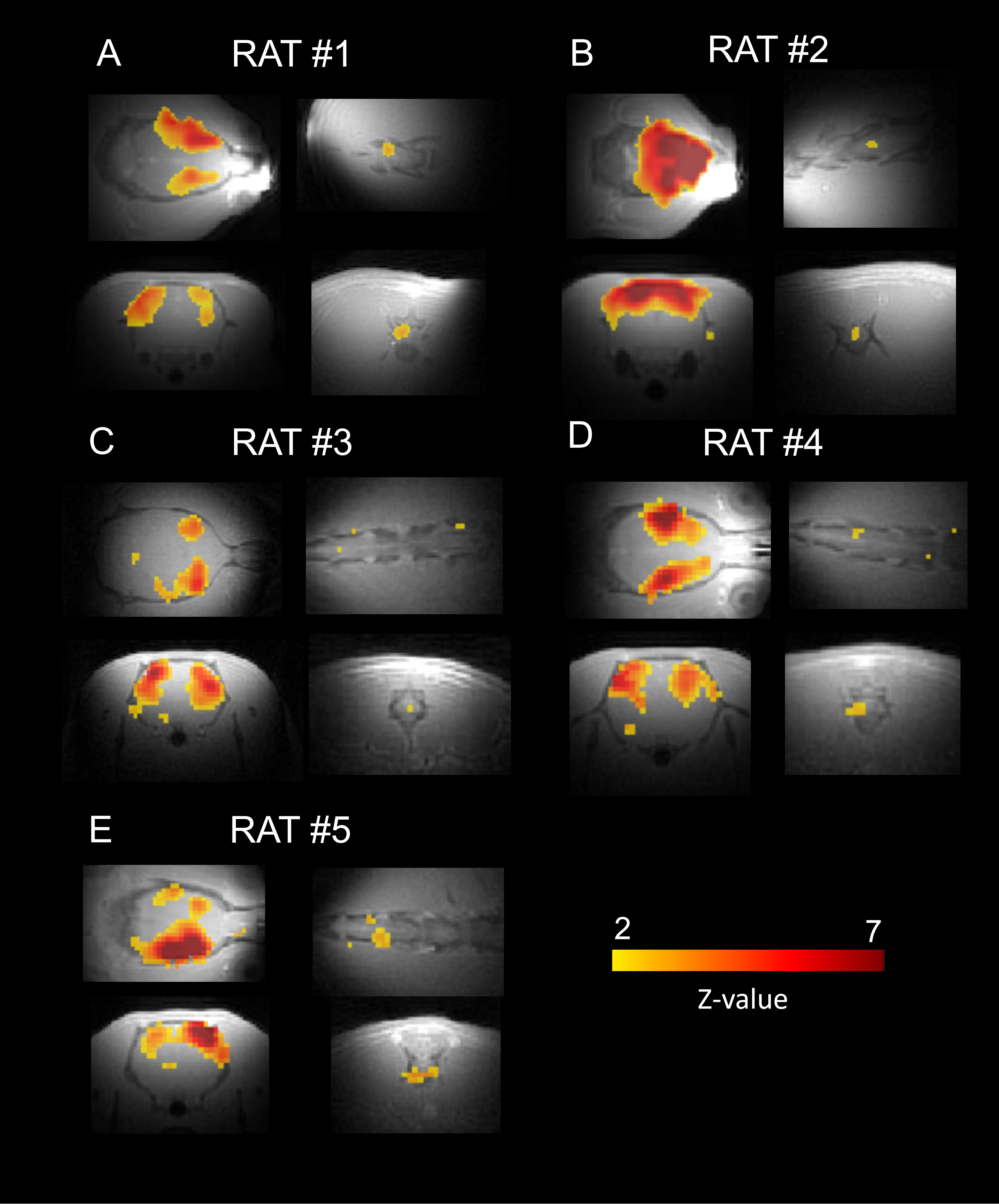
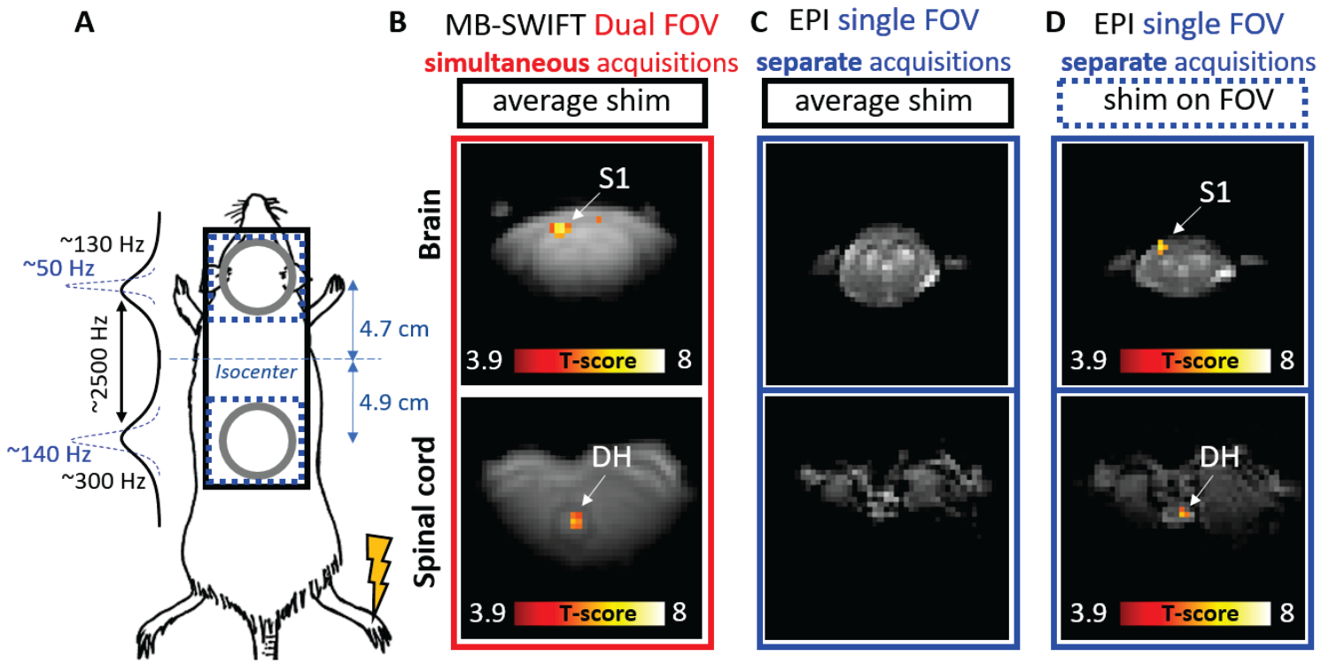
The independent component analysis (ICA) analysis of rs-fMRI data with dual-FOV MB-SWIFT shows consistent components in the somatosensory cortex connected with spinal cord around L1 level (Figure 2) in all 5 rats. In task-based fMRI, robust and specific activations were detected in both the brain and spinal cord during hind paw stimulation, whereas EPI failed to produce images of acceptable quality for fMRI analyses (Figure 3).Discussion
We have demonstrated that MB-SWIFT allows unprecedented fMRI studies of the CNS in distant sites of the brain and the spinal cord. The results of this work are also relevant to other zero or ultra-short echo time MRI approaches [10-13]. Our data prove the superiority of MB-SWIFT with dual-FOV capabilities vs EPI for CNS fMRI in the absence of dedicated shimming solutions.Conclusion
We developed and tested a novel approach of dual-FOV fMRI for studies of CNS networks. The approach involves the use of MB-SWIFT in conjunction with a DABSU to allow the excitation of two different FOVs using two coils independently from each other. The results demonstrate that MB-SWIFT enables unprecedented CNS functional imaging, and sets the stage for translation to human investigations.Acknowledgements
NIH grants R01NS129739, P41 EB027061, S10 OD032192, The Research Council of Finland 331955 (HL) and WM KECK Foundation.References
1. Idiyatullin, D., et al., Fast and quiet MRI using a swept radiofrequency. J Magn Reson, 2006. 181(2): p. 342-9.
2. Idiyatullin, D., C.A. Corum, and M. Garwood, Multi-Band-SWIFT. J Magn Reson, 2015. 251: p. 19-25.
3. Mangia, S., et al. Functional MRI with SWIFT. in 20th Scientific Meeting and Exhibition of the International Society for Magnetic Resonance in Medicine (ISMRM). 2012. Melbourne, VIC (Australia). 4. Lehto, L.J., et al., MB-SWIFT functional MRI during deep brain stimulation in rats. Neuroimage, 2017. 159: p. 443-448.
5. Paasonen, J., et al., Multi-band SWIFT enables quiet and artefact-free EEG-fMRI and awake fMRI studies in rat. Neuroimage, 2020. 206: p. 116338.
6. Paasonen, J., et al., Whole-brain studies of spontaneous behavior in head-fixed rats enabled by zero echo time MB-SWIFT fMRI. NeuroImage, 2022. In Press.
7. Laakso, H., et al., Spinal cord fMRI with MB-SWIFT for assessing epidural spinal cord stimulation in rats. Magn Reson Med, 2021. 86(4): p. 2137-2145.
8. Jenkinson, M., et al., Improved optimization for the robust and accurate linear registration and motion correction of brain images. 2002. 17(2): p. 825-841.
9. Beckmann, C.F. and S.M.J.I.t.o.m.i. Smith, Probabilistic independent component analysis for functional magnetic resonance imaging. 2004. 23(2): p. 137-152.
10. Weiger, M. and K.P. Pruessmann, Short-T2 MRI: Principles and recent advances. Prog Nucl Magn Reson Spectrosc, 2019. 114-115: p. 237-270.
11. Weiger, M., et al., ZTE imaging in humans. Magn Reson Med, 2013. 70(2): p. 328-32.
12. Kobayashi, N., et al., Gradient-Modulated PETRA MRI. Tomography, 2015. 1(2): p. 85-90.
13. Grodzki, D.M., P.M. Jakob, and B. Heismann, Ultrashort echo time imaging using pointwise encoding time reduction with radial acquisition (PETRA). Magn Reson Med, 2012. 67(2): p. 510-518.
Figures