3437
Assessing motion-associated tSNR of brainstem and spinal cord fMRI in a cohort with chronic hemiparetic stroke1Northwestern University, Chicago, IL, United States
Synopsis
Keywords: Task/Intervention Based fMRI, Spinal Cord, brainstem
Motivation: Motor-task fMRI is a critical modality for studying post-stroke neuronal changes in the brainstem and spinal cord, but data quality is reduced in these regions.
Goal(s): We aimed to anticipate the degree of head-motion confounds before a motor-task fMRI scan and assess how this head motion affects fMRI data quality.
Approach: Six post-stroke individuals performed a hand-grasp task during a head motion-capture session, then during cortical-brainstem and spinal-cord fMRI scans.
Results: Head motion in the lab was positively correlated with head and spinal-cord motion during fMRI. Head and spinal-cord motion were correlated with decreased tSNR in the cortex, brainstem, and spinal cord.
Impact: Head motion outside of the scanner is linked with head and spinal-cord fMRI motion and tSNR. With lab-based motion capture systems, we can anticipate motion and tSNR impacts in motor-task fMRI, useful as screening in clinical populations with increased movement.
Introduction
Motor-task functional MRI (fMRI) is a critical modality to study changes in neuronal activity after a stroke1. In particular, fMRI of deep structures such as the brainstem and spinal cord is key to uncovering how neural activation changes not just in the cortex, but throughout the nervous system2. However, one challenge of motor-task fMRI in a cohort with stroke is that participants exhibit higher head motion3, which can decrease data quality. This problem is compounded in the brainstem and spinal cord, which have lower data quality even in healthy participants4,5. Therefore, we aimed to (1) test a method to anticipate the degree of head motion in post-stroke participants before an MRI scan, and (2) assess how head motion affects fMRI data quality in the brain, brainstem, and spinal cord.Methods
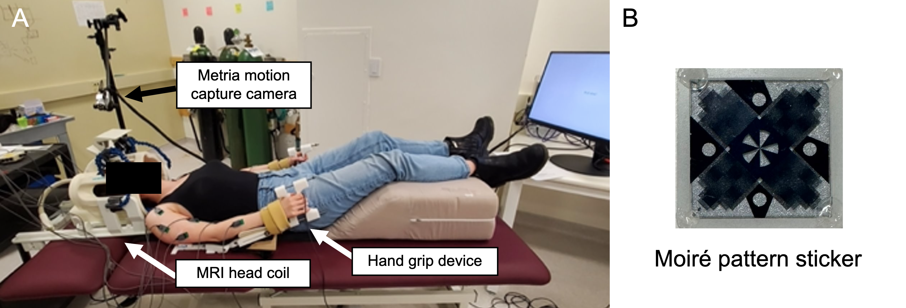
Data collection: Six individuals (62±7y, 6M) with chronic hemiparetic stroke and unilateral upper extremity motor impairment underwent three study sessions: one session outside the MRI scanner and two MRI scan sessions in a Siemens 3T Prisma MRI with a 64-channel head/neck coil. During each session, participants performed an isometric unilateral hand grasp task at 40% maximum grasp force: 10-s ‘squeeze’, 15-s ‘relax’, 11 trials/hand. During the first visit, participants lay on an exam table in an out-of-use head coil to simulate the MRI environment (Fig1). Grip force was sampled via two load cells (Interface) in a custom device. Head-motion data (6-DOF) were sampled at 66 Hz, using a camera to track a moiré-patterned marker on the subject's forehead (Metria Innovation Inc). Medical tape was placed across the forehead to reduce head motion6.During each MRI session, participants performed the hand-grasp task during a cortical-brainstem GRE EPI scan (TR=2.2s, TEs=13.4/39.5/65.6ms, FA=90°, MB factor=2, voxel size=1.731x1.731x4.0mm3) and a spinal cord (~C4-C7) GRE EPI scan with ZOOMit selective excitation (TR=2.13s, TE=30ms, FA=90°, voxel size=1x1x3mm3). Axial slices were aligned perpendicular to the base of the 4th ventricle or the longitudinal cord axis, respectively. Medical tape was similarly placed across the forehead. Structural T1- and T2-weighted scans were acquired for cortical-brainstem and spinal cord scans, respectively.
Lab session analysis: Motion data were downsampled to 0.5 Hz to approximate the fMRI TR, and Framewise Displacement (FD) was calculated as the sum of the difference in head motion between samples7.
Cortical-brainstem fMRI analysis: FSL and AFNI tools were used8,9. The first 10 fMRI volumes were removed to allow for steady-state magnetization, then scans were distortion-corrected. Head-motion realignment parameters were computed for the first echo with respect to the Single Band reference image, then applied to all echoes. An optimally combined image was calculated10. The hand-grasp task and motion were modeled out before mean tSNR was calculated in two ROIs: gray matter segmented from the T1-w scan, thresholded at 75%, and transformed to functional space; and brainstem, using the Harvard-Oxford subcortical structural atlas brainstem11 thresholded at 50% and transformed to functional space. FD was calculated using parameters from volume realignment.
Spinal cord fMRI analysis: FSL, AFNI, and Neptune tools were used8,9,12. 2D slicewise motion correction was performed and FD calculated using X and Y motion parameters. The spinal cord was manually segmented. FMRI models included task and motion regressors. Mean cord tSNR was calculated after regressors were modeled out.
Results
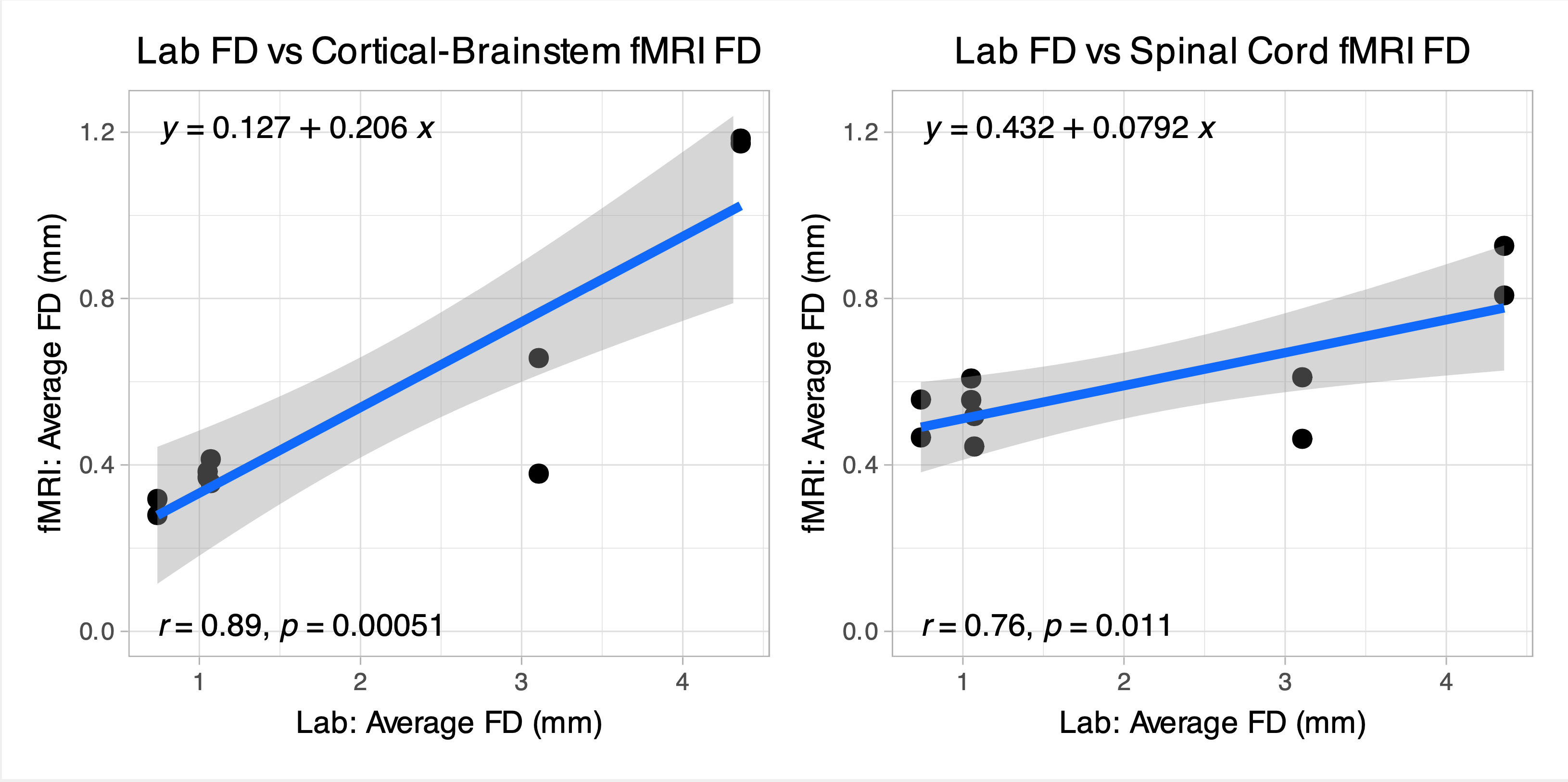
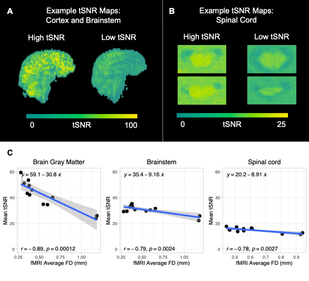
Head motion measured in the lab was significantly correlated with both head and spinal-cord motion (FD) during fMRI (Fig2; r=0.89, r=0.76). Head and spinal-cord motion was significantly correlated with reduction in mean tSNR in cortical, brainstem, and spinal-cord ROIs (Fig3; r=-0.89, r=-0.79, r=-0.78).Discussion
In cohorts expected to exhibit increased movement during motor-task fMRI, motion can be evaluated in a mock-MRI environment before scanning. With more data, it may be possible to define a threshold for head motion in the lab to achieve a desired tSNR for a given acquisition protocol. However, while the lab session is designed to simulate the MRI session, minor differences in setup still exist, causing variability in the relationship between motion in the lab vs. MRI.Even after motion correction and denoising, spinal-cord tSNR was lower in this stroke cohort compared to a similar dataset of hand grasping in the spinal cord13, indicating the importance of other denoising techniques. Implementing physiological regressors, such as RETROICOR14,15, end-tidal CO216, and/or CSF may be particularly beneficial to brainstem and spinal-cord areas that are highly susceptible to physiological noise. In addition, multi-echo independent component analysis in cortical-brainstem analysis may improve tSNR compared to typical single-echo methods17. Overall, our findings demonstrate that a motion-capture lab session can help anticipate and plan for potential decreases in tSNR in a clinical population.
Acknowledgements
This work was supported by the Center for Translational Imaging at Northwestern University, and NIH grants T32EB025766 (KJH, NAR) and F31NS134222 (KJH).References
- Cramer, S. C. (2004). Functional imaging in stroke recovery. Stroke, 35(11 SUPPL. 1), 2695–2698.
- Li, S., Chen, Y. T., Francisco, G. E., Zhou, P., & Rymer, W. Z. (2019). A Unifying Pathophysiological Account for Post-stroke Spasticity and Disordered Motor Control. Frontiers in neurology, 10, 468.
- Seto, E., Sela, G., McIlroy, W. E., Black, S. E., Staines, W. R., Bronskill, M. J., McIntosh, A. R., & Graham, S. J. (2001). Quantifying head motion associated with motor tasks used in fMRI. NeuroImage, 14(2), 284–297.
- Beissner F. (2015). Functional MRI of the Brainstem: Common Problems and their Solutions. Clinical neuroradiology, 25 Suppl 2, 251–257.
- Eippert, F., Kong, Y., Jenkinson, M., Tracey, I., & Brooks, J. C. W. (2017). Denoising spinal cord fMRI data: Approaches to acquisition and analysis. NeuroImage, 154, 255–266.
- Krause, F., Benjamins, C., Eck, J., Lührs, M., van Hoof, R., & Goebel, R. (2019). Active head motion reduction in magnetic resonance imaging using tactile feedback. Human Brain Mapping, 40(14), 4026–4037.
- Power, J. D., Barnes, K. A., Snyder, A. Z., Schlaggar, B. L., & Petersen, S. E. (2012). Spurious but systematic correlations in functional connectivity MRI networks arise from subject motion. NeuroImage, 59(3), 2142–2154.
- Jenkinson, M., Beckmann, C. F., Behrens, T. E. J., Woolrich, M. W., & Smith, S. M. (2012). FSL. NeuroImage, 62, 782–790.
- Cox R. W. (1996). AFNI: software for analysis and visualization of functional magnetic resonance neuroimages. Computers and biomedical research, an international journal, 29(3), 162–173.
- Dupre, E., Salo, T., Ahmed, Z., Bandettini, P. A., Bottenhorn, K. L., Caballero-Gaudes, C., Logan, T. D., Gonzalez-Castillo, J., Heunis, S., Kundu, P., Laird, A. R., Markello, R., Markiewicz, C. J., Moia, S., Staden, I., Teves, J. B., Uruñuela, E., Vaziri-Pashkam, M., Whitaker, K., & Handwerker, D. A. (2021). TE-dependent analysis of multi-echo fMRI with tedana. The Journal of Open Source Software, 6(59), 1–5.
- Desikan, R. S., Ségonne, F., Fischl, B., Quinn, B. T., Dickerson, B. C., Blacker, D., Buckner, R. L., Dale, A. M., Maguire, R. P., Hyman, B. T., Albert, M. S., & Killiany, R. J. (2006). An automated labeling system for subdividing the human cerebral cortex on MRI scans into gyral based regions of interest. NeuroImage, 31(3), 968–980.
- Deshpande, R., & Barry, R. (2022). Neptune: A toolbox for spinal cord functional MRI data processing and quality assurance [Abstract]. International Society for Magnetic Resonance in Medicine.
- Hemmerling, K. J., Hoggarth, M. A., Sandhu, M. S., Parrish, T. B., & Bright, M. G. (2023). Spatial distribution of hand-grasp motor task activity in spinal cord functional magnetic resonance imaging. Human Brain Mapping, 44(17), 5567–5581.
- Brooks, J. C., Beckmann, C. F., Miller, K. L., Wise, R. G., Porro, C. A., Tracey, I., & Jenkinson, M. (2008). Physiological noise modelling for spinal functional magnetic resonance imaging studies. NeuroImage, 39(2), 680–692.
- Harvey, A. K., Pattinson, K. T. S., Brooks, J. C. W., Mayhew, S. D., Jenkinson, M., & Wise, R. G. (2008). Brainstem functional magnetic resonance imaging: Disentangling signal from physiological noise. Journal of Magnetic Resonance Imaging, 28(6), 1337–1344.
- Reddy, N. A., Zvolanek, K. M., Moia, S., Caballero-Gaudes, C., & Bright, M. G. (2023). Denoising task-correlated head motion from motor-task fMRI data with multi-echo ICA. BioRxiv.
- Reddy, N. A., Clements R. G., Bright M. G. (2023). Multi-echo ICA denoising increases tSNR in combined cortical and brainstem task-fMRI [Abstract]. Organization for Human Brain Mapping Annual Meeting.
Figures