3436
Identification of cortical locations functionally connected to the hippocampus for personalized multi-target TMS neuromodulation1Physical Sciences Platform, Sunnybrook Research Institute, Toronto, ON, Canada, 2Department of Medical Biophysics, University of Toronto, Toronto, ON, Canada
Synopsis
Keywords: Functional Connectivity, fMRI (resting state), Transcranial Magnetic Stimulation
Motivation: Personalized TMS targeting based on functional connectivity has been proposed. However, the dispersion of personalized TMS targets remains unknown.
Goal(s): Characterize brain areas functionally connected to the hippocampus in a population.
Approach: Identify hippocampus-based memory networks using the resting-state fMRI data from the Human Connectome Project.
Results: The medial prefrontal cortex (mPFC) and lateral parietal cortex (PPC) were found to be connected to the hippocampus. The centroids of group-average results were separated from the atlas coordinates by about 11 mm. Individualized targets are dispersed around the group-average coordinates (mPFC: 11.1 +/- 3.8; PPC: 3.4 +/- 2.3 mm).
Impact: The functional connectivity analysis on the Human Connectome Project resting-state fMRI data with the seed at the hippocampus revealed that the medial prefrontal cortex (mPFC) and lateral parietal cortex (PPC) connected to the hippocampus significantly dispersed across individuals.
Introduction
Personalized neuromodulation targeting relies on the functional connectivity analysis of the resting-state fMRI data to reveal cortical regions showing correlated dynamics with the interested brain region. This strategy has been reported to be advantageous in guiding TMS pulse delivery to the parietal cortex for memory function preservation 1,2. Brain structural connectivity 3 and the activated brain regions subserving a given task 4 vary considerably across individuals. This highlights the importance of defining TMS targets based on an individual's brain.Here, we use the resting-state fMRI data from the Human Connectome Project 5 with 1,133 young adults to delineate the cortical regions functionally connected to the hippocampus. We hypothesize that the medial frontal cortex (mPFC) and posterior parietal cortex (PPC) are functionally connected to the hippocampus with variable patterns across individuals. The large-scale data permits the evaluation of distances between the standard TMS target and the most potent neuromodulation site for each individual.
Methods
The resting-state fMRI data for 1,133 young adults were downloaded from the Human Connectome Project database. The preprocessed data 6 had 2-mm isotropic spatial resolution and were sampled at the rate of 0.72 s per brain volume. The hippocampus was automatically segmented from an individual’s structural MRI using FreeSurfer 7. The time course of the hippocampus was averaged across image voxels within the segmented hippocampus. The functional connectivity to the hippocampus was calculated by calculating Pearson correlation coefficients. Group-level functional connectivity was calculated by testing if the transformed Z score was non-zero at each image voxel across all participants. False discovery rate 8 was controlled to suppress the inflation of type-I error in multiple comparisons. For each individual, we also calculated the centroids of the functional connectivity to the hippocampus at the medial frontal lobe and parietal lobe. These centroids were considered the personalized TMS targets. We also chose two atlas-based TMS targets for mPFC (0, 62, 4) and PPC (-42, -64, 48) derived from an automated meta-analysis (https://neurosynth.org) 9.Results
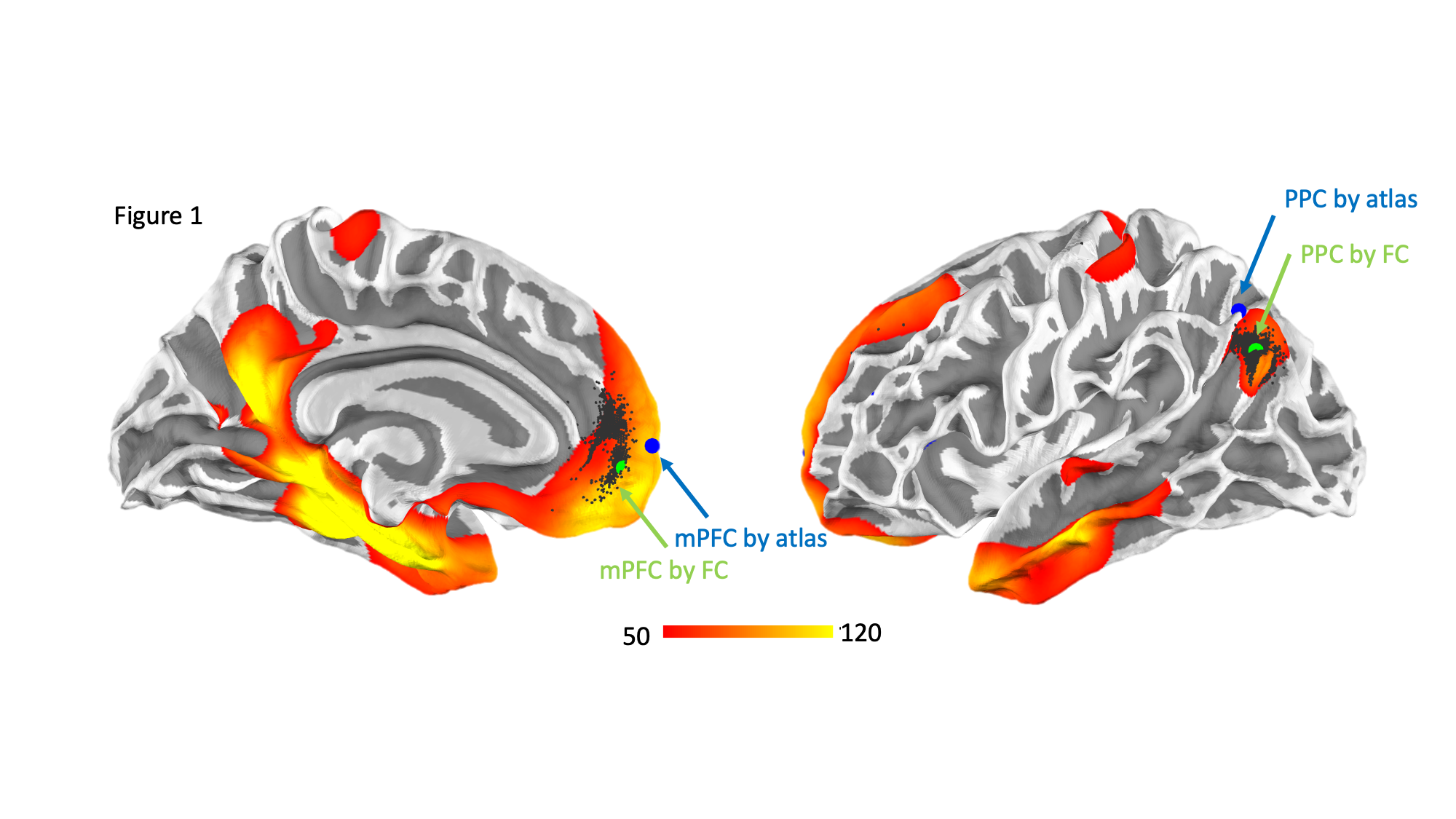
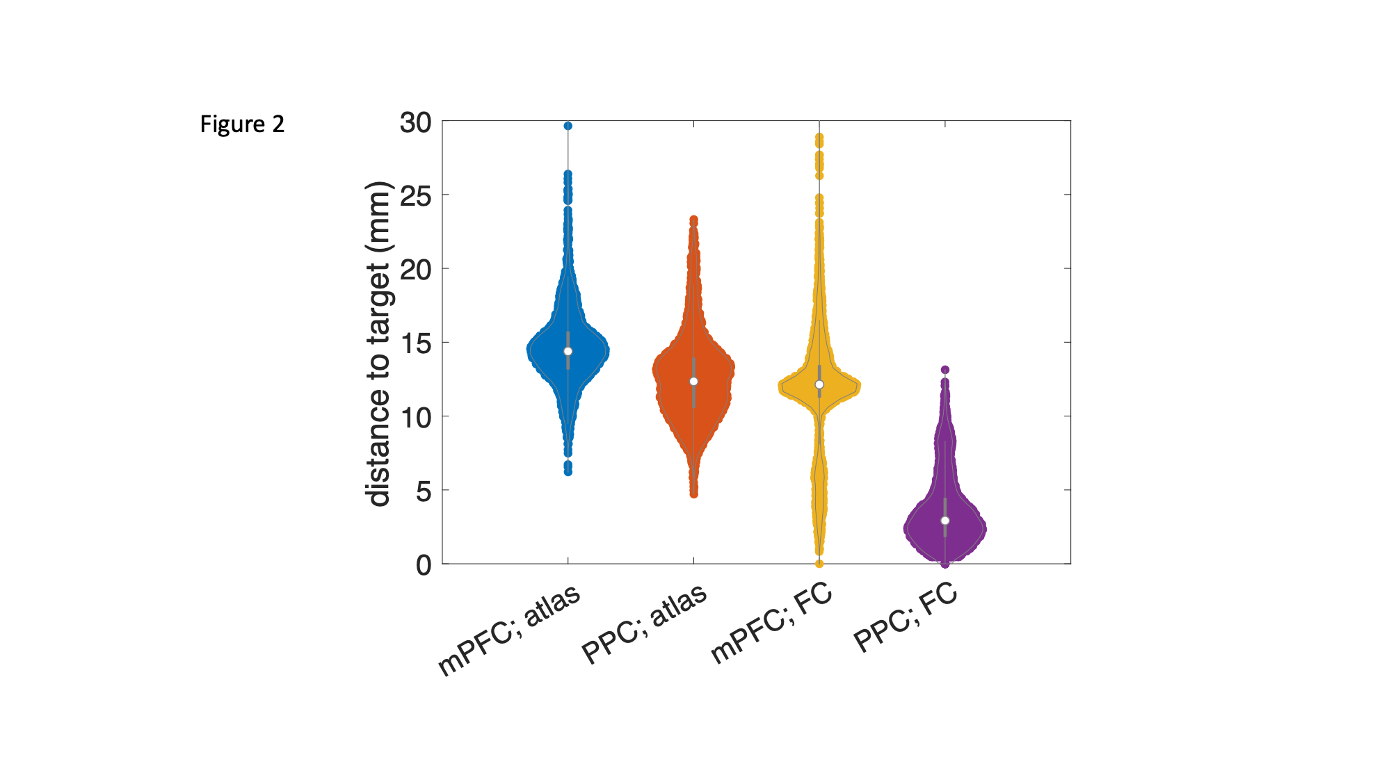
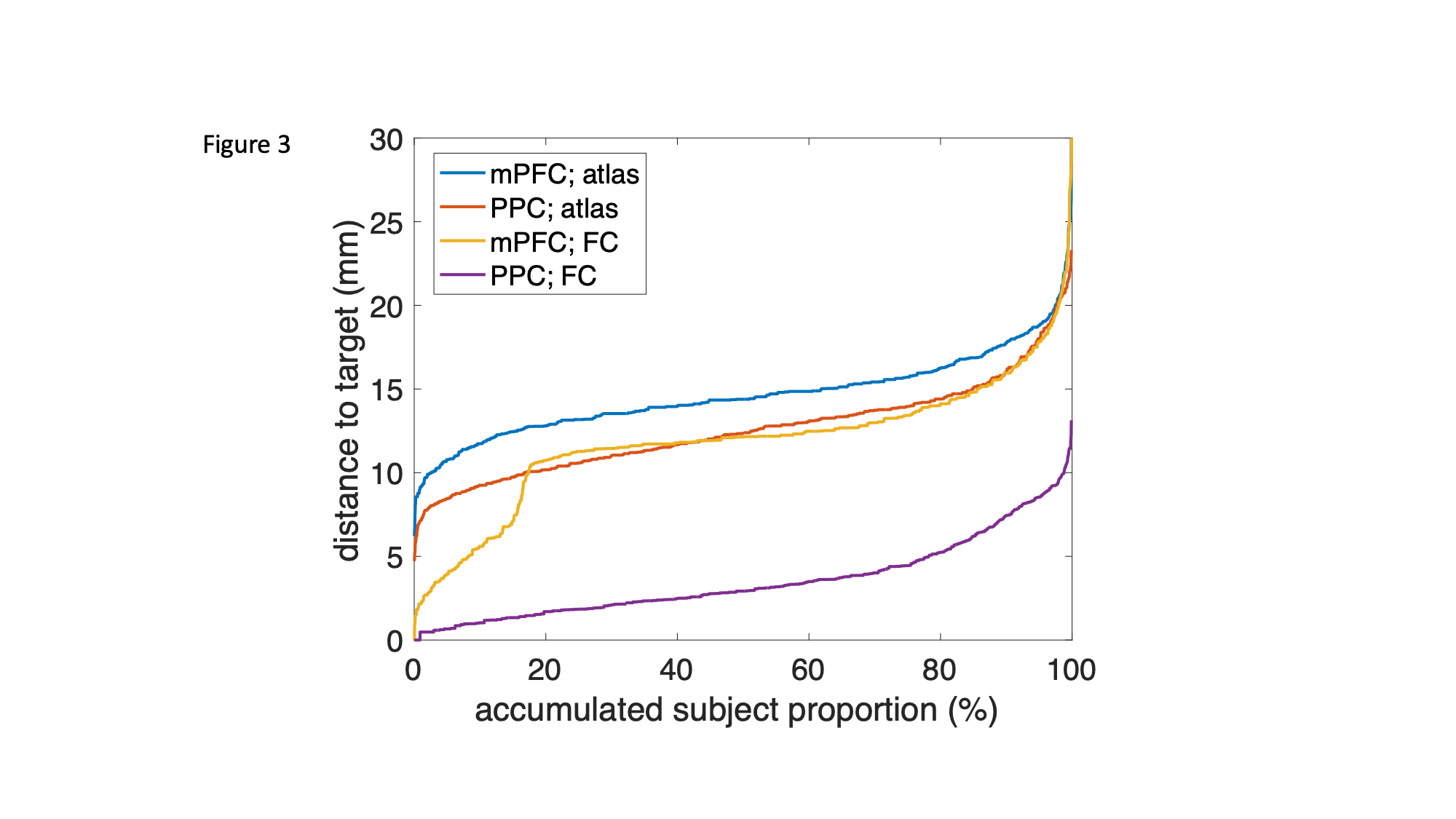
Figure 1 shows the group average of the mPFC and PPC functionally connected to the hippocampus. Significant functionally connected areas included the mPFC and PPC. In addition, the precuneus, left temporal poles, left sensory cortex and left auditory cortex had correlated hemodynamics to the hippocampus. Here, for the interest of personalized TMS targets for the memory function, we only focus on the analysis of mPFC and PPC.The centroids (green dots) were separated from the mPFC and PPC atlas coordinates (blue dots) by 10.8 mm and 12.8 mm, respectively. Individuals’ centroids at the mPFC and PPC are shown as black dots. We further calculated the individualized targets away from the atlas (blue and orange dots in the violin plot; Figure 2) or group average functional connectivity centroids (yellow and purple dots in the violin plot; Figure 2). The dispersion of the distance suggests a common target is sub-optimal (atlas targets: mPFC: 15.6 +/- 2.5; PPC: 12.6 +/- 2.8 mm; functional connectivity targets: mPFC: 11.1 +/- 3.8; PPC: 3.4 +/- 2.3 mm). Specifically, we sorted the distance metrics among 1,113 participants (Figure 3) and found that more than 50% of the individuals are 14.4 mm and 12.3 mm away from the atlas target for mPFC and PPC, respectively. Relative to the functional connectivity target, more than 50% of the individuals are still 11.4 mm and 2.9 mm away from mPFC and PPC, respectively.
Discussion
Using fMRI data from a large cohort, we show that cortical areas functionally connected to the hippocampus varied across individuals. This finding supports the recent view of using personalized TMS targets for the optimal modulatory effect. Importantly, regardless of the chosen target, a common TMS target can be away from the centroid of the functionally connected cortex for 5 mm or more. This discrepancy may cause remission of TMS due to inefficient stimulation strength because the induced magnetic field strength drops to 50 % or less at 5mm from the target 10.Our findings corroborate a potential strategy of stimulating the hippocampus using multi-focus TMS 11–13. Specifically, targeting both mPFC and PPC is likely to stimulate hippocampal activity. Separate TMS targeting at mPFC 14–17 and PPC 1,2,18,19 confirmed that hippocampal activity can be activated by non-invasive cortical stimulation. Applying TMS at two sites may synergically modulate hippocampal activity and memory function more effectively.
Acknowledgements
This work was supported by the Natural Sciences and Engineering Research Council of Canada (NSERC; RGPIN-2020-05927), Canada Foundation for Innovation (38913 and 41351), Canadian Institutes of Health Research (PJT 178345 and PJT 185882), MITACS (IT25405 and Global Link fellowship).References
- Cash, R. F. H. et al. Brain Stimul. 15, 1300–1304 (2022)
- Wang, J. X. et al. Science 345, 1054–1057 (2014)
- Heiervang, E. et al. Neuroimage 33, 867–877 (2006)
- Conner, C. R. et al. J. Neurosci. 31, 12855–12865 (2011)
- Smith, S. M. et al. Neuroimage 80, 144–168 (2013)
- Glasser, M. F. et al. Neuroimage 80, 105–124 (2013)
- Fischl, B. et al. Neuron 33, 341–355 (2002)
- Genovese, C. R. et al. Neuroimage 15, 870–878 (2002)
- Yarkoni, T. et al. Nat. Methods 8, 665–670 (2011)
- Lynch, C. J. et al. Neuron 110, 3263–3277.e4 (2022)
- Nieminen, J. O. et al. Brain Stimul. 15, 116–124 (2022)
- Brys, M. et al. Neurology 87, 1907–1915 (2016)
- Navarro de Lara, L. I. et al. Neuroimage 224, 117355 (2021)
- Yeh, N. et al. J. Neurosci. 41, 6273–6280 (2021)
- Bovy, L. et al. Cereb. Cortex 30, 3608–3616 (2020)
- Borgomaneri, S. et al. Curr. Biol. 30, 3672–3679.e4 (2020)
- Berkers, R. M. W. J. et al. Cortex 88, 42–52 (2017)
- Prime, S. L. et al. J. Neurosci. 28, 6938–6949 (2008)
- Hebscher, M. et al. Curr Opin Behav Sci 32, 35–42 (2020)
Figures