3431
Resting-state hypothalamic dysconnectivity associates with symptom severity in schizophrenia1Department of Radiology, and Functional and Molecular Imaging Key Laboratory of Sichuan Province, West China Hospital of Sichuan University, Chengdu, China, 2Huaxi MR Research Center (HMRRC), West China Hospital of Sichuan University, Chengdu, China, 3Research Unit of Psychoradiology, Chinese Academy of Medical Sciences, Chengdu, China, 4Departments of Psychiatry and of Neuroscience, Yale University School of Medicine, New Haven, CT, United States
Synopsis
Keywords: Functional Connectivity, fMRI (resting state)
Motivation: The Hypothalamic-Pituitary-Adrenal (HPA) axis dysfunction is crucial in stress response in schizophrenia. The hypothalamus is an essential part of the HPA axis. The hypothalamic connections with brain regions in schizophrenia.
Goal(s): This study examined hypothalamic resting-state functional connectivity (FC) in schizophrenia and how hypothalamic FCs related to symptom severity.
Approach: Resting-state functional magnetic resonance imaging data were obtained from 235 patients with schizophrenia and 222 healthy controls. We processed the imaging data with published routines and evaluated the results at a corrected threshold.
Results: Relative to healthy controls, schizophrenia showed lower hypothalamic FCs with mesocorticolimbic regions, which were connected with symptom severity.
Impact: Decreased hypothalamic functional connectivities, which are associated with symptomology, extend the understanding of functional connectivity alterations and suggest the potential pathophysiology of stress response dysfunction in schizophrenia, which needs further study.
Introduction
The hypothalamus is an essential part of the Hypothalamic-Pituitary-Adrenal axis and participates in emotion, motivation, and reward processing in schizophrenia (SCZ)1, 2. It is unknown how the hypothalamus connects with the cortical and subcortical areas in SCZ. This study examined hypothalamic resting-state functional connectivity (FC) in both antipsychotic-naïve first-episode schizophrenia (ANFES) and chronically treated schizophrenia (CTS) patients and how hypothalamic FCs related to symptom severity.Material and Methods
1. ParticipantsA total of 235 SCZ patients, including 159 ANFES and 76 CTS, were recruited in this study. Demographically matched healthy controls (HC) were recruited from local communities. The Ethics Committee on Biomedical Research of West China Hospital of Sichuan University approved this study. All participants provided written informed consent before the study.
2. Imaging acquisition and data pre-processing
All participants were scanned using a 3.0 T GE Signa EXCITE Scanner with an 8-channel head coil. The resting-state functional magnetic resonance imaging data (rs-fMRI) were acquired using a gradient-echo echo-planar imaging sequence with repetition time = 2000 ms, echo time = 30 ms, flip angle = 90°, matrix size = 64 × 64, slice thickness = 5 mm, and a field of view of 240 × 240 mm2, resulting in a voxel size of 3.75 × 3.75 × 5 mm3. High-resolution T1-weighted images were acquired using a three-dimensional spoiled gradient sequence (repetition time = 8.5 ms, echo time = 3.5 ms, inversion time = 400 ms, flip angle = 12°) with a 240 × 240 matrix in a field of view of 240 × 240 mm2.
3. Hypothalamus seed selection and seed-to-voxel FC analysis
Processing of rs-fMRI was performed using Data Processing Assistant for Resting-State fMRI (DPARSF Version 5.4, http://www.rfmri.org/content/dparsf ) 3, 4 based on Statistical Parametric Mapping software (SPM 12, https://www.fil.ion.ucl.ac.uk/spm/software/spm12/ ). The preprocessing procedures which including the removal of the first ten volumes, slice timing, realignment, coregistration and segmentation, normalization to the Montreal Neurologic Institute space, spatial smoothing, and temporal filtering.
4. Statistical analysis
4.1. Demographic and clinical data
The continuous and categorial variables were compared between overall SCZ and HC as well as between ANFES and CTS using two-sample t-tests and Chi-square tests, respectively.
4.2. Functional connectivity of the hypothalamus
We first compared the hypothalamus FC between overall SCZ and HC using analysis of covariance (ANCOVA) via SPM 12 controlling for age, sex, and mean framewise displacements (FD). The regions showing significantly altered hypothalamic FC were extracted and compared among the ANFES, CTS, and HC using ANCOVA with age, sex, and FD as covariates.
4.3. Correlation analysis

Pearson partial correlation analyses were done between the altered hypothalamic FC and symptom severity in overall SCZ, ANFES, and CTS, respectively, all with age and sex as covariates.
Results
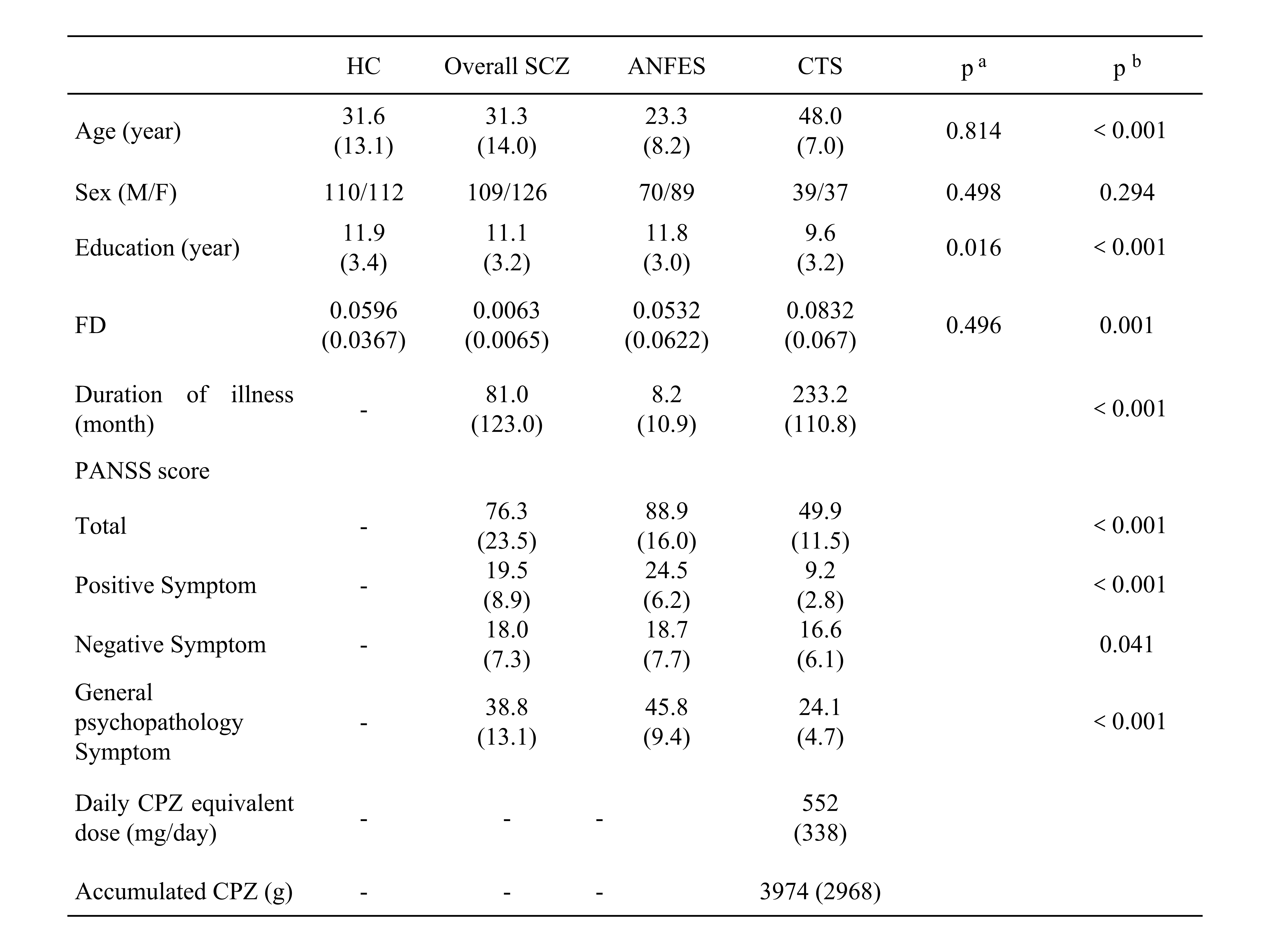
1. Demographic dataThe demographic and clinical characteristics of patients and HC are shown in Figure 1.
2. Functional connectivity of the hypothalamus
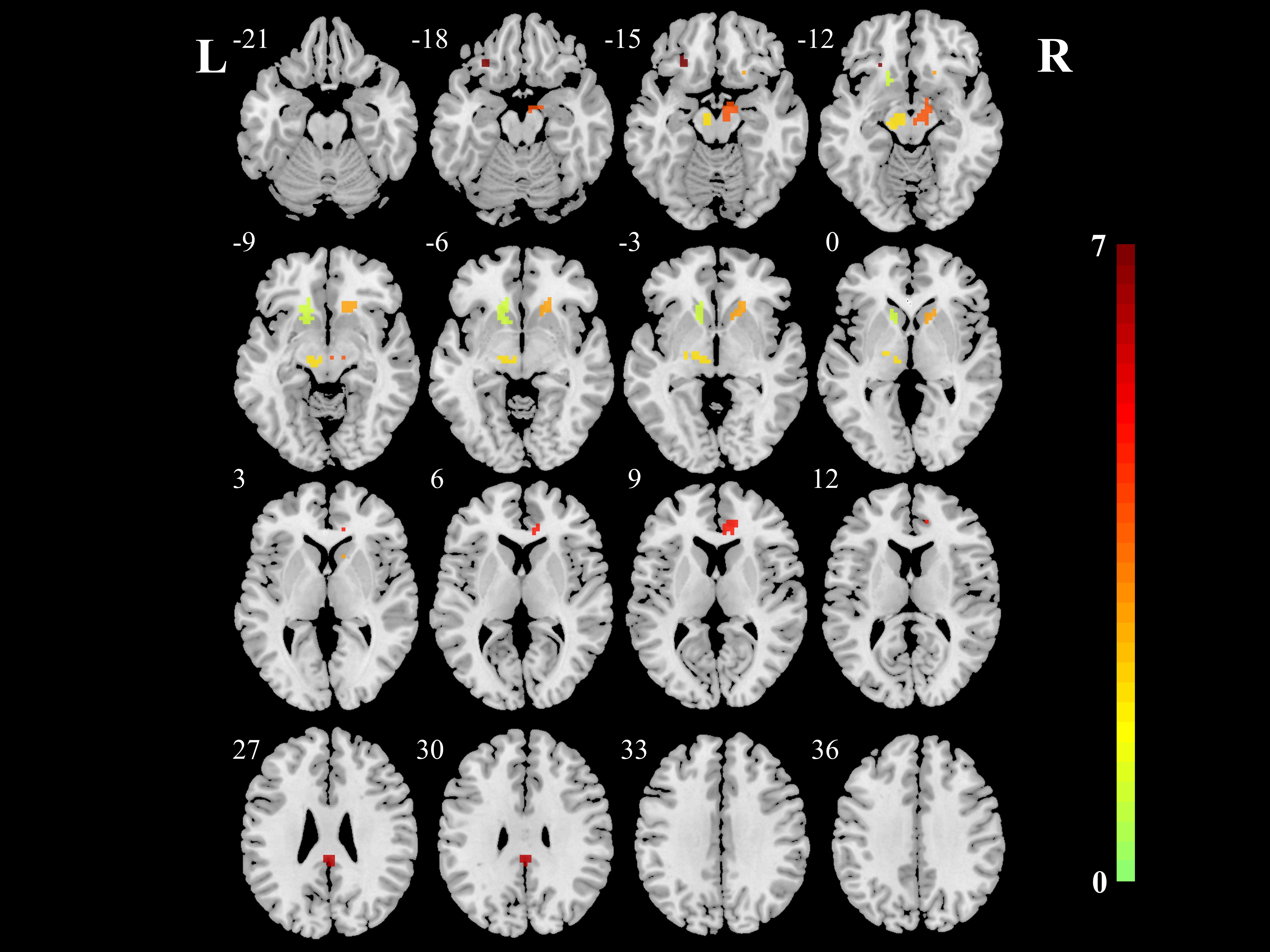
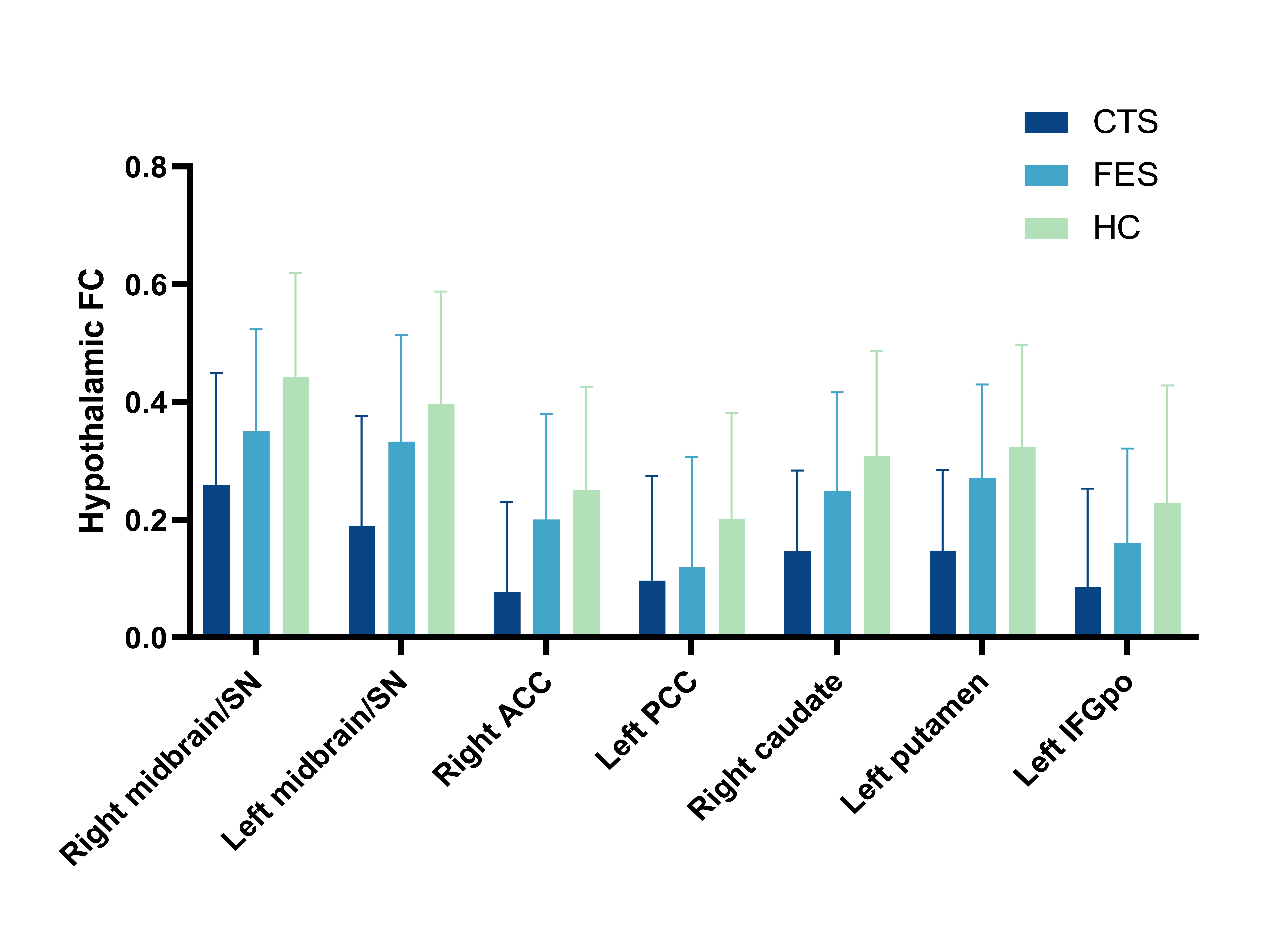
Compared with HC, the overall SCZ group showed significantly lower hypothalamic FCs with bilateral midbrain/substantia nigra (SN), right anterior cingulate cortex (ACC), left posterior cingulate cortex (PCC), right caudate, left putamen, and left inferior frontal gyrus, pars orbitalis (IFGpo) (Figure 2). In post hoc analyses, CTS presented significantly further decreased FCs compared with ANFES. (Figure 3).
3. Correlation analysis
In the overall SCZ group, the PANSS total scores, positive scores, and general psychopathology were positively correlated with altered hypothalamic FCs (Figure 4).
Discussion
In the present study, patients with SCZ showed disrupted hypothalamic FCs with the midbrain, striatum, and limbic system, particularly in chronic patients with long-term antipsychotic treatment. Furthermore, disrupted hypothalamic FCs with the midbrain, striatum, and ACC are positively associated with the severity of positive and general psychopathology symptoms. Our results are in line with previous studies 5, 6. Previous studies have demonstrated the hypothalamus receives dopamine projections from the mesolimbic system and immerses into the neuroendocrine, affective, and behavioral functions 7. Reduced midbrain FCs may imply dopamine pathway dysfunction 8. The disrupted functional connections between the hypothalamus and mesocorticolimbic regions, which are associated with symptomology may implicate in the psychopathology and symptomology of SCZ, especially in reward processing, and cognitive deficits, and further investigations are needed 9-11.Conclusion
The abnormal interactions between the hypothalamus and mesocorticolimbic regions, which are relatively stable and associated with symptomatology, extend our understanding of functional brain changes in SCZ, which might underpin the stress response after illness onset in affected individuals that might require further investigation.Acknowledgements
This study was supported by the National Natural Science Foundation of China (Project Nos. 82120108014, 82071908, and 82101998), the National Key R&D Program of China (Project Nos. 2022YFC2009901 and 2022YFC2009900), Sichuan Science and Technology Program (Project No. 2021JDTD0002), CAMS Innovation Fund for Medical Sciences (CIFMS) (Project No. 2021-I2M-C&T-A-022), Chengdu Science and Technology Office, major technology application demonstration project (Project Nos. 2022-YF09-00062-SN, 2022-GH03-00017-HZ). Dr. Su Lui acknowledges the support from Humboldt Foundation Friedrich Wilhelm Bessel Research Award and Chang Jiang Scholars (Program No. T2019069).References
1. Misiak, B., et al., A meta-analysis of blood and salivary cortisol levels in first-episode psychosis and high-risk individuals. Front Neuroendocrinol, 2021. 62: p. 100930.2. Bradley, A.J. and T.G. Dinan, A systematic review of hypothalamic-pituitary-adrenal axis function in schizophrenia: implications for mortality. J Psychopharmacol, 2010. 24(4 Suppl): p. 91-118.
3. Yan, C.G., et al., DPABI: Data Processing & Analysis for (Resting-State) Brain Imaging. Neuroinformatics, 2016. 14(3): p. 339-51.
4. Chao-Gan, Y. and Z. Yu-Feng, DPARSF: A MATLAB Toolbox for "Pipeline" Data Analysis of Resting-State fMRI. Front Syst Neurosci, 2010. 4: p. 13.
5. Dayan, E., M. Sklerov, and N. Browner, Disrupted hypothalamic functional connectivity in patients with PD and autonomic dysfunction. Neurology, 2018. 90(23): p. e2051-e2058.
6. Sudheimer, K., et al., Decreased hypothalamic functional connectivity with subgenual cortex in psychotic major depression. Neuropsychopharmacology, 2015. 40(4): p. 849-60.
7. Meltzer, H.Y. and S.M. Stahl, The dopamine hypothesis of schizophrenia: a review. Schizophr Bull, 1976. 2(1): p. 19-76.
8. Hadley, J.A., et al., Ventral tegmental area/midbrain functional connectivity and response to antipsychotic medication in schizophrenia. Neuropsychopharmacology, 2014. 39(4): p. 1020-30.
9. Catani, M., F. Dell'acqua, and M. Thiebaut de Schotten, A revised limbic system model for memory, emotion and behaviour. Neurosci Biobehav Rev, 2013. 37(8): p. 1724-37.
10. Nakamura, Y., et al., Distinctive alterations in the mesocorticolimbic circuits in various psychiatric disorders. Psychiatry Clin Neurosci, 2023. 77(6): p. 345-354.
11. Salamone, J.D. and M. Correa, The Neurobiology of Activational Aspects of Motivation: Exertion of Effort, Effort-Based Decision Making, and the Role of Dopamine. Annu Rev Psychol, 2023.
Figures