3429
Thalamic Connectivity and its Role in Resilience to Trauma: Insights from Ultra-High-Field MRI at 7T1Institute of Neuroscience and Medicine, Forschungszentrum Jülich GmbH, Jülich, Germany INM-4, Jülich, Germany, 2Department of Psychiatry, Psychotherapy and Psychosomatics, RWTH Aachen University, Aachen, Germany, Aachen, Germany, 3JARA – BRAIN – Translational Medicine, Aachen, Germany, Aachen, Germany, 4Institute of Neuroscience and Medicine, INM-11, Forschungszentrum Jülich GmbH, Jülich, Germany, Aachen, Germany, 5Department of Neurology, RWTH Aachen University, Aachen, Germany, Aachen, Germany, 6Center for Computational Life Sciences, RWTH Aachen, Germany, Aachen, Germany
Synopsis
Keywords: Functional Connectivity, fMRI (resting state), Resilience, Trauma, PTSD, Thalamus, UHF-MRI
Motivation: To discern why some individuals develop stress-related disorders post-trauma while others don't, focusing on the role of thalamus in emotional regulation and resilience.
Goal(s): Investigate thalamic functional connectivity differences in trauma-exposed individuals to identify neural mechanisms of resilience.
Approach: Acquired MRI data from 35 Syrian refugees using a 7T scanner, analyzed for trauma-related connectivity differences via seed-to-voxel thalamic analysis with the CONN toolbox, informed by RS-25 and HTQ questionnaires to distinguish between asymptomatic and symptomatic groups.
Results: Significant right thalamic connectivity differences were found, indicating potential neural resilience correlates and adaptive changes in sensory-motor processing related to PTSD symptom severity.
Impact: This study enhances our understanding of trauma's neural basis and resilience, potentially directing new therapeutic strategies targeting thalamic connectivity to prevent stress-related disorders, thereby improving trauma care and mental health outcomes. Ref.
Introduction:
Exposure to trauma can lead to a wide array of psychological impacts. Resilience, the capacity to withstand or quickly recover from such events, is a significant variable in the psychological trajectory post-trauma 1. The variability in individual responses ranges from debilitating stress-related disorders to complete asymptomatic resilience 2. The thalamus acts as an important gateway due to its role in sensory processing and emotional regulation. It acts as a relay station, modulating the signals that underpin our reactions to traumatic experiences 3,4. Understanding its function is, therefore, crucial to unraveling the neural basis of trauma and resilience.Methods:
- Data Acquisition: Using a 7T MAGNETOM Terra scanner by Siemens Healthineers, the MRI data we acquired from 35 Syrian refugees. The group was categorized based on their symptomatic response to trauma into 18 asymptomatic individuals (mean age 25.1, SD 5.2, including six females) and 17 symptomatic individuals (mean age 28.6, SD 9.4, also including six females).
- Psychological Questionnaires: Resilience and trauma symptoms were quantified using RS-25 and HTQ, respectively 5,6.
- Functional MRI: Resting-state fMRI data were collected with TE/TR of 25 ms/2000 ms across 305 volumes within 10 minutes, using a 168 x 168 image matrix and a 220 x 220 mm² FOV, resulting in 1.3 mm isotropic resolution. Participants were instructed to relax with closed eyes without engaging in directed thought. Structural MRI: The MP2RAGE sequence captured structural brain images with TR/TE of 4500ms/1.99ms at 0.75 mm isotropic resolution.
- MRI Data Analysis: Analysis was performed using the CONN toolbox (v.22.a) 7, supported by SPM 12, and executed in MATLAB R2023a. The bilateral thalamus, crucial for sensory and emotional processing, was the seed region for our seed-to-voxel analysis 8. A between-subject design factoring in-group status, age, and gender were employed, with a -1 1 0 0 contrast vector to compare functional connectivity (FC) between the asymptomatic and symptomatic groups. This approach aimed to uncover neural connectivity patterns linked to trauma response and resilience.
Results:
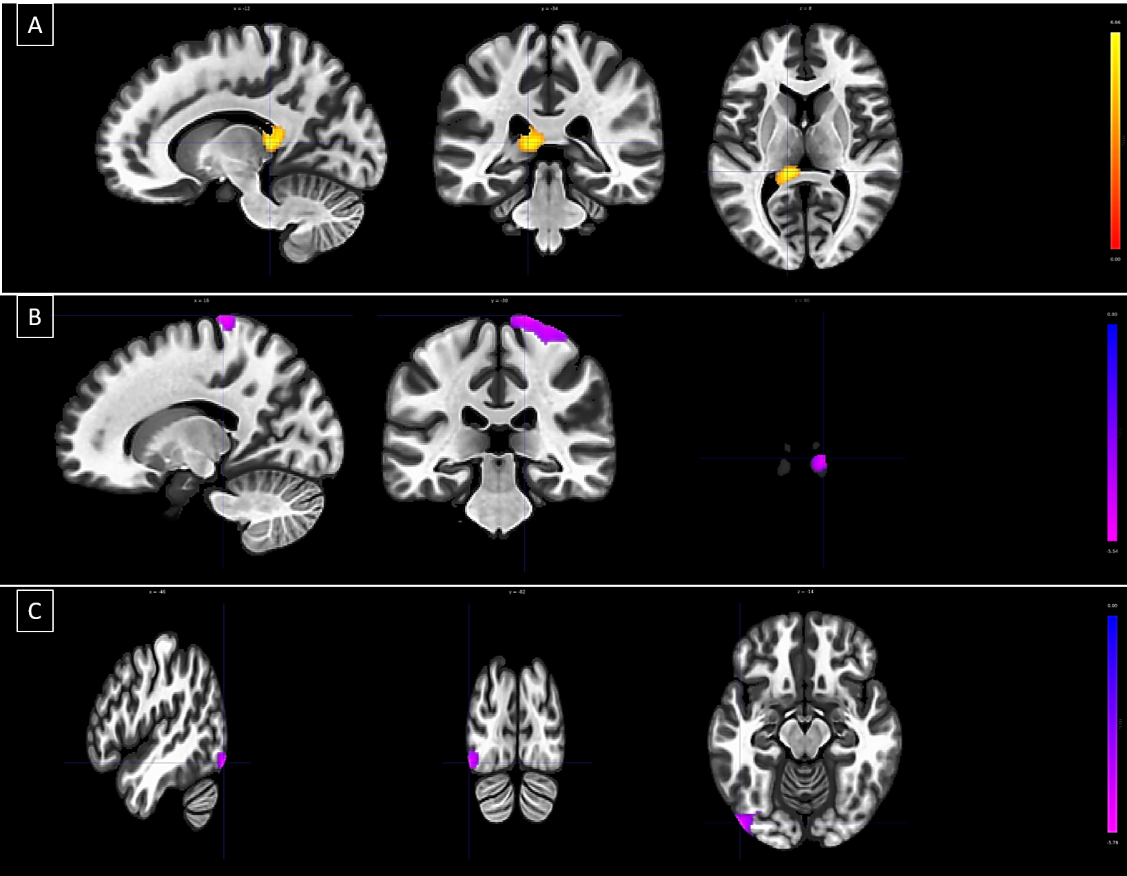
Significant FC alterations were observed in the right thalamus across three clusters. The asymptomatic group showed increased connectivity in Cluster 1 with the left thalamus and reduced connectivity with sensory-motor areas, including the right postcentral and precentral gyrus, and the left inferior lateral occipital cortex. A significant inverse correlation was noted between Cluster 1's FC and PTSD symptom severity, as measured by the Harvard Trauma Questionnaire, suggesting a potential link to resilience mechanisms.Discussion:
The differentiated patterns of thalamic connectivity between the trauma-exposed asymptomatic and symptomatic groups offer compelling insights into the neural basis of trauma response and resilience. In the asymptomatic group, the increased connectivity between the bilateral thalami suggests a potential neural mechanism for resilience. This is characterized by enhanced inter-thalamic communication that may support efficient sensory integration and emotional regulation. Conversely, the decreased connectivity observed between the right thalamus and the right postcentral gyrus in the same group could reflect an adaptive modulation of sensory and motor responses, possibly affording a protective effect against developing PTSD symptoms. Furthermore, the significant inverse relationship between Cluster 1's functional connectivity (FC) and Harvard Trauma Questionnaire PTSD scores underlines the thalamus's pivotal role in PTSD symptomatology. Higher PTSD symptoms were associated with lower FC in the thalamus, possibly indicative of a disruption in the neural pathways typically involved in processing traumatic experiences. These findings suggest that thalamic dysregulation may contribute to the manifestation of PTSD, and interventions aimed at normalizing thalamic function may have therapeutic benefits. Additionally, the reduced connectivity with the left inferior lateral occipital cortex, a part of Cluster 3, suggests that visual processing, often a trigger for traumatic memories, may be modulated differently in trauma-exposed individuals who remain asymptomatic. This could represent a neural strategy to minimize the re-experiencing of traumatic events.Conclusion:
The significance of the thalamus in the neurobiological underpinnings of trauma and resilience is underscored by these results. The distinct FC patterns observed could potentially serve as biomarkers for susceptibility or resistance to stress-related disorders following trauma exposure. Future research is warranted to explore the potential of modulating thalamic connectivity to enhance resilience and mitigate the adverse effects of trauma. This should include focusing on the interactions of the thalamus with sensory, motor, and visual processing regions. The current study adds to the growing body of evidence that emphasizes the need for a nuanced approach to understanding and treating the complex sequelae of trauma exposure.Acknowledgements
The authors extend their gratitude to Elke Bechholz, Anita Köth, and Petra Engels, for their invaluable technical support during MRI sessions, and to Raghad Kiwan for her contributions to the participant recruitment efforts.References
1. Luthar, S. S., Cicchetti, D., & Becker, B. (2000). The construct of resilience: A critical evaluation and guidelines for future work. Child Development. https://doi.org/10.1111/1467-8624.00164 2. Yehuda, R., & LeDoux, J. (2007). Response variation following trauma: A translational neuroscience approach to understanding PTSD. Neuron, 56(1), 19-32. 3. Sherman, S. M. (2007). The thalamus is more than just a relay. Current Opinion in Neurobiology, 17(4), 417-422. 4. Etkin, A., & Wager, T. D. (2007). Functional neuroimaging of anxiety: A meta-analysis of emotional processing in PTSD, social anxiety disorder, and specific phobia. The American Journal of Psychiatry, 164(10), 1476-1488. 5. Wagnild, G. M., & Young, H. M. (1993). Development and psychometric evaluation of the Resilience Scale. Journal of Nursing Measurement, 1(2), 165-178. 6. Mollica, R. F., Caspi-Yavin, Y., Bollini, P., Truong, T., Tor, S., & Lavelle, J. (1992). The Harvard Trauma Questionnaire: Validating a cross-cultural instrument for measuring torture, trauma, and posttraumatic stress disorder in Indochinese refugees. The Journal of Nervous and Mental Disease, 180(2), 111-116. 7. Nieto-Castanon, A., & Whitfield-Gabrieli, S. (2022). CONN functional connectivity toolbox: RRID SCR_009550, Release 22. https://doi.org/10.56441/hilbertpress.2246.5840 8. Nieto-Castanon, A. (2020). Handbook of functional connectivity magnetic resonance imaging methods in CONN. https://doi.org/10.56441/hilbertpress.2207.6598Figures