3427
Comparing functional connectivity of small nucleus in brainstem with multi- and single-echo fMRI: A resting state study1Ruijin Hospital, Shanghai Jiao Tong University School of Medicine, Shanghai, China, 2Philips Healthcare, Xi'an, China, 3Philips Healthcare, Shanghai, China, 4Radiology, Ruijin Hospital, Shanghai Jiao Tong University School of Medicine, Shanghai, China, 5College of Health Science and Technology, Shanghai Jiao Tong University School of Medicine, Shanghai, China
Synopsis
Keywords: Functional Connectivity, fMRI
Motivation: Efforts to characterize the function of DR, MR and LC in humans using functional magnetic resonance imaging (fMRI) have been hampered by their small size and location near a large source of noise.
Goal(s): The aim of this study is to compare the functional connectivity (FC) of small nucleus in brainstem with multi- (ME) and single-echo (SE) resting state fMRI.
Approach: The whole-brain FC of LC, DR and MR were generated by using ME-fMRI and SE-fMRI.
Results: Precuneus was correlated with LC in both methods. Meanwhile, both methods shown cerebellum was correlated with DR, the cingulum was correlated with MR.
Impact: It suggests that ME-fMRI may also provide significant advantages over single echo fMRI approaches when investigating the FC of small nucleus of brainstem. ME-fMRI offers substantial advantages over single echo fMRI.
Introduction
The serotonergic system is mainly derived from the dorsal (DR) and median (MR) raphe nuclei, meanwhile, the locus coeruleus (LC) is main part of noradrenergic system, which is very important in the pathological process of neurodegenerative diseases.[1,2] Efforts to characterize the function of DR, MR, and LC in humans using functional magnetic resonance imaging (fMRI) have been hampered by their small size and location near a large source of noise. Previous studies utilized single-echo fMRI (SE-fMRI) to analyze brainstem nucleus. However, SE-fMRI was influenced by non-signal noise, signal distortion, and signal drop-out[3]. There is no unified pipeline for denoising SE-fMRI, a method for denoising such as independent components analysis was needed during the data analysis.[4,5] The multi-echo (ME) acquisition strategy collects three or more images per repetition time (TR). ME-fMRI has two advantages compared to SE-fMRI.[6] First, different echoes can be aggregated into a single “combined echo”, enhancing blood oxygenation level-dependent (BOLD) contrast and reducing susceptibility artifacts by giving greater weight to echoes near the estimated average T2* decay rate at each voxel. Second, signal decay patterns across echoes can be leveraged during denoising to distinguish between signals of interest (T2*-dependent or 'BOLD-like') and various forms of noise (non-T2*-dependent or 'non-BOLD-like'). To date, there are few studies exploring the small brain nucleus’ functional connectivity (FC) by using ME-fMRI. Therefore, the aim of this study is to compare the functional connectivity (FC) of small nucleus in brainstem with multi- and single-echo resting state fMRI.Methods
In this study, 12 young healthy adults were recruited from Ruijin Hospital and all of them underwent the MR examination by a 3.0T scanner (Elition, Philips Healthcare, the Netherlands) with a 32-channel head coil. The ME-fMRI protocol was as follows: TR = 2000 ms, TE = 15/35/50 ms, multiband sense factor = 2, number of slices = 50, spatial resolution = 3 x 3 x 3 mm3, time points = 240, total scan time = 480 seconds. Besides, we also collected 1 mm isotropic 3D-T1 images for subsequent data analysis. For ME-fMRI, data were processed by AFNI (https://afni.nimh.nih.gov/). The second echo (TE = 35 ms) fMRI data was extracted for SE-fMRI analysis. LC was defined according to the atlas based on 7T neuromelanin-sensitive MRI analysis.[7] The center of positions for DR and MR were based on MNI space coordinates.[8] For each region of interest (ROI), a whole-brain FC was obtained for each participant by using the DPARSF6.0 toolbox (Data Processing Assistant for Resting-State fMRI, version 7.0) (http://www.restfmri.net). The correlation coefficients were converted to z-scores using Fisher's z transformation to improve the normality. The one-sample t-test was applied to the group to determine the connectivity profile of each ROI. The significant level for the within-group test was set at a Gaussian random field (GRF correction with 0.001 voxel P-value and 0.05 cluster P-value).Results
Table 1 showed the demographic characteristics of the enrolled participants. Figure 1 showed the schematic diagram of SE- and ME-fMRI data processing procedures. ME-fMRI could separate BOLD signal from non-BOLD signal, which SE-fMRI could not. The SE-fMRI and ME-fMRI results are shown in Figure 1. Precuneus was correlated with LC both in ME-fMRI and SE-fMRI. Meanwhile, for DR, the cerebellum was correlated with DR in both methods. For MR, the cingulum was the nuclei which both methods showed. Compared with ME-fMRI, SE-fMRI has a larger area of noise.Discussion
In our study, the SE-fMRI was extracted from ME-fMRI which made this comparison with less bias and individual variability. ME-fMRI is designed to provide a better signal-noise ratio compared to SE-fMRI.[9] The ME-fMRI data can provide more accurate and reliable measurements, which might result in narrower and more stable FC patterns. In contrast, SE-fMRI could lead to more variable and less precise measurements, resulting in wider and unstable FC patterns. In addition, ME-fMRI data may benefit from improved motion correction and artifact removal techniques, resulting in cleaner and more accurate data for small brainstem nuclei. The brainstem nucleus, especially LC, DR, and MR is crucial for neurodegenerative diseases such as Parkinson’s disease and Alzheimer’s disease. Further study will need to investigate the brainstem functional network changes and FC in patients with neurodegenerative disease.Conclusion
It suggests that suggest that ME-fMRI may also provide significant advantages over single echo fMRI approaches when investigating the FC of the nucleus of brainstem. ME-fMRI offers substantial advantages over single echo fMRI.Acknowledgements
This work was supported, in part, by the National Natural Science Foundation of China (grant number: 82271954, 81971576); Chinese National Science & Technology Pillar Program (grant number: 2022YFC2009900/2022YFC2009905) and the Innovative Research Team of High-level Local Universities in Shanghai.References
[1] K.G. Commons, Ascending serotonin neuron diversity under two umbrellas, Brain Struct Funct. 221 (2016) 3347–3360. https://doi.org/10.1007/s00429-015-1176-7.
[2] M.J. Betts, E. Kirilina, M.C.G. Otaduy, D. Ivanov, J. Acosta-Cabronero, M.F. Callaghan, C. Lambert, A. Cardenas-Blanco, K. Pine, L. Passamonti, C. Loane, M.C. Keuken, P. Trujillo, F. Lüsebrink, H. Mattern, K.Y. Liu, N. Priovoulos, K. Fliessbach, M.J. Dahl, A. Maaß, C.F. Madelung, D. Meder, A.J. Ehrenberg, O. Speck, N. Weiskopf, R. Dolan, B. Inglis, D. Tosun, M. Morawski, F.A. Zucca, H.R. Siebner, M. Mather, K. Uludag, H. Heinsen, B.A. Poser, R. Howard, L. Zecca, J.B. Rowe, L.T. Grinberg, H.I.L. Jacobs, E. Düzel, D. Hämmerer, Locus coeruleus imaging as a biomarker for noradrenergic dysfunction in neurodegenerative diseases, Brain. 142 (2019) 2558–2571. https://doi.org/10.1093/brain/awz193.
[3] K.-J. Bär, F. de la Cruz, A. Schumann, S. Koehler, H. Sauer, H. Critchley, G. Wagner, Functional connectivity and network analysis of midbrain and brainstem nuclei, Neuroimage. 134 (2016) 53–63. https://doi.org/10.1016/j.neuroimage.2016.03.071.
[4] J. Sun, J. Ma, L. Gao, J. Wang, D. Zhang, L. Chen, J. Fang, T. Feng, T. Wu, Disruption of locus coeruleus-related functional networks in Parkinson’s disease, NPJ Parkinsons Dis. 9 (2023) 81. https://doi.org/10.1038/s41531-023-00532-x.
[5] J. Wang, J. Sun, L. Gao, D. Zhang, L. Chen, T. Wu, Common and unique dysconnectivity profiles of dorsal and median raphe in Parkinson’s disease, Hum Brain Mapp. (2022). https://doi.org/10.1002/hbm.26139.
[6] P. Kundu, V. Voon, P. Balchandani, M.V. Lombardo, B.A. Poser, P.A. Bandettini, Multi-echo fMRI: A review of applications in fMRI denoising and analysis of BOLD signals, Neuroimage. 154 (2017) 59–80. https://doi.org/10.1016/j.neuroimage.2017.03.033.
[7] R. Ye, C. Rua, C. O’Callaghan, P.S. Jones, F.H. Hezemans, S.S. Kaalund, K.A. Tsvetanov, C.T. Rodgers, G. Williams, L. Passamonti, J.B. Rowe, An in vivo probabilistic atlas of the human locus coeruleus at ultra-high field, NeuroImage. 225 (2021) 117487. https://doi.org/10.1016/j.neuroimage.2020.117487.
[8] K.-J. Bär, S. Köhler, F. de la Cruz, A. Schumann, F.D. Zepf, G. Wagner, Functional consequences of acute tryptophan depletion on raphe nuclei connectivity and network organization in healthy women, Neuroimage. 207 (2020) 116362. https://doi.org/10.1016/j.neuroimage.2019.116362.
[9] H.B. Turker, E. Riley, W.-M. Luh, S.J. Colcombe, K.M. Swallow, Estimates of locus coeruleus function with functional magnetic resonance imaging are influenced by localization approaches and the use of multi-echo data, NeuroImage. 236 (2021) 118047. https://doi.org/10.1016/j.neuroimage.2021.118047.
Figures
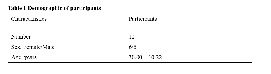
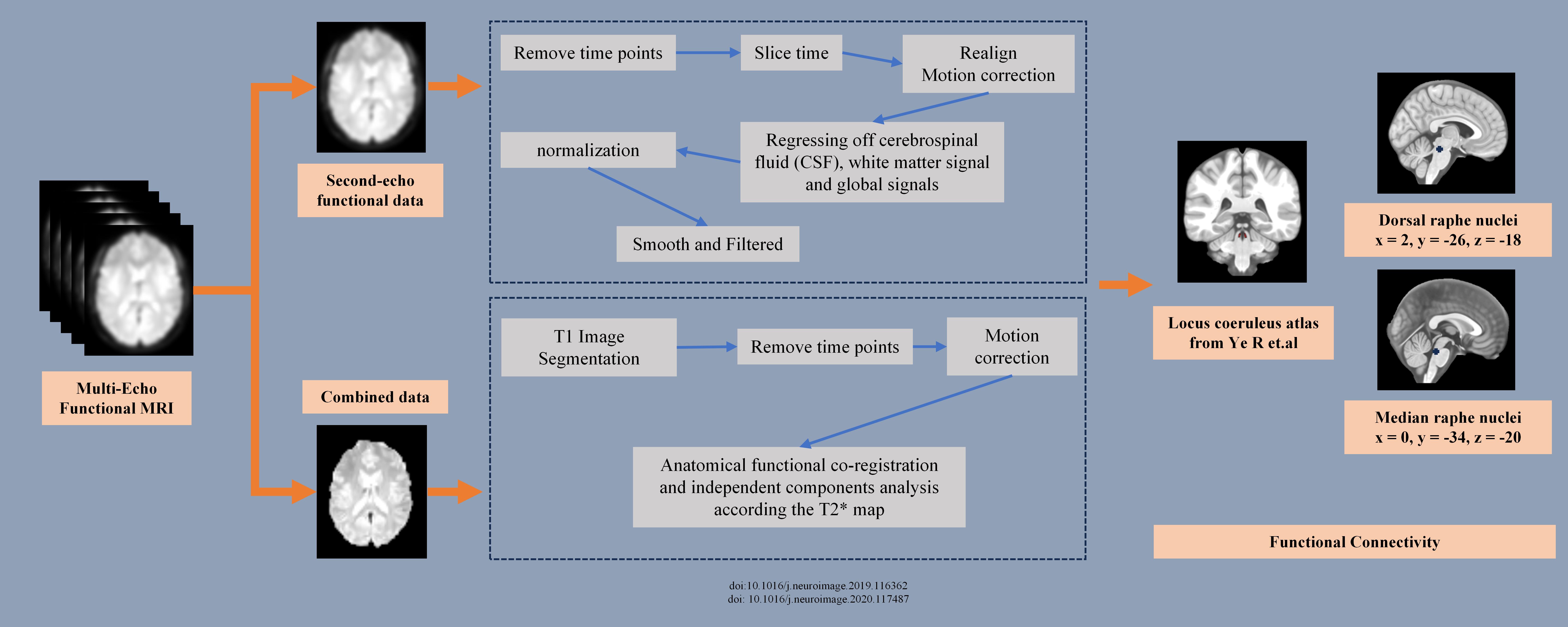
Figure 1: Workflow of preprocessing and functional connectivity analysis.
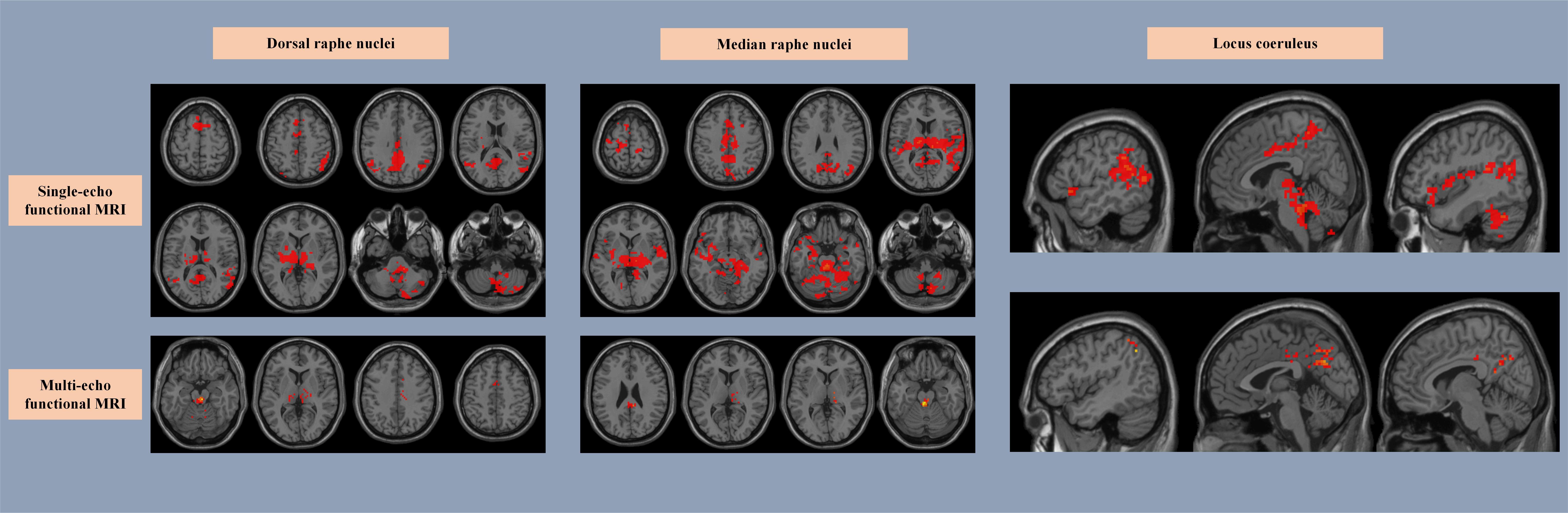
Figure 2: Functional connectivity patterns of three brainstem nuclei between two methods.