3421
Submillimeter Whole-Brain VASO fMRI using a View-Sharing with Temporal Random Walk at 7 Tesla1Chonnam National University, Gwangju, Korea, Republic of, 2University of California, Berkeley, Berkeley, CA, United States, 3Advanced MRI Technologies, Sebastopol, CA, United States, 4Siemens Medical Solutions USA, Inc, Berkeley, CA, United States
Synopsis
Keywords: fMRI Acquisition, Brain, functional MRI
Motivation: Cerebral blood volume (CBV) fMRI has inherent limitations regarding its imaging efficiency, restricting the spatial volume coverage and spatiotemporal resolution
Goal(s): To improve imaging efficiency for whole-brain VASO fMRI with high temporal resolution
Approach: we introduce a view sharing approach combined with flexible temporal encoding
Results: Proposed method achieves whole-brain VASO and BOLD imaging in 8.3 seconds, while resulting in higher VASO and BOLD activations with high sensitivity
Impact: We confirmed that 1) a view-sharing approach allows VASO and BOLD signal detection without contrast confounding and 2) the random encoding coupled with view sharing significantly improves the imaging efficiency by achieving an 8.3 second whole-brain VASO imaging.
Introduction
Cerebral blood volume (CBV) fMRI has the potential to overcome several specific limitations of BOLD fMRI1. This allows direct physiological interpretation of the data and promises superior localization specificity at ultra-high magnetic fields. Despite the advantages of non-invasive CBV fMRI, there are inherent limitations regarding imaging efficiency, restricting the spatial volume coverage and spatiotemporal resolution. Recent advances in acceleration and reconstruction have mitigated this limitation, such as SMS 2D-EPI or 3D EPI in combination with CAIPI field-of-view shifting. To further improve imaging efficiency, we developed a novel whole-brain VASO fMRI imaging method by introducing view sharing approach combined with flexible temporal encoding4. Experiments confirm the advantages in volume coverage, SNR, sensitivity of the proposed acquisition method: 1) view-sharing approach between VASO and BOLD imaging increases the spatial coverage up to the whole-brain or slab coverage (e.g., 0.64mm-iso and 0.48mm-iso resolutions while maintaining TR in the 5 to 8 second range), and 2) flexible temporal encoding provides higher VASO and BOLD activations with over 9-fold accelerations.Methods
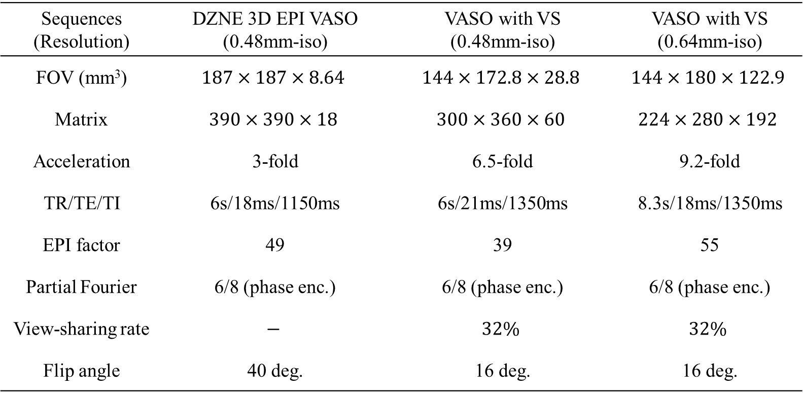
MRI hardware: All fMRI data were collected on the Berkeley Next generation 7T MAGNETOM Terra scanner5 (Siemens Healthcare, Erlangen, Germany) equipped with the investigational Impulse gradient system (200 mT/m and 900 T/m/s gradient system) and a 64-channel head receiver coil (MR CoilTech Ltd, Glasgow, UK). Imaging parameters are summarized in Table 1.Pulse sequence: The pulse sequence diagram and the associated z-magnetization of blood and gray matter are depicted in Fig. 1. Two contrasts of VASO and BOLD are continuously acquired with an inversion pulse. To acquire blood-nulled images for VASO contrast, a nonselective hyperbolic secant adiabatic pulse is employed by putting the center of k-space around the expected blood-nulling time TI. The acquisition of the second volume is acquired without a preceding inversion pulse for not-nulled BOLD contrast. Based on the fact that the central k-space is responsible for the overall image contrast while the peripheral k-space is responsible for image details, the outer k-space is shared between the two imaging volumes when the steady-state is reached to ensure that signal transition in the longitudinal direction has minor influence of the volume sharing. To further improve the imaging efficiency across time, the sampling pattern is designed by combining a variable density CAIPI encoding with temporal random walk under the framework of EPI acquisition introduced in our previous work.
Image reconstruction: The acquired data is reconstructed using coil sensitivity and temporal priors to exploit random undersampled spatiotemporal data structure. For self-calibration, the coil sensitivity was calculated by using an averaging k-t data. For temporal redundancy, dynamic compressed sensing was used using low rank and sparse priors6.
Stimulation timing and image analysis: Brain activation was measured using a flashing checkerboard task (18s rest followed by 10 blocks of 30s stimulation and 30s rest) presented using PsychoPy and a 7T compatible projector with extra long throw lens (VPixx Technologies, Inc). Data were sorted by CBV and oxygen level contrast, motion corrected using AFNI and corrected for BOLD contamination (LayNii). Voxel-wise task activation was estimated using a conventional GLM analysis (FSL FEAT), with high-pass filtering (0.008 Hz) and correction for temporal autocorrelations.
Results and Conclusion
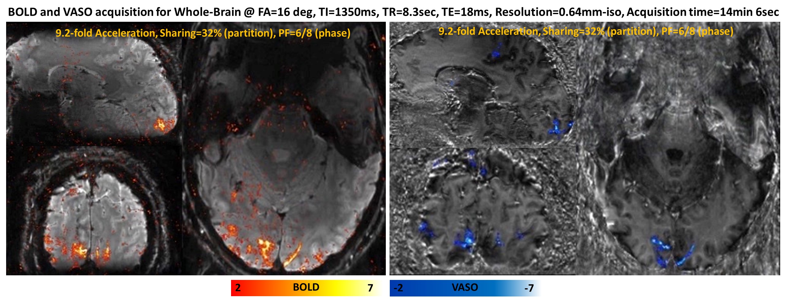
Fig. 2 shows high image quality in whole-brain 3D volumes for VASO and BOLD contrasts with 0.64mm-iso resolution according to different sharing rates of 25% and 37.5%. Based on the observation, we set the sharing rate to 32% along the partition direction. Fig. 3 shows functional VASO and BOLD activations between DZNE 3D EPI VASO and our proposed VASO sequence for 0.48mm-iso resolution. Note that our proposed view-sharing and temporal random encoding shows higher sensitivities to BOLD and VASO. Fig. 4 shows the feasibility of our proposed VASO sequence for whole-brain application with 0.64mm-iso resolution using TR=8.3sec and 9.2-fold acceleration. We confirmed that 1) a view-sharing approach allows VASO and BOLD signal detection without contrast confounding and 2) the random encoding coupled with view sharing significantly improves the imaging efficiency by allowing whole-brain VASO imaging compared to longer acquisition of DZNE 3D EPI VASO imaging.Acknowledgements
This project is supported in part by the NIH BRAIN Initiative (R44-MH129278. , U01EB025162), 1R44MH129278 (to Feinberg), and NRF/MSIT No. 2021R1C1C1013603 (to Park).
This research was supported by the MSIT(Ministry of Science and ICT), Korea, under the ICAN(ICT Challenge and Advanced Network of HRD) program(IITP-2023-RS-2022-00156385) supervised by the IITP(Institute of Information & Communications Technology Planning & Evaluation).
This research was supported by the MSIT(Ministry of Science and ICT), Korea, under the Innovative Human Resource Development for Local Intellectualization support program(IITP-2023-RS-2022-00156287) supervised by the IITP(Institute for Information & communications Technology Planning & Evaluation).
References
[1] Huber L, Ivanov D, Handwerker DA, Marrett S, Guidi M, Uludağ K, Bandettini PA, Poser BA. Techniques for blood volume fMRI with VASO: From low-resolution mapping towards sub-millimeter layer-dependent applications. Neuroimage. 2018; 164:131-143.
[2] Le Ster C, Moreno A, Mauconduit F, Gras V, Stirnberg R, Poser BA, Vignaud A, Eger E, Dehaene S, Meyniel F, Boulant N. Comparison of SMS-EPI and 3D-EPI at 7T in an fMRI localizer study with matched spatiotemporal resolution and homogenized excitation profiles. Plos one. 2019;14(11):e0225286.
[3] Stirnberg R, Stöcker T. Segmented K‐space blipped‐controlled aliasing in parallel imaging for high spatiotemporal resolution EPI. Magn Reson Med. 2021; 85(3):1540-51
[4] Suhyung P, Alexander B, Suvi H, Samantha M. Whole-brain Sub-millimeter Resolution fMRI using 3D EPI Accelerated with Temporal Random Walk. In Proceedings of the international society of magnetic resonance in medicine 2023; p.3295.
[5] Feinberg DA, Dietz P, Liu C, Setsompop K, Mukherjee P, Wald LL, Vu AT, Beckett A, Insua IG, Schröder M, Stocker S, Bell PH, Rummert E, Davids M. Design and Development of a Next-Generation 7T human brain scanner with high-performance gradient coil and dense RF arrays. In Proceedings of the international society of magnetic resonance in medicine 2021; p.0562.
[6] Trémoulhéac, B, Nikolaos D, David A, and Simon RA. Dynamic MR Image Reconstruction–Separation From Undersampled k-t Space via Low-Rank Plus Sparse Prior." IEEE Trans Med Imaging 2014; 33: 1689-1701.
Figures
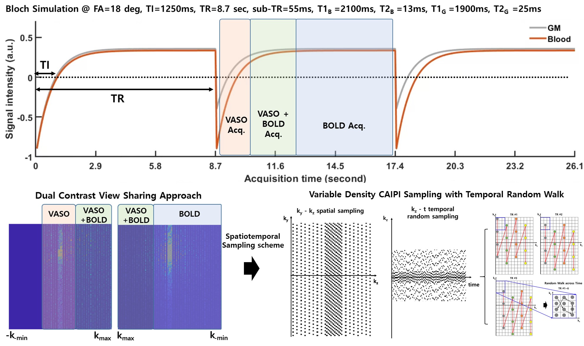
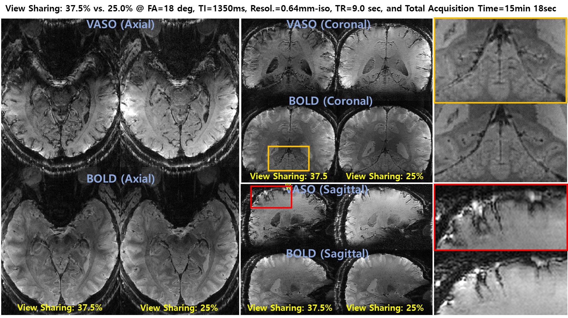
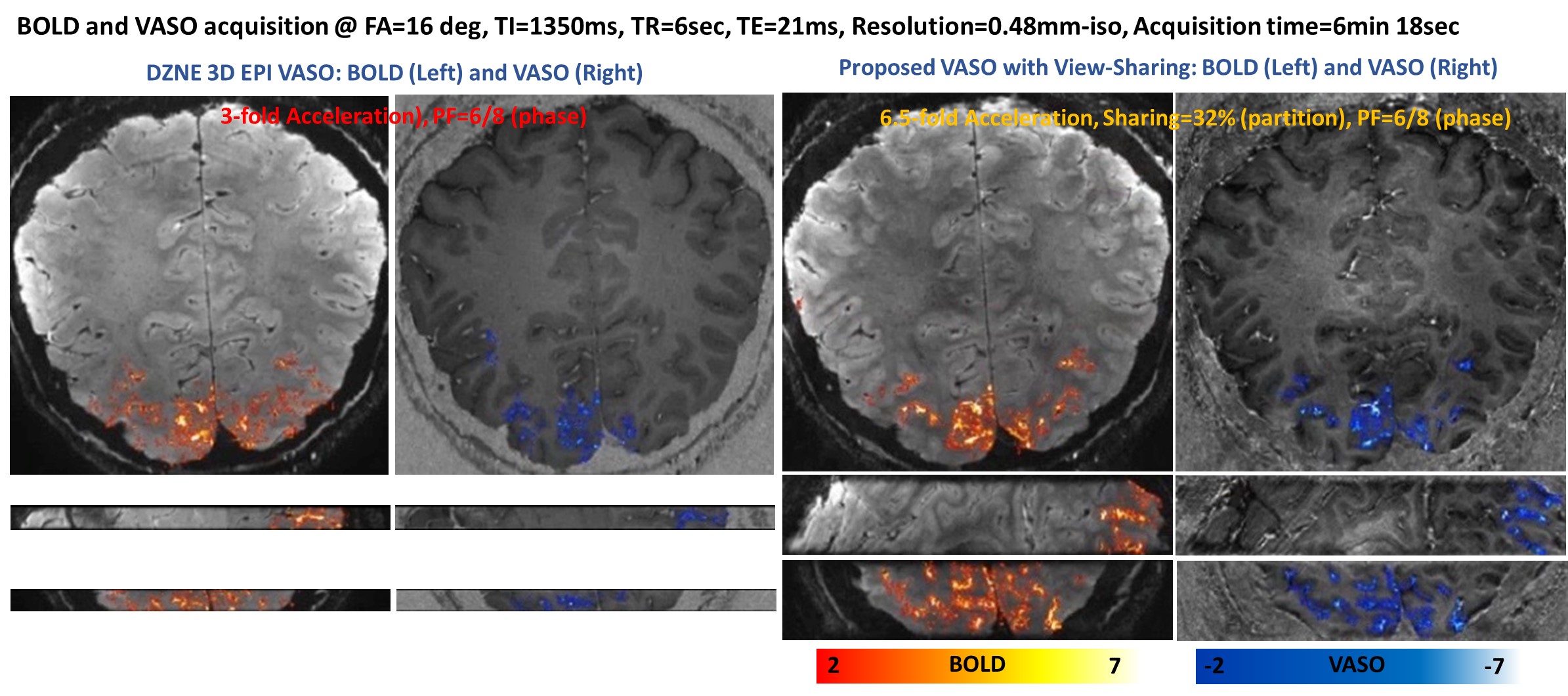
Fig 3. Comparison of BOLD and VASO activations using DZNE 3D EPI VASO (left) and proposed VASO with view-sharing and random encoding (right) for 0.48mm-iso resolution. Note that the proposed method achieves much greater slab volume coverage of brain with view-sharing and random k-space encoding.