3420
Achieving high temporal resolution using a sliding-window approach for SPARKLING fMRI data: A simulation study1Université Paris-Saclay, CEA, NeuroSpin, CNRS, Gif-sur-Yvette, France, Gif-sur-Yvette, France, 2Université Paris-Saclay, Inria, MIND, Palaiseau, France, Palaiseau, France
Synopsis
Keywords: fMRI Acquisition, fMRI, sliding-window, high temporal resolution, BOLD
Motivation: Higher temporal resolution in fMRI has been increasingly sought after, primarily because of a greater interest in the high-frequency components of BOLD, but also because high temporal resolution is essential to get rid of physiological artifacts.
Goal(s): In light of this mounting interest, we explore the sliding-window approach to improve the temporal resolution, specifically using SPARKLING fMRI.
Approach: A simulation study is conducted for 2D BOLD fMRI at realistic SNR.
Results: It demonstrates the possibility through the sliding window approach to detect oscillations beyond 0.2Hz in the BOLD response and separate the physiological noise from the neural activity.
Impact: Such a strategy can be extended to 3D imaging in a straightforward manner, thereby making whole-brain high spatiotemporal resolution fMRI feasible.
Introduction
As an indirect measure of neural activity, blood oxygenation level-dependent (BOLD) effect1 has slower dynamics than the neural activity itself, partly due to the data acquisition process and partly due to the sluggishness of the hemodynamic response function (HRF)2. However, while the slowness of BOLD with regard to neural events is widely acknowledged, its inherent temporal resolution remains ambiguous. Despite some precursor interest3,4, the temporal dimension received less attention than the spatial dimension, as fMRI was primarily used as a brain mapping tool. Nevertheless, this has changed in recent years primarily because of a greater interest in the high-frequency components of BOLD5,6,7 but also because high temporal resolution is essential to get rid of physiological artifacts related to breathing or heart rate8,9. Most current attempts achieve high temporal resolution at the expense of either diminished brain coverage10,11 or a loss in spatial resolution12,13. The assumption of repeatability has also been used to enhance temporal resolution14 as well as sharing data between different frames15,16,17. In this context, we re-investigate the sliding-window approach introduced in15 and demonstrate its ability detect oscillations beyond 0.2Hz and to disentangle physiology from neural activity in simulations of 2D BOLD fMRI using non-Cartesian In-Out SPARKLING18,19 sampling pattern (characterized by the fact that each shot crosses the center of k-space at the echo time) in a scan-and-repeat mode.Methods
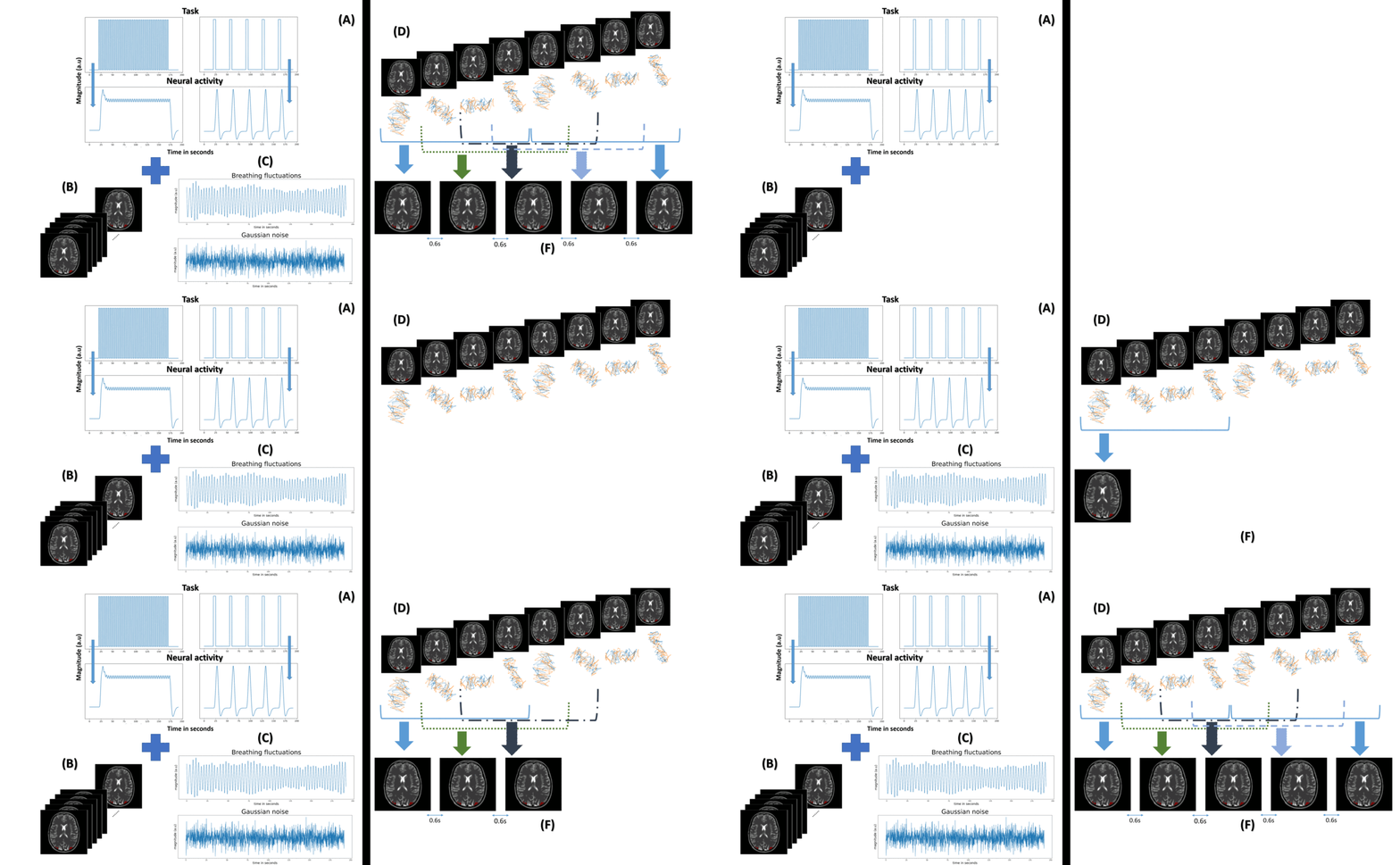
To study the potential benefit of the sliding-window approach applied to 2D SPARKLING BOLD fMRI, simulated data was synthesized according to the pipeline presented in Fig.1.(A)Highly temporally-resolved simulated neural activity: Two types of neural events are convolved with the Glover HRF to produce the ground truth or reference for slow and fast BOLD responses at a temporal resolution of 0.1s.
(B)Highly temporally-resolved ground truth of 2D fMRI frames: The BOLD responses in (A) are used to modulate the magnitude of the voxels of a chosen region of interest (ROI) in a sequence of a 2D (1mm2 and (192x192)mm2 FOV) numerical brain phantom.
(C)Highly temporally-resolved physiological and thermal noises: Gaussian noise and breathing fluctuations are emulated over all voxels. A given signal-to-noise ratio (SNR) is chosen, and the maximum intensity change due to breathing fluctuations is defined as 0.2% of the baseline magnitude. The SNR here equals the temporal SNR.
(D)Retrospective k-space sampling: The k-space data is synthesized by sampling each 2D frame using 2 consecutive shots (from a complete sampling pattern of 48 shots), meaning at a pace of 0.1s since each shot corresponds to TRshot=50ms. Therefore, every 24-period spaced frames are sampled using the same trajectories.
(E)Sequential fMRI image reconstruction at the native TR (TRnative=2.4s): The k-space data generated in (D) is gathered to form 48-shot blocks. Such a large number of shots (and therefore TRnative) is not necessary for a single MRI slice and reflects more a 3D acquisition setup. Here, we chose, however, to proceed in 2D for the sake of simplicity, computational efficiency, and demonstration. The reconstruction is performed using a simple zero-filled adjoint.
(F)Sliding-window fMRI image reconstruction at the effective TR (TReff=0.6s): A sliding-window approach of width and stride of 48 and 12 shots, respectively, is used to reconstruct consecutive frames from overlapping k-space data.
Results
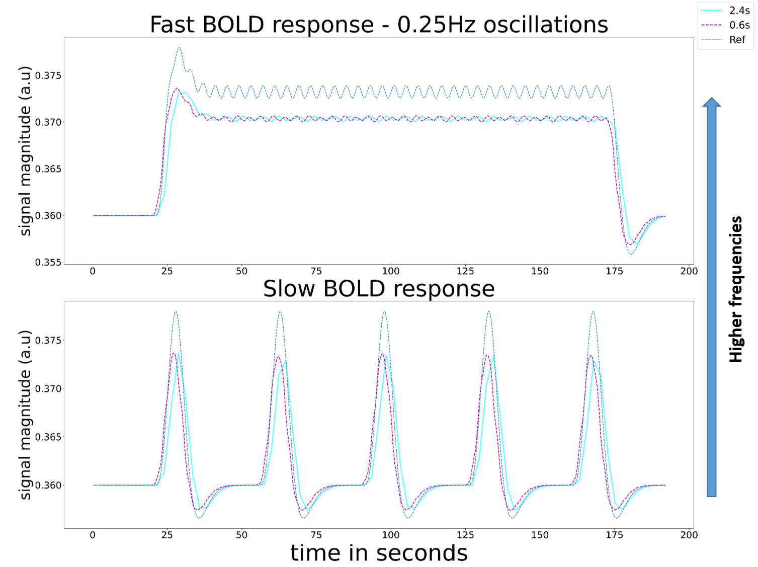
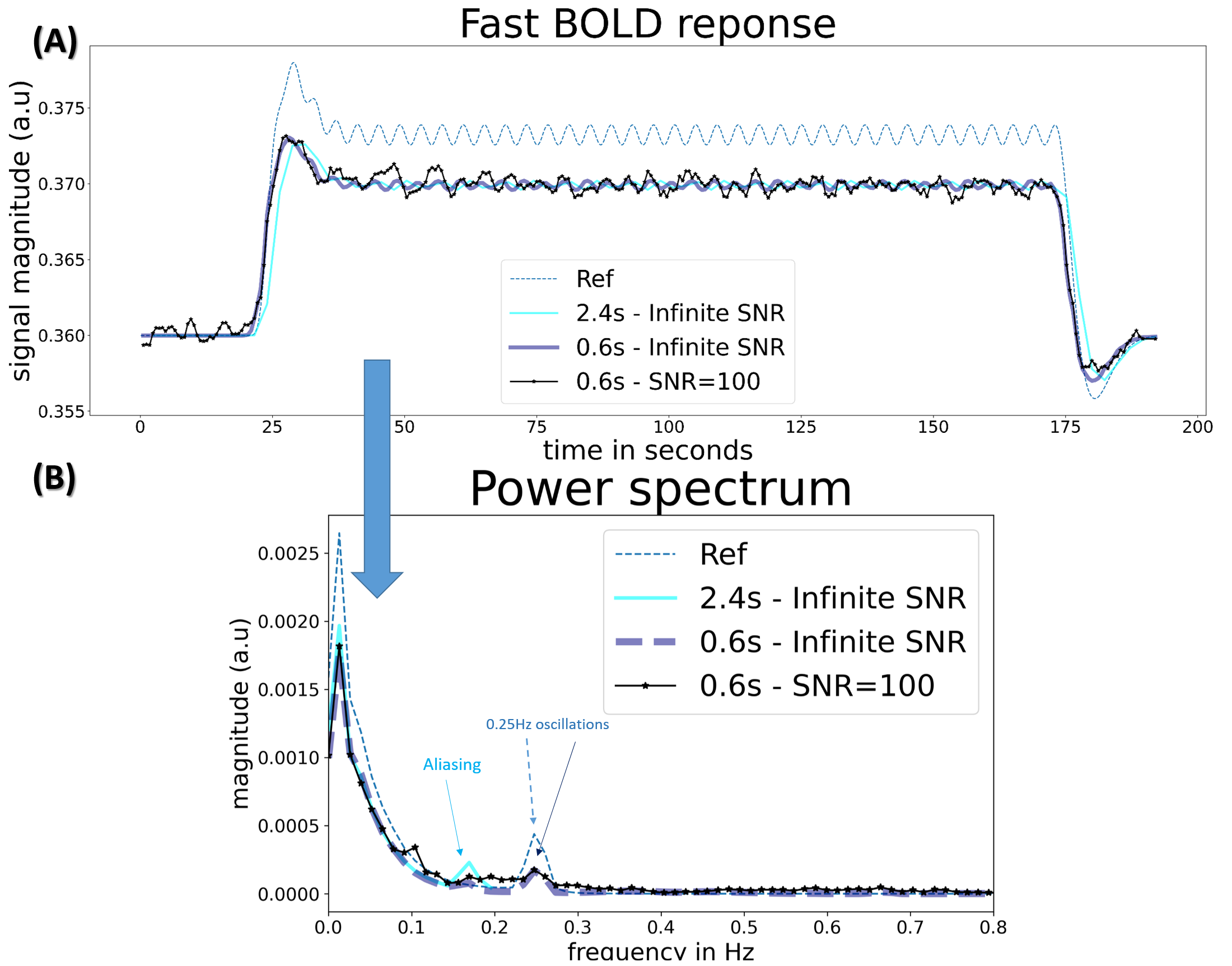
Through two scenarios, the first characterized by a fast BOLD response and the second by a slow one, Fig.2 showcases the ability of the sliding-window approach to reveal some high-frequency neural activity/dynamics as it enables the tracking of rapid fluctuations.Fig.3 demonstrates that it is possible to track BOLD fluctuations at 0.25Hz for an SNR=100 (leading to realistic tSNR).
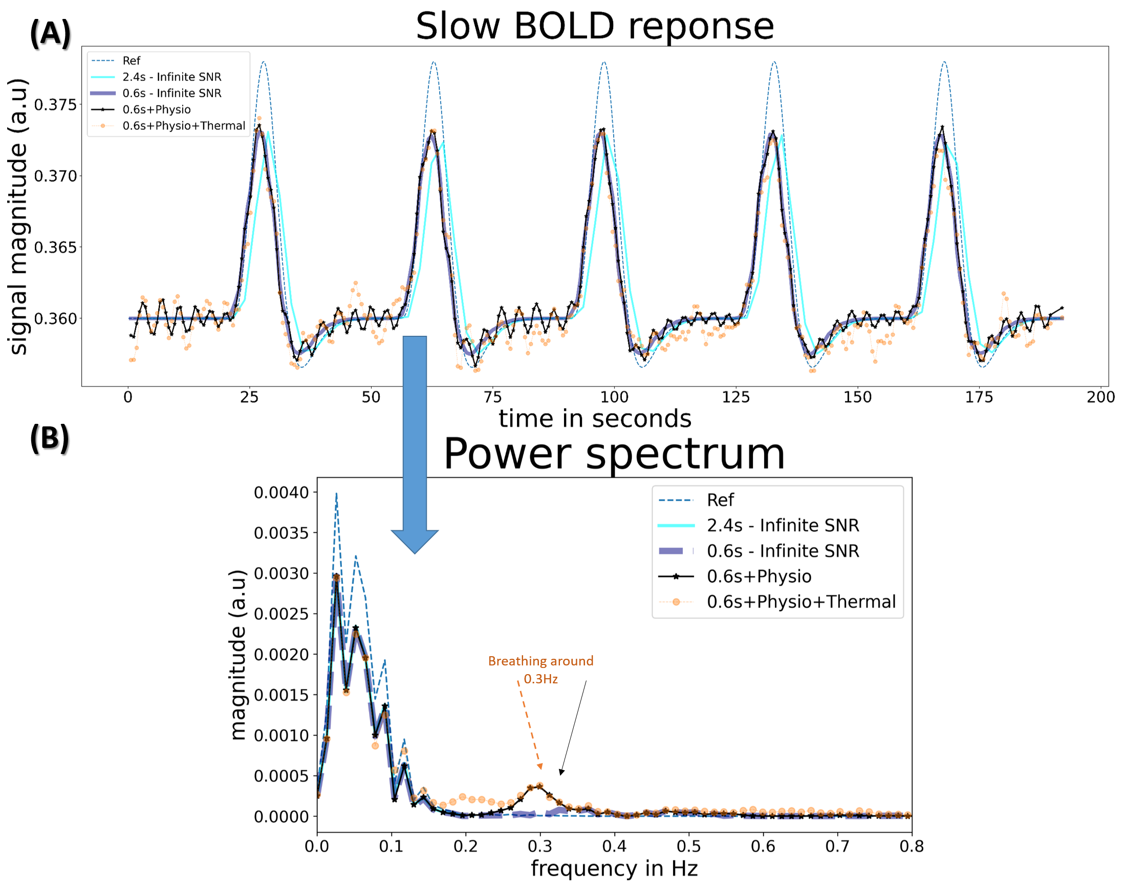
Fig.4 shows that it's possible to effectively resolve and separate physiological noise from neural activity using the sliding-window technique.
Discussion
As neural events become faster, the convolution with the conventional HRF model2 is expected to produce highly attenuated oscillations in the BOLD response that can be easily drowned in thermal noise. This is why we chose to stick to 0.25Hz BOLD oscillations for the sake of demonstration. TReff=0.6s is sufficiently short to detect such oscillations. However, shorter TReff are achievable. Furthermore, in5, the authors demonstrate that fast BOLD oscillations detected in vivo have higher amplitudes than those expected theoretically. The ability of the sliding-window approach to detect high-frequency oscillations in BOLD responses in vivo ought to be further investigated.Conclusion
This simulation-based work demonstrates that a sliding-window reconstruction method allows for the tracking of BOLD oscillations beyond 0.2Hz and separation of physiological noise from neural activity at realistic SNR/tSNR in 2D. Such a strategy can be extended to 3D imaging in a straightforward manner, thereby making whole-brain high spatiotemporal resolution fMRI feasible.Acknowledgements
No acknowledgement found.References
1S. Ogawa et al., “Brain magnetic resonance imaging with contrast dependent on blood oxygenation,” Proceedings of the National Academy of Sciences of the United States of America, vol. 87, no. 24, pp. 9868–9872, 1990.
2K. J. Friston et al., “Analysis of fMRI Time-Series Revisited,” NeuroImage, vol. 2, no. 1, pp. 45–53, 1995.
3P. A. Bandettini, “The spatial, temporal, and interpretive limits of functional MRI,” Neuropsychopharmacology: The Fifth Generation of Progress: An Official Publication of the American College of Neuropsychopharmacology, 2002.
4R. L. Savoy et al., “Pushing the temporal resolution of fMRI: Studies of very brief visual stimuli, onset variability and asynchrony, and stimulus-correlated changes in noise,” Program of SMR presented at 3d Annual Meeting, 1995.
5L. D. Lewis et al., “Fast fMRI can detect oscillatory neural activity in humans,” The Proceedings of the National Academy of Sciences, vol. 113,no. 43, E6679–E6685, 2016.
6J. E. Chen et al., “Investigating mechanisms of fast BOLD responses: The effects of stimulus intensity and of spatial heterogeneity of hemodynamics,” NeuroImage, vol. 245, p. 100-288, 2021.
7J. R. Polimeni and L. D. Lewis, “Imaging faster neural dynamics with fast fMRI: A need for updated models of the hemodynamic response,” Progress in neurobiology, vol. 207, p. 102 174, 2021.
8H. Jahanian et al., “Advantages of short repetition time resting-state functional MRI enabled by simultaneous multi-slice imaging,” Journal of Neuroscience Methods, vol. 311, pp. 122–132, 2019.
9N. Huotari, “Sampling Rate Effects on Resting State fMRI Metrics,” Frontiers in Neuroscience, vol. 13, p. 279, 2019.
10L. Raimondo et al., “A line through the brain: Implementation of human line-scanning at 7T for ultra-high spatiotemporal resolution fMRI,” Journal of Cerebral Blood Flow and Metabolism, vol. 41, no. 11, pp. 2831–2843, 2021.
11L. Raimondo et al., “Robust high spatio-temporal line-scanning fmri in humans at 7T using multi-echo readouts, denoising and prospective motion correction,” Journal of Neuroscience methods, vol. 384, p. 109746, 2023.
12A. P. Leynes et al., Extreme looping star: Quiet fMRI at high spatiotemporal resolution, Proceedings of the 29th Scientific Meeting of the International Society for Magnetic Resonance in Medicine 2021.
13S. Bianchi et al., “T-hex spirals for whole-brain fMRI at 5 frames per second: Image quality and bold sensitivity,” in Proceedings of the 30th Scientific Meeting of the International Society for Magnetic Resonance in Medicine, 2022.
14M. Dziadosz et al., “High temporal resolution blood oxygen level dependent functional MRI,” Proceedings of the 31rst Scientific Meeting of the International Society for Magnetic Resonance in Medicine, 2023.
15M. Kramer et al., “Functional magnetic resonance imaging using PROPELLER-EPI,” Magnetic Resonance in Medicine, vol. 68, no. 1, pp. 140–51, 2012.
16S. D. Yun et al., “Parallel imaging acceleration of EPIK for reduced image distortions in fMRI,” Neuroimage, vol. 73, pp. 135–43,1262013.
17S. D. Yun et al., “Evaluating the utility of EPIK in a finger tapping fMRI experiment using bold detection and effective connectivity,” Scientific Reports, vol. 9, p. 10978, 2019.
18C. Lazarus et al., “SPARKLING: variable-density k-space filling curves for accelerated T2*-weighted MRI,” Magnetic Resonance in Medicine, vol. 81, no. 6, pp. 3643–3661, 2019.
19GR Chaithya et al., “Optimizing full 3D SPARKLING134trajectories for high-resolution Magnetic Resonance imaging,” IEEE Transactions on Medical135Imaging, vol. 41, no. 8, pp. 2105–2117, 2022.
Figures