3418
Multi-echo fMRI at 7T in infants1Masonic Institute for the Developing Brain, University of Minnesota, Minneapolis, MN, United States, 2Institute of Child Development, University of Minnesota, Minneapolis, MN, United States, 3Center for Magnetic Resonance Research, University of Minnesota, Minneapolis, MN, United States, 4Department of Neuroscience, University of Minnesota, Minneapolis, MN, United States, 5Department of Pediatrics, University of Minnesota, Minneapolis, MN, United States
Synopsis
Keywords: fMRI Acquisition, High-Field MRI, Infants, multi-echo EPI, 7T, functional connectivity, developmental neuroimaging
Motivation: Imaging infants with traditional voxel sizes is suboptimal due to their smaller brains. 7T imaging allows for higher spatial resolutions, while multi-echo fMRI provides optimal contrast across the brain.
Goal(s): Establish the feasibility of high-resolution multi-echo fMRI in infants at 7T.
Approach: Acquire data in the same infant at 3T/7T within the same week, allowing for direct comparisons, while also considering unique safety guidelines for 7T infant imaging.
Results: Initial data demonstrate 3T/7T multi-echo fMRI in the same infant, establishing the feasibility of high-resolution ME-fMRI at 7T in infants with high contrast, sensitivity, and stronger functional connections.
Impact: This first demonstration of multi-echo fMRI at 7T, including the use of voxel volumes (1.6 mm3) that are half the volume of what is normally used in 3T infant fMRI (2.0 mm3), promises opportunities to advance developmental neuroimaging.
INTRODUCTION
Important insight into brain structure and function during early development can be gained from fMRI in infants. However, a number of methodological challenges arise when working with this age group that go beyond practical considerations. The lack of commercially available head coils optimized for developmental populations necessitates the use of head coils designed for adults, resulting in sub-optimal signal-to-noise ratios and increases in partial voluming because of the smaller brain sizes relative to the standard voxel sizes1. Higher spatial resolutions can be achieved by moving to higher field strengths (i.e. > 3T). However, despite the availability of FDA approved 7T MRI scanners, they are rarely used in infants because it requires additional safety considerations2. Further, the most popular 7T system, the Siemens Magnetom Terra, is currently not FDA-approved for subjects as light as infants (<30kg). Independent of field strength, finding optimal acquisition protocols, which consider the tissue properties of the developing brain, is an additional challenge. We recently showed that multi-echo (ME) fMRI at 3T could be a promising tool to account for this in a developmental population3. Up to now, the application of ME fMRI sequences have not been tested in infants at 7T. We show initial results from a high resolution 7T ME-fMRI acquisition.METHODS
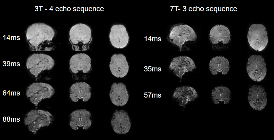
The example infant presented here was a healthy full-term infant, seven weeks old (48 weeks postmenstrual age) from whom we acquired 3T and 7T data within a four day period. To make 7T acquisition possible, we developed an in-house system to assess the safe operating power limits for a newborn infant. Functional data at 3T was acquired using a four-echo sequence (14ms, 39ms, 64ms, 88ms, TR = 1.761s, 2mm resolution, MB factor = 6, flip angle = 68°). Functional data at 7T was acquired using a three-echo sequence (14ms, 35ms, 57ms, TR = 1.768s, 1.6mm resolution, MB factor = 4, flip angle = 60°). T2w and T1w anatomical references were acquired at 3T (T2: TR = 42.3s, TE = 323ms, resolution = 0.8 x 0.8 x 0.8 mm, flip angle = 120°; T1: TR = 2.4s, TE = 2.2ms, resolution = 0.8 x 0.8 x 0.8 mm, flip angle = 8°). All data acquisition was performed during natural sleep. We preprocessed data using NORDIC4 for thermal denoising, BIBSnet5 for creating segmentations of the anatomical data, Nibabies6 with its multi-echo preprocessing workflow and XCP-D7 for functional connectivity processing. Functional connectivity matrices were calculated using low motion data (framewise displacement < 0.3mm) only.RESULTS
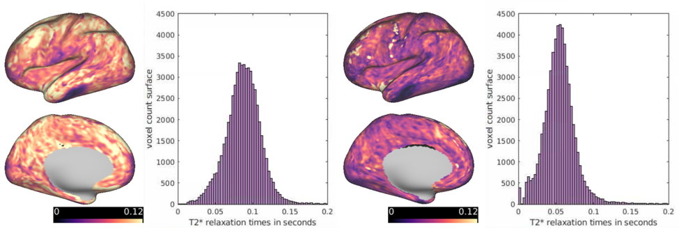
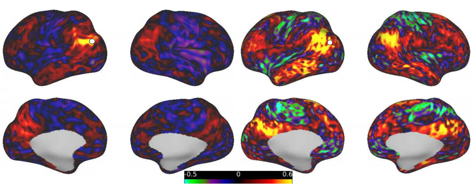
All ME data acquired at 3T and 7T showed high functional tissue contrast, indicating that despite halving the voxel volume at 7T, sensitivity was still not limited (Figure 1). As expected, T2* relaxation times for the same infant across both field strengths were shorter in 7T compared to 3T (Figure 2). At both field strengths, T2* across the cortex revealed a similar variance and showed similar areas with shorter T2*s due to susceptibility artifacts, highlighting the benefits of ME imaging. Functional connectivity matrices showed similar correlation patterns between brain regions for data acquired at 3T and 7T. However, the absolute magnitude of functional connections was higher in data acquired at 7T (Figure 3).DISCUSSION
Our initial results, using data from the same infant acquired at 3T and 7T within a short time span, show that ME fMRI in infants at 7T is not only feasible but feasible with much higher resolutions, resulting in data with high specificity and sensitivity. Investigation of T2* relaxation times points towards similar advantages of ME fMRI in infants in 7T as previously shown in 3T3. The observed increase in functional connectivity strength is consistent with the adult literature8, however, it is notable that the 7T voxels are half the volume. Overall, the smaller voxel size achievable at 7T is more suitable for the size of an infant brain and could be very promising for developmental neuroimaging. Moving forward, further benefits for functional connectivity research that go beyond the absolute strength of connections can be explored as well as even smaller voxel sizes.CONCLUSION
Initial results of 7T fMRI in a seven week old infant look very promising and show multiple advantages compared to the same infant’s 3T data. Given careful safety considerations and suitable acquisition protocols, 7T imaging could be established as a promising tool for developmental neuroimaging.Acknowledgements
This work was supported by the following grants: NIBIB P41 EB027061 and NINDS R01NS115180.References
1. Dubois, J., Alison, M., Counsell, S. J., Hertz-Pannier, L., Hüppi, P. S., & Benders, M. J. N. L. (2021). MRI of the Neonatal Brain: A Review of Methodological Challenges and Neuroscientific Advances. Journal of Magnetic Resonance Imaging: JMRI, 53(5), 1318–1343.
2. Malik, S. J., Hand, J. W., Carmichael, D. W., & Hajnal, J. V. (2022). Evaluation of specific absorption rate and heating in children exposed to a 7T MRI head coil. Magnetic Resonance in Medicine: Official Journal of the Society of Magnetic Resonance in Medicine / Society of Magnetic Resonance in Medicine, 88(3), 1434–1449.
3. Moser, J., Koirala, S., Madison, T., Labonte, A. K., Carrasco, C. M., Feczko, E., Moore, L. A., Ahmed, W., Myers, M. J., Yacoub, E., Trevo-Clemmens, B., Larsen, B., Laumann, T. O., Nelson, S. M., Vizioli, L., Sylvester, C. M., & Fair, D. A. (2023). Multi-echo Acquisition and Thermal Denoising Advances Infant Precision Functional Imaging. In bioRxiv (p. 2023.10.27.564416). https://doi.org/10.1101/2023.10.27.564416
4. Vizioli, L., Moeller, S., Dowdle, L., Akçakaya, M., De Martino, F., Yacoub, E., & Uğurbil, K. (2021). Lowering the thermal noise barrier in functional brain mapping with magnetic resonance imaging. Nature Communications, 12(1), 5181.
5. Hendrickson, T. J., Reiners, P., Moore, L. A., Perrone, A. J., Alexopoulos, D., Lee, E. G., Styner, M., Kardan, O., Chamberlain, T. A., Mummaneni, A., Caldas, H. A., Bower, B., Stoyell, S., Martin, T., Sung, S., Fair, E., Uriarte-Lopez, J., Rueter, A. R., Rosenberg, M. D., … Feczko, E. (2023). BIBSNet: A Deep Learning Baby Image Brain Segmentation Network for MRI Scans. In bioRxiv (p. 2023.03.22.533696). https://doi.org/10.1101/2023.03.22.533696
6. Goncalves, M., Markiewicz, C. J., Esteban, O., Feczko, E., Poldrack, R. A., & Fair, D. A. (2023). NiBabies: a robust preprocessing pipeline for infant functional MRI. https://doi.org/10.5281/zenodo.7562647
7. Adebimpe, A., Bertolero, M., Mehta, K., Salo, T., Murtha, K., Cieslak, M., Meisler, S., Madison, T., Sydnor, V., Covitz, S., Fair, D., & Satterthwaite, T. (2023). XCP-D : A Robust Postprocessing Pipeline of fMRI data. https://doi.org/10.5281/zenodo.7640010
8. Vu, A. T., Jamison, K., Glasser, M. F., Smith, S. M., Coalson, T., Moeller, S., Auerbach, E. J., Uğurbil, K., & Yacoub, E. (2017). Tradeoffs in pushing the spatial resolution of fMRI for the 7T Human Connectome Project. NeuroImage, 154, 23–32.
Figures