3417
Simultaneous high-resolution BOLD and fiber photometry of awake mice whisking shows neuromodulation response of acetylcholine1A.A. Martinos Center for Biomedical Imaging, Department of Radiology, Massachusetts General Hospital, Boston, MA, United States, 2Department of Radiology, Harvard Medical School, Boston, MA, United States
Synopsis
Keywords: fMRI Analysis, fMRI, preclinical fMRI, awake fMRI, multimodal imaging, whisker stimulation, whisker tracking
Motivation: Anesthesia causes alterations in brain functional data and internal brain states affect sensory perception.
Goal(s): We sought to investigate how the Blood Oxygen Level Dependence (BOLD) response from spontaneous and stimulated whisker motion in awake mice is related to cerebral arousal state monitored by acetylcholine.
Approach: Mice with implanted RF coils and fiber optic cable were simultaneously imaged and acetylcholine signals were monitored to measure correlation between BOLD and acetylcholine signals.
Results: We found stimulating the whiskers produced the expected contralateral BOLD response in awake animals but also an acetylcholine spike in the ipsilateral side indicating whole-brain dynamic activation occurs in awake mice.
Impact: This work shows feasibility of simultaneous high-resolution EPI-based functional imaging and fiber photometry of acetylcholine response to whisker stimulation in awake mice as a multimodal method for investigating brain state from external sensory stimulation using implantable RF coils.
Introduction
Functional magnetic resonance imaging (fMRI) can be challenging in preclinical settings. Motion from animals causes imaging artifacts. Additionally, anesthesia can cause alterations in brain functional data and internal brain states affect sensory perception. This study seeks to investigate if the Blood Oxygen Level Dependance (BOLD) response from spontaneous and stimulated whisker motion in awake mice can be related to cerebral arousal state by utilizing lightweight implantable RF coils. Cholinergic axons are known to increase activity in the barrel cortex during whisking and provide the primary source of acetylcholine to the cerebral cortex. Data acquisition was accomplished using a 14T horizontal bore scanner and 3D printed animal cradle with mounted camera to measure whisking in awake (non-anesthetized) mice.Materials and Methods
Animal Model: Male C57BL/6J mice (25-30g) underwent an implantation of 600MHz single loop RF coils and fiber optic cable for head fixation and record acetylcholine signal. Genetically encoded reporter AAV-hsyn-Ach4.3(Ahc3.0) expressed by the AAV9 virus was injected into the left barrel cortex. 3 weeks post-injection, an optical fiber (200µm) was inserted into the barrel cortex expressing the fluorescent biosensors and secured with 2-part dental cement. The coil was positioned over the target area 1mm above the exposed skull and attached using cyanoacrylate glue and 2-part dental cement.MR Techniques: High resolution functional images were acquired in awake mice at 14T. fMRI data was acquired with a multi-slice 2D EPI scan with 2 segments to reduce distortions. In-plane resolution of 100x100µm, matrix size of 144x96, slice thickness of 200µm, repetition time of 1 s, echo time of 6.2 ms, and 205 repetitions were used resulting in an acquisition time of 6 minutes 50 seconds (2 s/repetition). A small camera was positioned in front of the mouse (at a distance to eliminate any susceptibility distortions of the image) to visualize its whiskers during experiments.
Anatomical images for EPI localization were acquired using multi-slice T1-weighted 2D gradient echo Fast Low Angle Shot (FLASH). Mice were imaged with an identical resolution and geometry of the EPI. Repetition time of 475ms, echo time of 3ms, 30o flip angle, and 2 averages were used for an approximate acquisition time of 3 minutes.
ACH Recording: Acetylcholine signals were recorded for the duration of each EPI scan using BIOPAC
Data Analysis: fMRI data was processed using Analysis of Functional Neuroimages (AFNI)1. Simultaneous videos of whisking were used to pinpoint the times of BOLD response during scans. Bruker 2dseq images were converted to AFNI format using ‘to3d’ and resampled to the proper coordinate system before masking and aligning the dataset to a template. Data was then despiked, blurred, and scaled, before running motion correction, and linear regression. A clustering threshold was set at 200 voxels and the Pearson correlation values were limited to p<0.05.
Results and Discussion
The implanted coils act as a head fixation apparatus(Figure 1) to limit motion during fMRI scan acquisition. Acetylcholine signal from resting state (spontaneous whisking) and stimulated scans was acquired showing the corresponding spikes aligned with the BOLD signal. In the resting state, the camera monitors the whisker motion and can be used to determine the time point of the corresponding BOLD signals (Figure 2). BOLD activation is seen in the barrel cortex, medial dorsal nucleus, and the retrosplenial area; all somatosensory associated regions. Corresponding acetylcholine spikes at rest are seen associated with the brain-wide negative BOLD (Figure 3), which may be relevant to the typical arousal state changes coupled with negative BOLD signals2. Stimulated BOLD response shows a strong acetylcholine spike and associated barrel cortex activation (Figure 4). Interestingly, acetylcholine signal evoked by whisker stimulation shows strong BOLD signal correlation not only in the BC, but also in the basal forebrain nuclei (medial septal area), hypothalamus, and cingulate cortex compared to spontaneous whisking indicating the potential for different pathways possibly associated with active investigation and stimulus response. Ultimately this can be a useful tool to help improve translational studies by investigating functional alterations that occur in neurodegenerative or other disease models.Conclusions
Acetylcholine is an important neuromodulator associated with brain arousal states. Whisker activation produces simultaneous BOLD and acetylcholine signal changes. By implanting lightweight surface coils which can also be used as a head fixation apparatus, fiber optics can be inserted through the center to facilitate multimodal imaging studies involving awake animals. Additionally, this work will help to expand the application of fMRI and fiber photometry especially in translational studies by allowing for more robust imaging of conscious awake animals, even those exhibiting neurodegenerative diseases.Acknowledgements
This work was supported by the National Science Foundation (DMR-1644779) and the US NIH (R01-NS120594, RF1-NS113278, R21-NS121642, RF1-NS124778, R01-NS122904)
References
1. Cox, RW. AFNI: Software for analysis and visualization of functional magnetic resonance neuroimages. Computers and Biomedical Research, 29:162-173, 1996.
2. Chang, C., Leopold, D. A., Schölvinck, M. L., Mandelkow, H., Picchioni, D., Liu, X., Ye, F.Q., Turchi, J.N. & Duyn, J. H. (2016). Tracking brain arousal fluctuations with fMRI. Proceedings of the National Academy of Sciences, 113(16), 4518-4523.
Figures
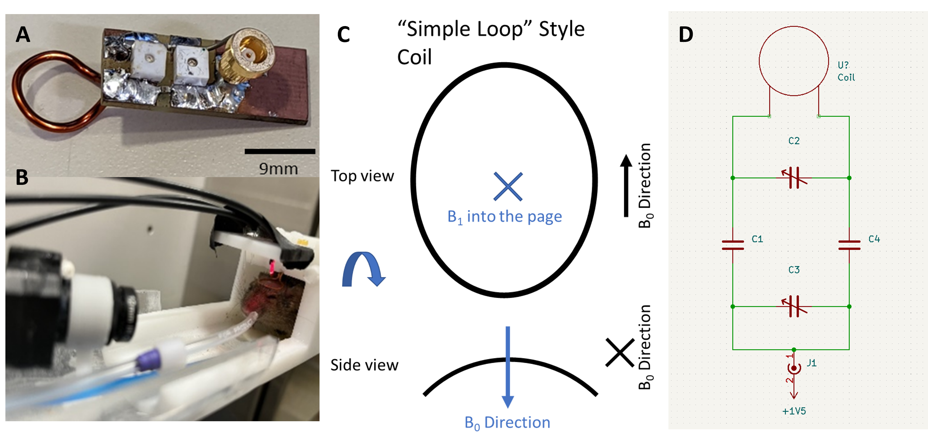
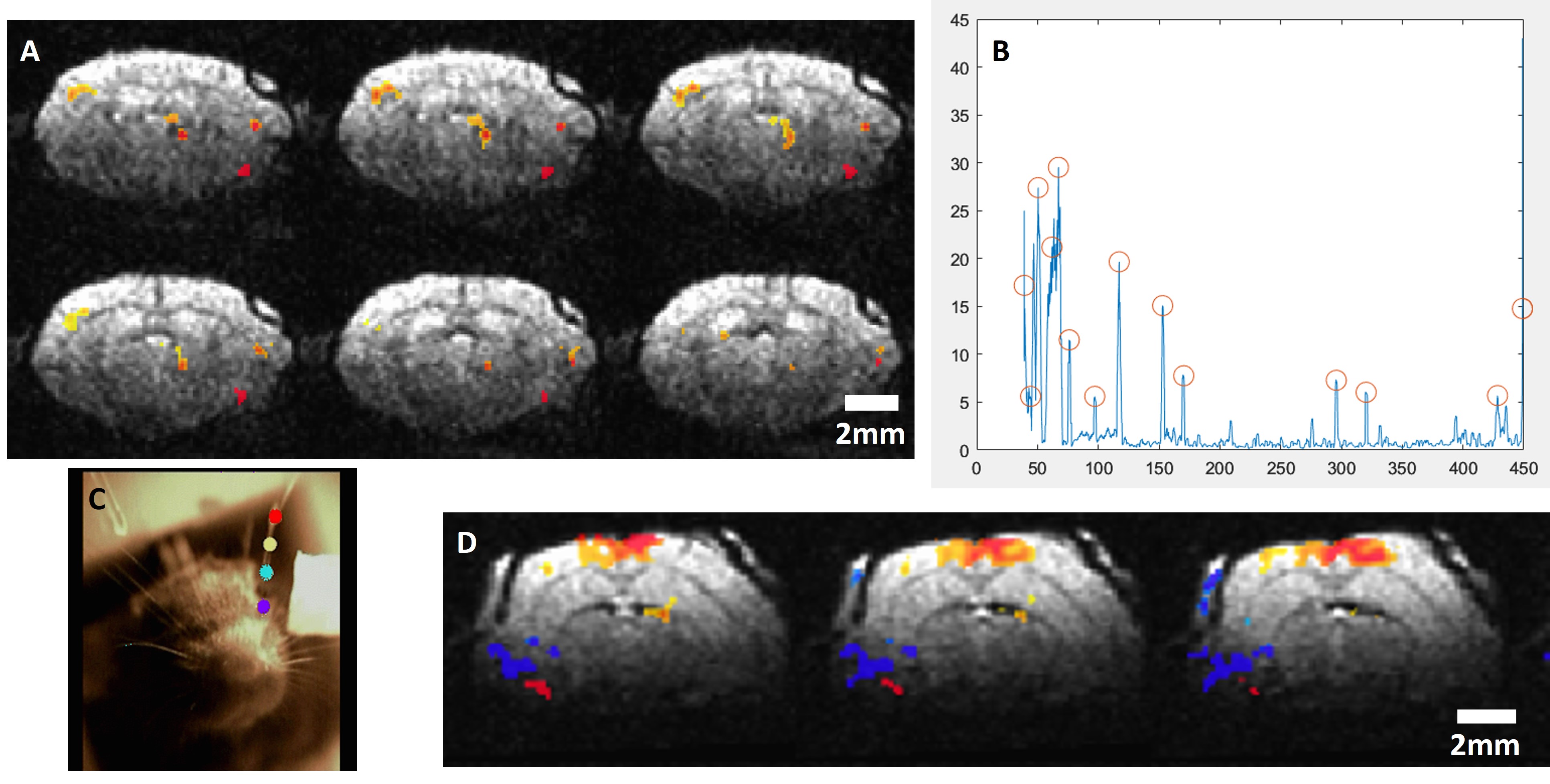
Figure 2: BOLD activation from spontaneous whisking. A) BOLD response seen in the barrel cortex and medial dorsal areas. B) Whisker motion graph derived from video tracking a single whisker providing time points to classify BOLD response. C) Still image showing the “in-scanner” camera view of the whiskers with whisker tracking points visualized. D) BOLD response seen in the retrosplenial area following a spontaneous whisking event.
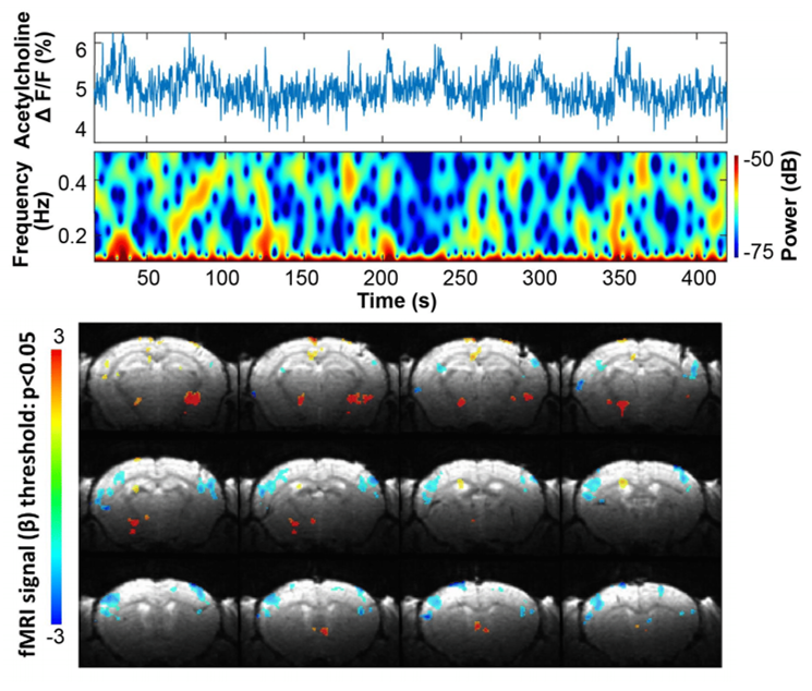
Figure 3: Time course of acetylcholine during a resting state (non-stimulated/spontanious whisking) scan showing acetylchoine spikes as well as BOLD activation.
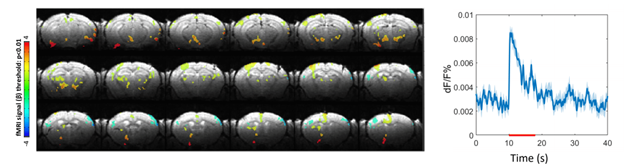
Figure 4: Averaged acetylcholine signal and BOLD response from 10sec whisker stimulation during fMRI scan showing activations in the the barrel cortex, lateral dorsal nucleus, ventral trigeminal area, and the lateral geniculate areas of the brain.