3416
High Temporospatial-Resolution fMRI in a Focused Brain Volume with (k, t)-Space Undersampling and Temporal Priors1Center for Magnetic Resonance Research, University of Illinois at Chicago, Chicago, IL, United States, 2Department of Biomedical Engineering, University of Illinois at Chicago, Chicago, IL, United States, 3Departments of Radiology and Neurosurgery, University of Illinois at Chicago, Chicago, IL, United States
Synopsis
Keywords: fMRI Acquisition, fMRI, Undersampling, Temporal Priors
Motivation: Three-dimension reduced field-of-view imaging with (k, t)-space undersampling (k-t 3D-rFOVI) can be used for high temporospatial-resolution fMRI acquisition in a focused brain volume.
Goal(s): This study aims at developing an image reconstruction method for k-t 3D-rFOVI to improve the fMRI data quality.
Approach: Timing information about stimulation tasks was integrated into image reconstruction of k-t 3D-rFOVI. Simulation and human fMRI experiments with a 6-fold acceleration factor were conducted to demonstrate the performance of the reconstruction method.
Results: The proposed reconstruction method substantially increased the sensitivity of k-t 3D-rFOVI in detecting fMRI activations and the accuracy in recovering the time course of fMRI signals.
Impact: By incorporating stimulus timing information into image reconstruction, high-quality fMRI data can be obtained using k-t 3D-rFOVI with high temporal (480 ms) and spatial (1.5 mm) resolutions over a reduced field-of-view.
Introduction
Using (k, t)-space undersampling and reconstruction methods based on partial separability (PS),1-3 3D reduced field-of-view imaging (3D-rFOVI)4 has been developed for acquiring fMRI data with high temporal (~800 ms) and spatial (~1.5 mm) resolutions in a focused brain volume.5 In task fMRI experiments, stimulus timing information is known prior to data acquisition. This study aims at improving fMRI data quality of k-t 3D-rFOVI by incorporating temporal priors from stimulation paradigms into PS reconstruction. The performance of k-t 3D-rFOVI using the new reconstruction strategy is demonstrated by using simulation data and human visual fMRI experiments.Methods
Image reconstruction: In k-t 3D-rFOVI, the central k-space region is fully sampled along the through-slab direction (kz,c) for all time frames while the outer regions (kz,o) are randomly undersampled at different frames.5 The k-space data can be reconstructed with a PS method, which models time series (C) as multiplication of spatial (Us) and temporal (Vt) subspace matrices2. In conventional PS with no priors (PS-NP), Vt is estimated by applying singular value decomposition (SVD) to the MR signals in kz,c (Sk,c) without any prior information. To more accurately estimate Vt, we propose a PS method with temporal priors (PS-TP) from stimulation task paradigms: Firstly, Sk,c can be expressed as a summation of BOLD (SB) and non-BOLD (SNB) signals (e.g., head motions) and noise (σ) (Sk,c=SB+SNB+σ). To estimate SB, a finite input response (FIR) filter is constructed according to the stimulation paradigm used in the fMRI experiment and then fitted to Sk,c.6,7 SNB is obtained by subtracting the estimated SB from Sk,c. Vt of BOLD (Vt,B) and non-BOLD (Vt,NB) components are calculated using SVD (order = 8) of SB and SNB, respectively. Lastly, images are reconstructed using PS with a combined Vt = [Vt,B, Vt,NB].Simulation and human fMRI experiments: Simulated k-t 3D-rFOVI data were generated by replicating 500 copies of one complex image volume (36 slices). Simulated BOLD signals and white noise were added to ROIs and all the voxels in the images, respectively. The synthesized data were transformed to k-space and undersampled with 2/4 kz-planes in the kz,c/kz,o region, corresponding to an acceleration factor (R) of 6. Images were reconstructed with PS-TP and PS-NP separately, followed by spatial smoothing (FWHM=1.5 mm). Detected activations were considered true/false positives if they were located inside/outside the ROIs. For in vivo demonstrations, visual fMRI experiments were carried out on healthy subjects at 3T (GE MR750) with a 32-channel head coil, using interleaved 16s/16s ON/OFF flashing checkerboard stimuli. Images focusing on the visual cortex were acquired with the k-t 3D-rFOVI sequence: FOV=19.6×10.8×5.4 cm3, acquisition matrix=72×72×36, volume TR/TR/TE = 480/80/30 ms, flip angle=20°. The fMRI data from both simulations and experiments were analyzed in AFNI.
Results
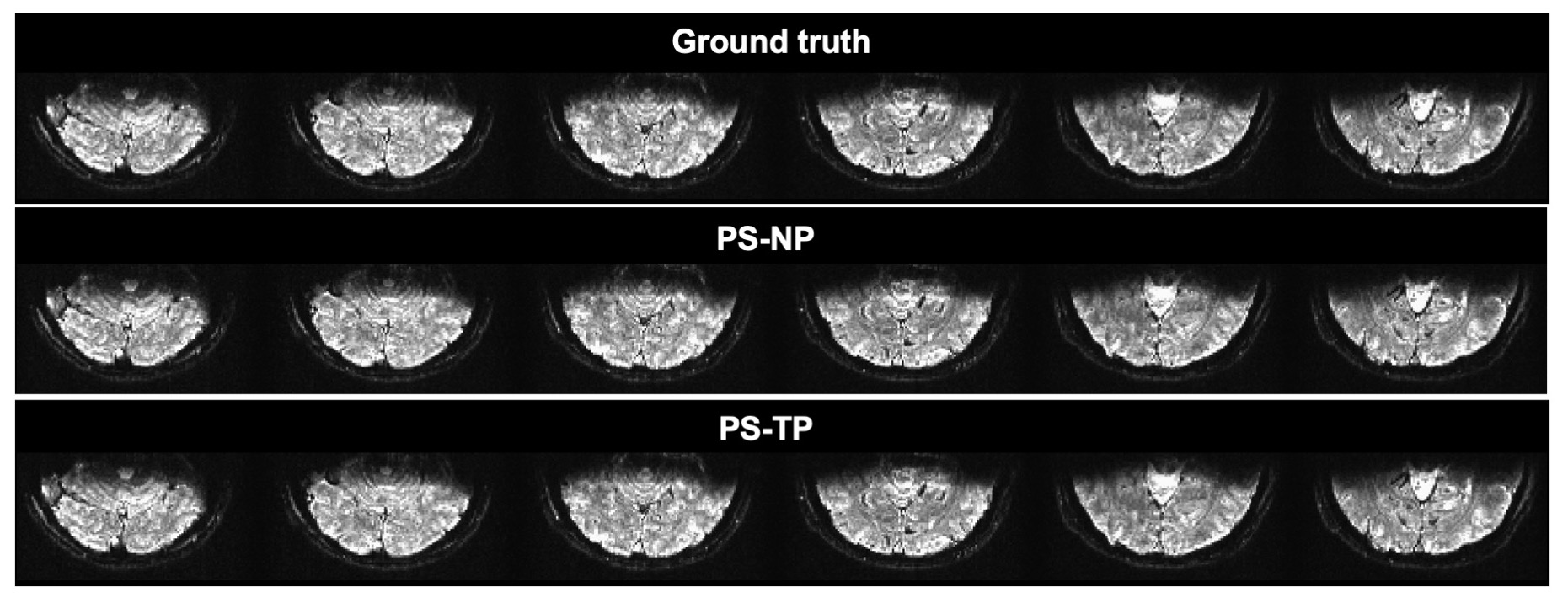
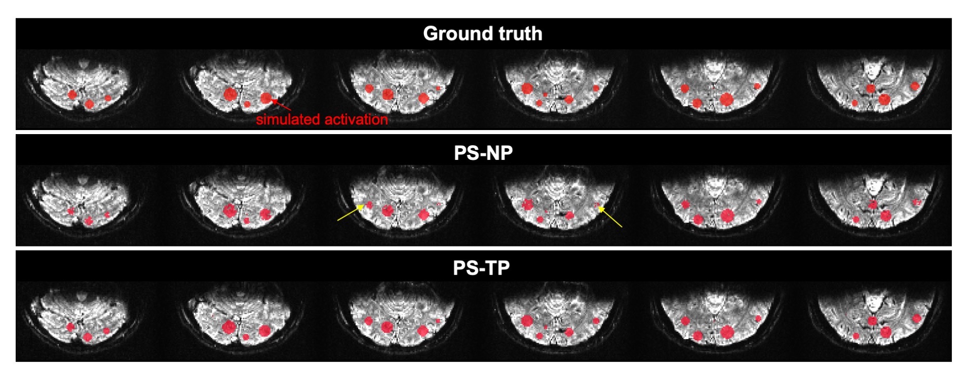
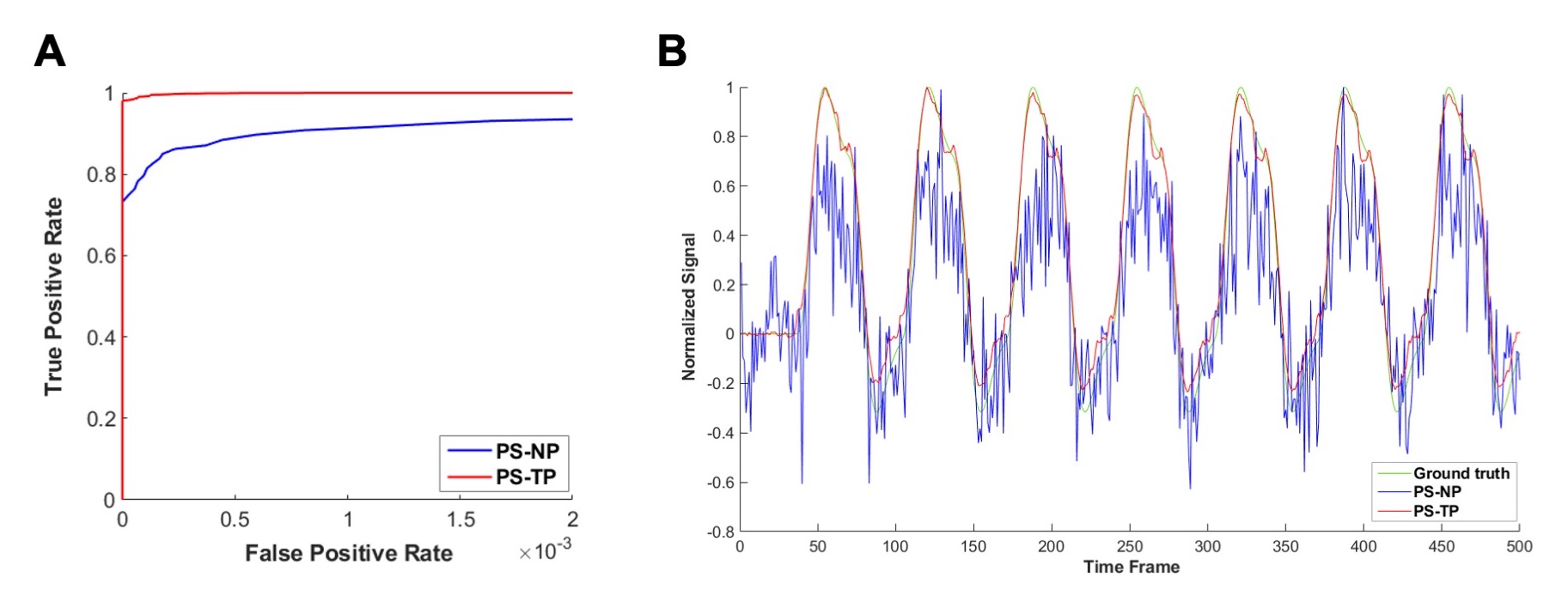
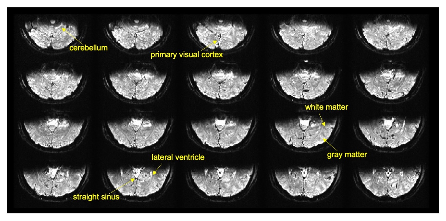
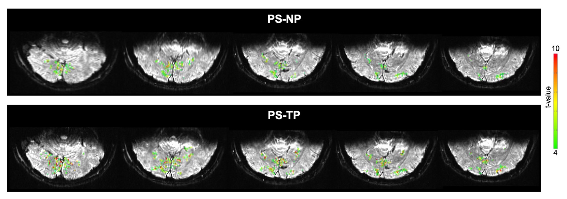
Figure 1 illustrates k-t 3D-rFOVI images reconstructed separately with PS-NP and PS-TP from the simulation data. The two image sets showed the same structural similarity index (SSIM = 0.88) relative to the ground truth. However, compared to the PS-NP data, more true activations were detected in the PS-TP data at a given false positive rate (FPR) (Fig. 2). At FPR=0.0001, the PS-TP data showed a true positive rate (TPR) of 0.991 while the TPR (0.815) in the PS-NP data was notably lower (Fig. 3A). The correlation coefficient between the average time course of activated voxels and the ground truth was 0.997 in PS-TP data, which was higher than that in the PS-NP images (0.860) (Fig. 3B).In the human experiments (Fig. 4), images reconstructed with PS-TP provided excellent quality for visualizing and localizing different brain structures (e.g., primary visual cortex and lateral ventricle) and tissue types (i.e., gray and white matters). Significant BOLD activations were detected in the visual cortex (Fig. 5). The number of activated voxels in the images reconstructed with PS-TP increased by approximately 64% relative to the PS-NP reconstruction, and the average t-value elevated from 5.7 to 6.4.
Discussion and Conclusions
In this study, we developed a reconstruction method (PS-TP) for k-t 3D-rFOVI by incorporating stimulus timing information into the reconstruction. Our simulation data indicated that PS-TP boosted fMRI detection sensitivity by about 22% compared to the conventional method (PS-NP). Although PS-TP and PS-NP images showed the same structural similarity to the ground truth, PS-TP offered a higher accuracy in recovering fMRI signal time courses, which directly affected the sensitivity to detecting brain activations. The superior performance of PS-TP was further demonstrated by the human fMRI experiments at 480-ms temporal and 1.5-mm spatial resolutions. In conclusion, k-t 3D-rFOVI with PS-TP reconstruction provides a robust strategy for high temporospatial-resolution fMRI in a focused brain volume.Acknowledgements
This work was supported in part by the National Institutes of Health (Grant No. 5R01EB026716-01, 1S10RR028898-01, and 1R03EB034480-01). The content is solely the responsibility of the authors and does not necessarily represent the official views of the National Institutes of Health.References
1. Liang Z-P. Spatiotemporal imaging with partially separable functions. Proc. IEEE Int’l Symposium on Biomedical Imaging. 2007; pp. 988-991.
2. Zhao B, Haldar JP, Christodoulou AG, Liang Z-P. Image reconstruction from highly undersampled (k, t)-space data with joint partial separability and sparsity constraints. IEEE Trans Med Imaging. 2012;31:1809-1820.
3. Lam F, Zhao B, Liu Y, Liang Z-P, Weiner M, Schuff N. Proc ISMRM Annual Meeting. 2013; S2620.
4. Sun K, Zhong Z, Dan G, Karaman M, Luo Q, Zhou XJ. Three-dimensional reduced field-of-view imaging (3D-rFOVI). Magn Reson Med. 2022;87:2372-2379.
5. Luo Q, Sun K, Scotti A, Dan G, Karaman M, Zhou XJ (2022). Fast 3D fMRI acquisition over a small field-of-view with (k, t)-space undersampling. Proc ISMRM Annual Meeting. 2022; S3328.
6. Goutte C, Nielsen FA, Hansen LK. Modeling the haemodynamic response in fMRI using smooth FIR filters. IEEE Trans Med Imag. 2000;19:1188–1201.
7. Lindquist MA, Wager TD. Validity and power in hemodynamic response modeling: a comparison study and a new approach. Hum Brain Mapp. 2007;28:764-784.
Figures