3414
Simultaneous EEG-fMRI reveals spontaneous neural oscillatory activity in cingulate cortex underlying transient rsfMRI network dynamics1Laboratory of Biomedical Imaging and Signal Processing, The University of Hong Kong, Hong Kong SAR, China, 2Department of Electrical and Electronic Engineering, The University of Hong Kong, Hong Kong SAR, China, 3Department of Diagnostic Radiology, The University of Hong Kong, Hong Kong SAR, China, 4School of Biomedical Sciences, The University of Hong Kong, Hong Kong SAR, China
Synopsis
Keywords: fMRI Acquisition, Multimodal, fMRI (resting state), functional connectivity, neuroscience
Motivation: rsfMRI network dynamics are essential for cognitive processes, however, their underlying neural bases remain unclear.
Goal(s): Here, we aim to examine the neural oscillatory events underlying the dynamic patterns of rsfMRI networks across the entire brain.
Approach: We employed simultaneous EEG-fMRI to record brain-wide rsfMRI and neural signals at default mode network. Further, EEG events were identified and temporally matched with dynamic rsfMRI network states derived from a data-driven model.
Results: Our results demonstrated robust associations between rsfMRI network dynamics and neural oscillatory events, especially slow oscillation-coupled spindle, gamma events and slow oscillation.
Impact: Our results demonstrated the different effects of spontaneous neural oscillatory events (e.g., slow oscillation, spindle, and gamma) in default mode network underlying the dynamics of rsfMRI networks.
Introduction
Normal brain functions are supported by distinct neural oscillatory events resulting from synchronized activity in neural circuits, such as slow oscillation (SO), spindle (SP), and gamma (G) events which are associated with sleep1-5, arousal6,7, and cognition5,7,8. Previous studies from our laboratory and others revealed that low frequency (<10Hz) and/or high frequency (>20Hz) oscillatory activities constrained and elicited resting-state fMRI (rsfMRI) connectivity9-11 during anaesthesia, sleep, and waking states. Meanwhile, converging studies indicate the importance of second-level transient states in subserving rsfMRI networks dynamics12-14. However, the specific contribution of spontaneous neural oscillatory events to the rsfMRI networks dynamics remains unresolved.In this study, our aim is to examine the neural bases underlying the dynamic patterns of rsfMRI networks across the entire brain. We employed simultaneous electroencephalography and fMRI (EEG-fMRI) to record and detect spontaneous neural oscillatory events (i.e., SO, SP, and G) in an important node of default mode network-cingulate cortex (Cg). These events were then used to align and average the time courses of transient network states derived from a data-driven model15.
Method
Simultaneous EEG recording and fMRI acquisition: Customized MR-compatible electrodes were placed above Cg of SD rats (10-12 weeks, N=4, Figure 1A). Each animal underwent 2-3 hours rsfMRI scans (600s/trial). EEG recordings were simultaneously acquired with rsfMRI using a multi-channel electrophysiology recording system (TDT).fMRI data analysis: Standard fMRI preprocessing and concatenation of all rsfMRI scans (N=17 animals, n=90 trials) were performed. The Gaussian hidden Markov model (HMM) generated 18 states based on the PCA time courses (70% variance explained). Voxel-based mean activation map was obtained by averaging the preprocessed BOLD signal change when each state was activated.
Electrophysiological signal processing: MRI gradient artifacts in the electrophysiological signals were removed using a conventional algorithm16. Local field potential (LFP) signal of distinct events such as SO, SP and G were extracted based on previous studies2,4,11.
Results
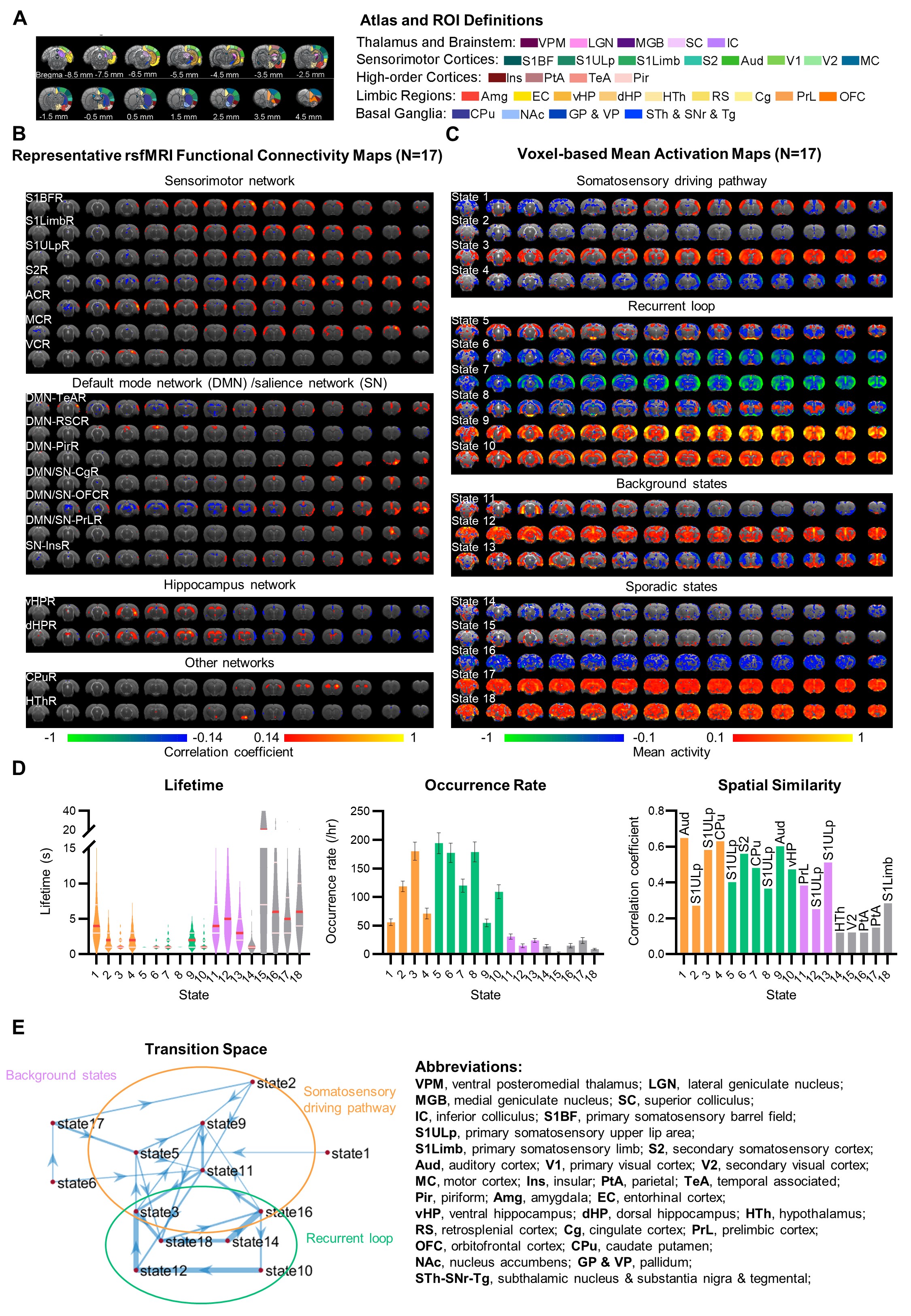
Transient states and transition pathways in rsfMRI networks (Figure 2):HMM network states resembled the combination of rsfMRI networks computed with seed-based analysis, such as homotopic default mode (DMN), salience (SN), and basal forebrain (BF) networks. Note that, sporadic states (states 14-18) with long lifetime, low occurrence rate, and low spatial similarity to RSN were excluded. States 1-13 exhibited short lifetime (mostly 1-6s medians) and high occurrence rate (>20/hr), indicating their transient and repeatedly occurring properties. Such transient states can be divided into three groups based on the state transition space encompassing the 90th percentile transitions. Specifically, states in somatosensory and Cg-driving pathways (SCDP) showed neural activity transition from positive S1 and negative Cg (states 1, 9) to long-range areas (state 3), then to reversed counterpart (state 5). Recurrent loop (RL) showed a high transition probability between states involving brain-wide regions, especially DMN, SN, BF, HTh, and sensorimotor networks. Further, RL was marked by network state transitions between positive and negative mean activations wherein the polarity switching first occurred at BF (states 5, 6). Background states consisted of HP network (state 11) or DMN (states 12, 13) exhibited directional efferent but no directional afferent transitions.
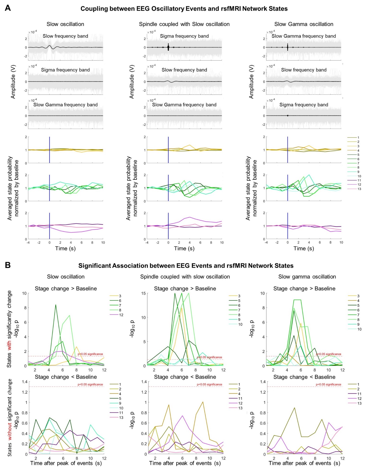
Different spontaneous neural events exhibit distinct coupling with rsfMRI network dynamics (Figure 3):
Both SO-coupled SP (SP+SO) and G events were synchronized with SO activity showed by the peak-aligned mean LFP change at the slow frequency band (0.2-1Hz). Note that, Gamma was also locked with sigma frequency activity (7-15Hz). Importantly, all events displayed distinct but robust association with transient rsfMRI network state dynamics. Specifically, Gamma events were coupled with most of the rsfMRI network states among all detected events and significantly present in all RL states and other states with positive Cg activity in SCDP (states 3, 4). SP+SO were associated with similar states to Gamma except state 4. In contrast, SO was only coupled to RL states with negative Cg activity (states 6-8), and two other SCDP and background states with positive DMN and CPu (states 3, 12).
Discussion and Conclusion
In conclusion, our findings demonstrate the distinct contributions of cingulate spontaneous neural oscillatory events (e.g., SO, SP, and G) to the rsfMRI networks dynamics. Our study suggested the important role of SO, SP, and Gamma events in DMN, which could be essential for internally oriented cognitive processes13,17-19. We found that spindle and gamma oscillations significantly locked with all RL states connecting/integrating DMN and other networks, such as BF and SN. Taken together, we highlighted gamma-band activity-associated interaction between BF and DMN activity, which may have implications for the treatment of DMN dysfunction in conditions such as epilepsy or depression19,20.Acknowledgements
This work was supported in part by Hong Kong Research Grant Council (HKU17112120, HKU17127121, HKU17127022 and HKU17127523 to E.X.W; and HKU17104020, HKU17127021, HKU17127723 to A.T.L.L.), Lam Woo Foundation, and Guangdong Key Technologies for AD Diagnostic and Treatment of Brain (2018B030336001) to E.X.W.References
1. Yu, Y., et al. Sleep fMRI with simultaneous electrophysiology at 9.4 T in male mice. Nature Communications 14(2023).
2. Staresina, B.P., Niediek, J., Borger, V., Surges, R. & Mormann, F. How coupled slow oscillations, spindles and ripples coordinate neuronal processing and communication during human sleep. Nat Neurosci 26, 1429-1437 (2023).
3. Sela, Y., Vyazovskiy, V.V., Cirelli, C., Tononi, G. & Nir, Y. Responses in Rat Core Auditory Cortex are Preserved during Sleep Spindle Oscillations. Sleep 39, 1069-1082 (2016).
4. Nir, Y., et al. Regional slow waves and spindles in human sleep. Neuron 70, 153-169 (2011).
5. Weber, F.D., et al. Coupling of gamma band activity to sleep spindle oscillations - a combined EEG/MEG study. Neuroimage 224, 117452 (2021).
6. Bai, Y., et al. Spontaneous transient brain states in EEG source space in disorders of consciousness. Neuroimage 240, 118407 (2021).
7. Lewis, L.D., Setsompop, K., Rosen, B.R. & Polimeni, J.R. Fast fMRI can detect oscillatory neural activity in humans. Proc Natl Acad Sci U S A 113, E6679-E6685 (2016).
8. Kim, J., Gulati, T. & Ganguly, K. Competing Roles of Slow Oscillations and Delta Waves in Memory Consolidation versus Forgetting. Cell 179, 514-526 e513 (2019).
9. Wang, X., Leong, A.T.L., Chan, R.W., Liu, Y. & Wu, E.X. Thalamic low frequency activity facilitates resting-state cortical interhemispheric MRI functional connectivity. Neuroimage 201, 115985 (2019).
10. Ma, Y., et al. Resting-state hemodynamics are spatiotemporally coupled to synchronized and symmetric neural activity in excitatory neurons. Proc Natl Acad Sci U S A 113, E8463-E8471 (2016).
11. Wang, X., et al. Functional MRI reveals brain-wide actions of thalamically-initiated oscillatory activities on associative memory consolidation. Nat Commun 14, 2195 (2023).
12. Favaretto, C., et al. Subcortical-cortical dynamical states of the human brain and their breakdown in stroke. Nat Commun 13, 5069 (2022).
13. Menon, V., et al. Optogenetic stimulation of anterior insular cortex neurons in male rats reveals causal mechanisms underlying suppression of the default mode network by the salience network. Nat Commun 14, 866 (2023).
14. Allen, E.A., et al. Tracking whole-brain connectivity dynamics in the resting state. Cereb Cortex 24, 663-676 (2014).
15. Stevner, A.B.A., et al. Discovery of key whole-brain transitions and dynamics during human wakefulness and non-REM sleep. Nat Commun 10, 1035 (2019).
16. Allen, P.J., Josephs, O. & Turner, R. A method for removing imaging artifact from continuous EEG recorded during functional MRI. Neuroimage 12, 230-239 (2000).
17. Clancy, K.J., et al. Transcranial stimulation of alpha oscillations up-regulates the default mode network. Proceedings of the National Academy of Sciences 119(2021).
18. Kaefer, K., Stella, F., McNaughton, B.L. & Battaglia, F.P. Replay, the default mode network and the cascaded memory systems model. Nat Rev Neurosci 23, 628-640 (2022).
19. Nair, J., et al. Basal forebrain contributes to default mode network regulation. Proc Natl Acad Sci U S A 115, 1352-1357 (2018).
20. Gusnard, D.A., Akbudak, E., Shulman, G.L. & Raichle, M.E. Medial prefrontal cortex and self-referential mental activity: relation to a default mode of brain function. Proceedings of the National Academy of Sciences 98, 4259-4264 (2001).
Figures
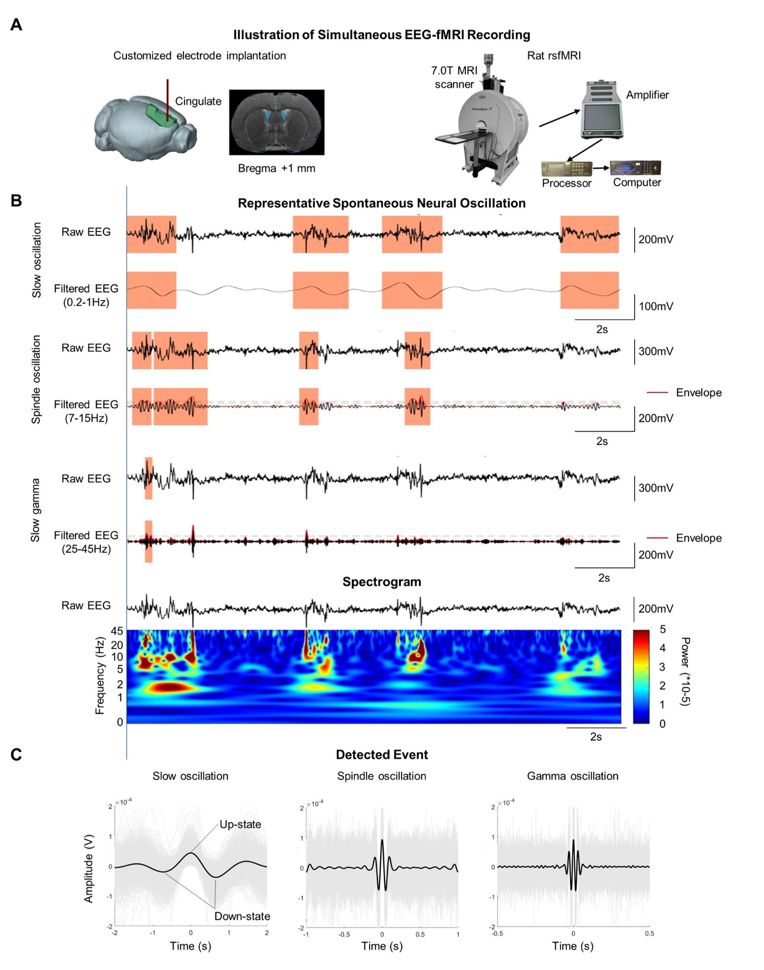
Figure 1 Experimental set up and detected events. (A) Illustration of simultaneous EEG-fMRI experimental set up; (B) Example of spontaneous neural event detection. (C) Event identifications of slow oscillation, spindle and slow gamma oscillation. fMRI data were acquired on 7T Bruker scanner using GE-EPI (FOV=32×32mm2, matrix=64×64, α=56°, TE/TR=20/1000ms, and 16 contiguous slices with 1mm thickness).