3413
Inverted BOLD signal changes in the transient phase of passband balanced SSFP1High Field Magnetic Resonance, Max Planck Institute for Biological Cybernetics, Tübingen, Germany, 2Department of Biomedical Magnetic Resonance, University of Tübingen, Tübingen, Germany, 3Center for MR Research, University Children's Hospital, Zurich, Switzerland
Synopsis
Keywords: fMRI Acquisition, Contrast Mechanisms
Motivation: Efficient, distortion-free, and selective to microvessels: these attributes make bSSFP an attractive candidate for fMRI. 2D multi-slice scans in the transient state are increasingly employed as an alternative to 3D steady-state imaging to improve localization.
Goal(s): To evaluate the BOLD sensitivity of bSSFP during the transient phase as compared to the steady state.
Approach: bSSFP acquisitions in transient and steady state are utilized for task-based fMRI. Monte Carlo simulations and in vitro microsphere experiments are conducted to verify the in vivo results.
Results: An inversion of the BOLD contrast with a trend towards negative BOLD in the early transient phase can be observed.
Impact: The bSSFP BOLD contrast undergoes an inversion during the transient phase. Consequently, the BOLD response appears suppressed in transient-state imaging with a tendency to become negative, which has important implications when designing 2D multi-slice fMRI experiments with high temporal resolution.
Introduction
Balanced steady-state free precession (bSSFP) fMRI (1-4) is characterized by a pronounced selectivity to microvessels similar to the spin echo (SE), yielding BOLD-related signal changes, which may be closer to the neuronal event as compared to gradient echo (GE) (5,6). As 3D imaging can be suboptimal for localized fMRI applications and parallel imaging acceleration options on pre-clinical MR systems are often lacking, 2D multi-slice bSSFP is increasingly used to provide high temporal resolution and sufficient spatial coverage (7,8). Because of the short scan time to acquire a single slice, paired with a recovery period resulting from the sequential excitation of multiple slices, 2D multi-slice imaging is performed in the transient phase rather than in steady state. The BOLD sensitivity of transient-state bSSFP is not yet understood and thus explored here.Methods
In vivo fMRI setup and analysisThree different passband bSSFP acquisition schemes were implemented for task-based fMRI at 3T: 1) 3D steady-state imaging (Fig. 1A), 2) partition-segmented 3D transient-state imaging (Fig. 1B), and 3) 2D multi-slice transient-state imaging (Fig. 1C). A flip-angle-ramp preparation module consisting of eight RF pulses was applied as sketched in Figure 1. BOLD fMRI data were acquired based on a block paradigm with alternating resting and task conditions using a flickering checkerboard visual stimulus (10 Hz) during the task period. The duration of each condition was matched to the acquisition time of 10 volumes in case of schemes 1) and 3) as well as to 1 volume in case of scheme 2). For schemes 1) and 3), rest-task blocks were repeated eight times, resulting in the acquisition of 160 volumes, whereas for scheme 2), rest-task blocks were repeated 25 times, resulting in the acquisition of 50 volumes (cf. Fig. 1). The relevant bSSFP scan parameters are summarized in Table 1.
The acquired bSSFP data were denoised using NORDIC (9) and motion corrected using FSL (10). Activated voxels were detected with the FEAT package of FSL (10). BOLD signal change maps in percentage were calculated according to [(<Stask>–<Srest>)/<Srest>]∙100 with <Stask> and <Srest> referring to the average signal in the task and resting condition, respectively. In case of schemes 1) and 3), only the volumes acquired in the second half of the task and resting condition were averaged.
Monte Carlo simulations and microsphere experiments
The extravascular bSSFP BOLD signal change ΔM at the passband was quantified based on a Monte Carlo approach and monitored after an initial α/2-preparation pulse during the transition towards steady state. The vessels of the vascular network were modeled as artificial cylinders with different radii (6). The simulation parameters were as follows: B0=3T, TR=5 ms, α=30°, T1/T2=1400 ms/77 ms (gray matter), Δχ=0.1 ppm, blood volume=2%. In vitro MR measurements on 2.5% (v/v) precision polystyrene microspheres suspended in an aqueous gel doped with 2 mM Dy-DTPA of varying radii and Δχ=0.1 ppm relative to water were performed at 3T. The transient phase of passband bSSFP was sampled after an initial α/2-preparation pulse using a segmented 2D single-slice multi-contrast sequence (TR=7 ms, α=30°, recovery time Trec=8 s). Signal differences of tubes containing microspheres (Sm) relative to a reference tube without microspheres (S0) were calculated as ΔS/S0 with ΔS=S0−Sm.
Results
Typical activation maps overlaid on an anatomical MPRAGE contrast, identifying positive (red) and negative (blue) z-scores, are displayed in Figure 2. It is apparent that the number of activated voxels and maximal achievable z-scores are greatly reduced for transient-state bSSFP imaging (Figs. 2B-D) in comparison to a steady-state acquisition (Fig. 2A). The corresponding BOLD signal change maps in Figure 3 show a similar behavior. A direct comparison of 3D transient-state imaging without (Fig. 3B) and with (Fig. 3C) parallel imaging acceleration indicates that the BOLD response shows a tendency to become negative for the accelerated acquisition (see arrows in Fig. 3C) where the signal is acquired earlier in the transient phase. These in vivo results are corroborated by the Monte Carlo simulations (Fig. 4A) and microsphere experiments (Fig. 4B), which reveal that the BOLD sensitivity as well as vessel size specificity is reduced in the transient state and approaches negative values.Discussion and Conclusion
Our results demonstrate that transient-state imaging considerably affects the bSSFP BOLD sensitivity, which has important implications when designing 2D multi-slice bSSFP acquisition schemes for fMRI. In particular, an inversion of the BOLD contrast during the approach to steady state becomes apparent, which yields negative BOLD in the early transient phase and a suppression of the BOLD response at later stages of the transient phase until maximum positive BOLD is reached in the steady state.Acknowledgements
No acknowledgement found.References
1. Scheffler K, Seifritz E, Bilecen D, Venkatesan R, Hennig J, Deimling M, Haacke EM. Detection of BOLD changes by means of a frequency-sensitive trueFISP technique: preliminary results. NMR Biomed 2001;14(7-8):490-496.
2. Kim TS, Lee J, Lee JH, Glover GH, Pauly JM. Analysis of the BOLD Characteristics in Pass-Band bSSFP fMRI. Int J Imaging Syst Technol 2012;22(1):23-32.
3. Miller KL. FMRI using balanced steady-state free precession (SSFP). Neuroimage 2012;62(2):713-719.
4. Scheffler K, Ehses P. High-resolution mapping of neuronal activation with balanced SSFP at 9.4 Tesla. Magn Reson Med 2016;76(1):163-171.
5. Baez-Yanez MG, Ehses P, Mirkes C, Tsai PS, Kleinfeld D, Scheffler K. The impact of vessel size, orientation and intravascular contribution on the neurovascular fingerprint of BOLD bSSFP fMRI. Neuroimage 2017;163:13-23.
6. Scheffler K, Heule R, Baez-Yanez MG, Kardatzki B, Lohmann G. The BOLD sensitivity of rapid steady-state sequences. Magn Reson Med 2019;81(4):2526-2535.
7. Freches GB, Chavarrias C, Shemesh N. BOLD-fMRI in the mouse auditory pathway. Neuroimage 2018;165:265-277.
8. Reynaud O, da Silva AR, Gruetter R, Jelescu IO. Multi-slice passband bSSFP for human and rodent fMRI at ultra-high field. Journal of magnetic resonance 2019;305:31-40.
9. Vizioli L, Moeller S, Dowdle L, Akcakaya M, De Martino F, Yacoub E, Ugurbil K. Lowering the thermal noise barrier in functional brain mapping with magnetic resonance imaging. Nature communications 2021;12(1):5181.
10. Smith SM, Jenkinson M, Woolrich MW, Beckmann CF, Behrens TEJ, Johansen-Berg H, Bannister PR, De Luca M, Drobnjak I, Flitney DE, Niazy RK, Saunders J, Vickers J, Zhang YY, De Stefano N, Brady JM, Matthews PM. Advances in functional and structural MR image analysis and implementation as FSL. Neuroimage 2004;23:S208-S219.
Figures
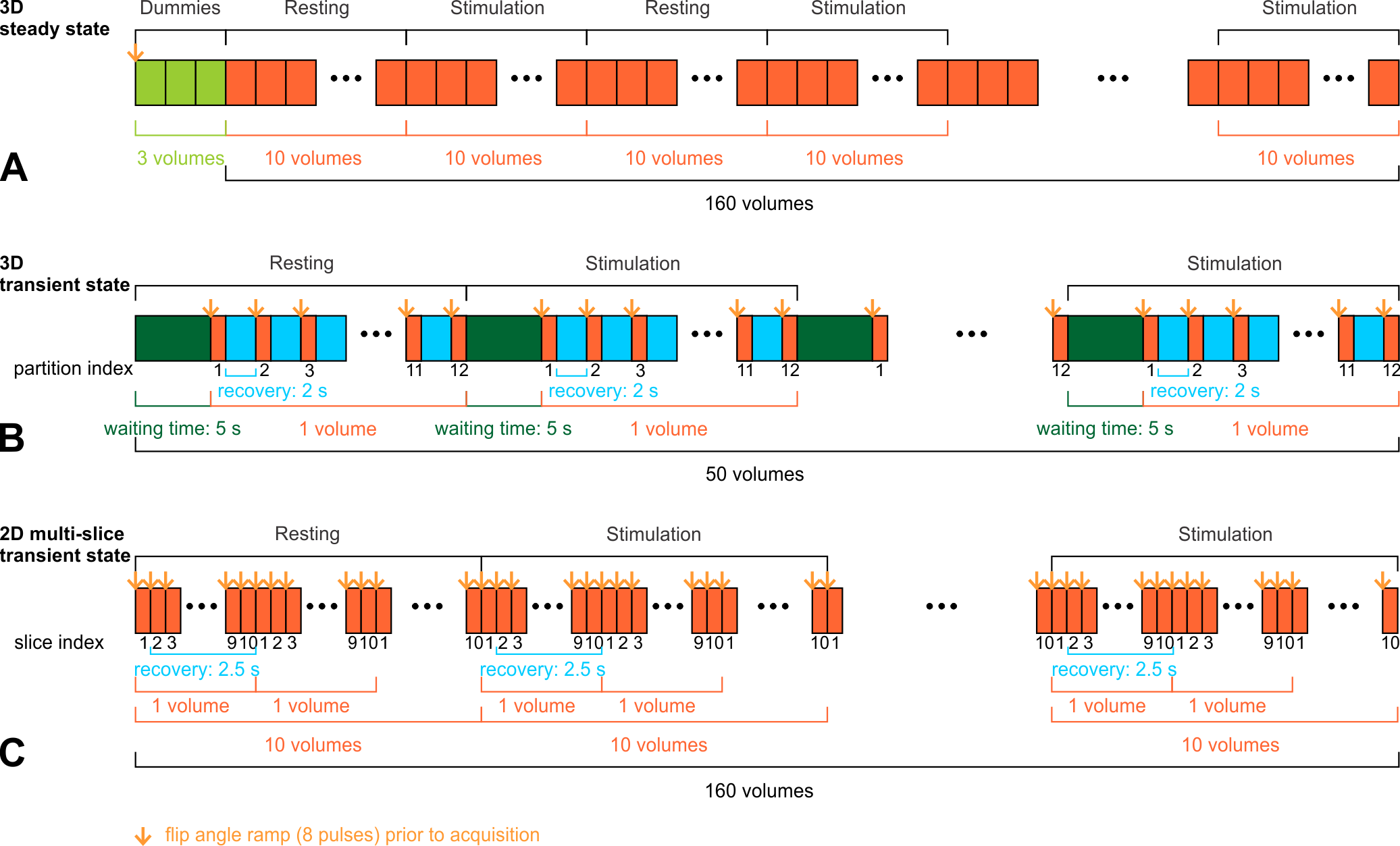
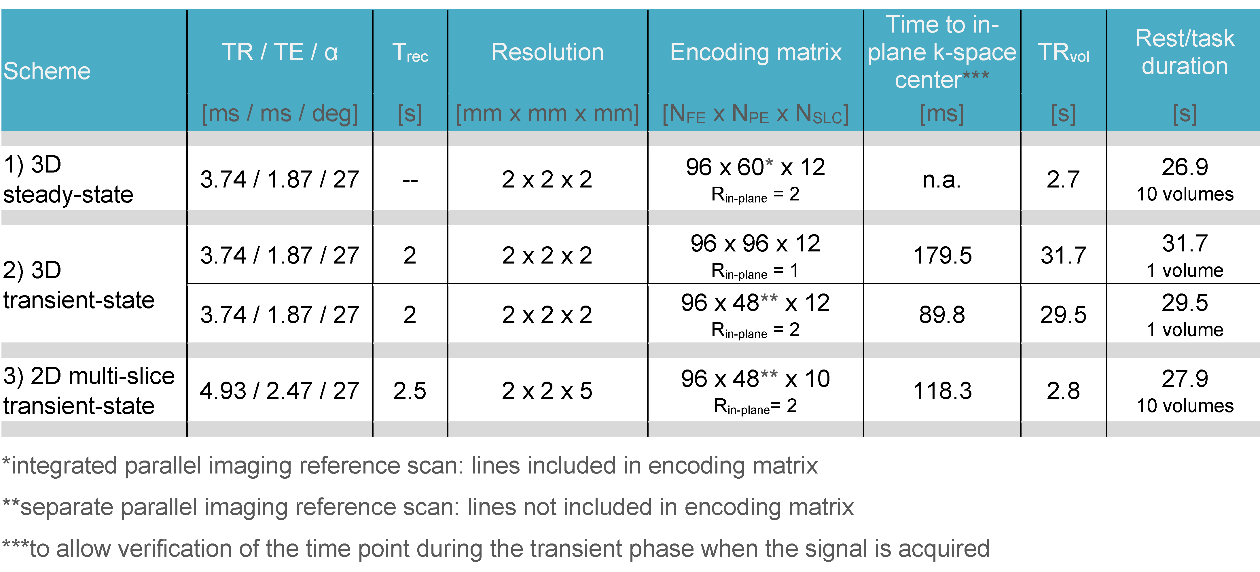
Table 1. Acquisition and fMRI paradigm parameters of the three different passband bSSFP sequences set up at 3T: repetition time (TR), echo time (TE), flip angle (α), recovery time (Trec), resolution, encoding matrix (NFE: frequency encoding steps, NPE: phase-encoding steps, NSLC: number of partitions (3D)/slices (2D), Rin-plane: in-plane parallel imaging acceleration factor), time to in-plane k-space center, volume TR (TRvol), rest/task duration.
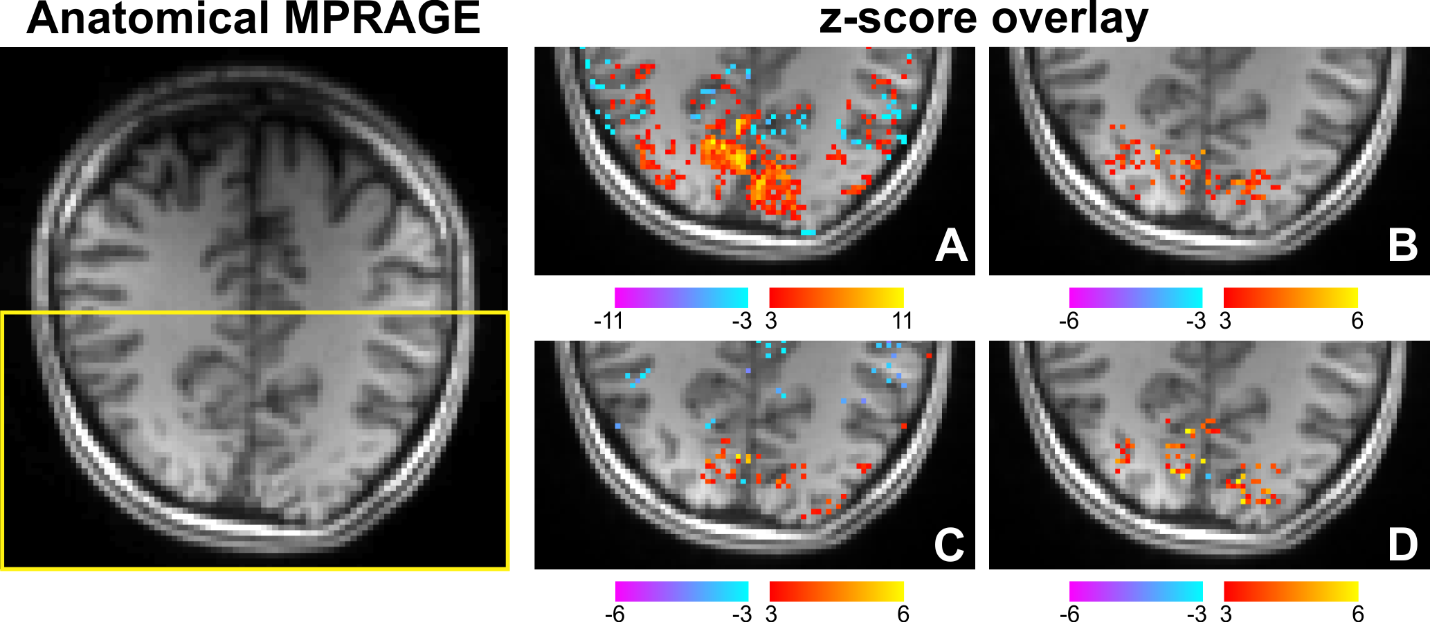
Figure 2. Anatomical MPRAGE on the left and z-score overlays on the right showing activated voxels for a representative slice obtained in a visual task experiment at 3T. A: 3D steady-state imaging, B: 3D transient-state imaging without parallel imaging acceleration (time to in-plane k-space center: 179.5 ms), C: 3D transient-state imaging with a parallel imaging acceleration factor of 2 (time to in-plane k-space center: 89.8 ms), and D: 2D multi-slice transient-state imaging (time to in-plane k-space center: 118.3 ms).
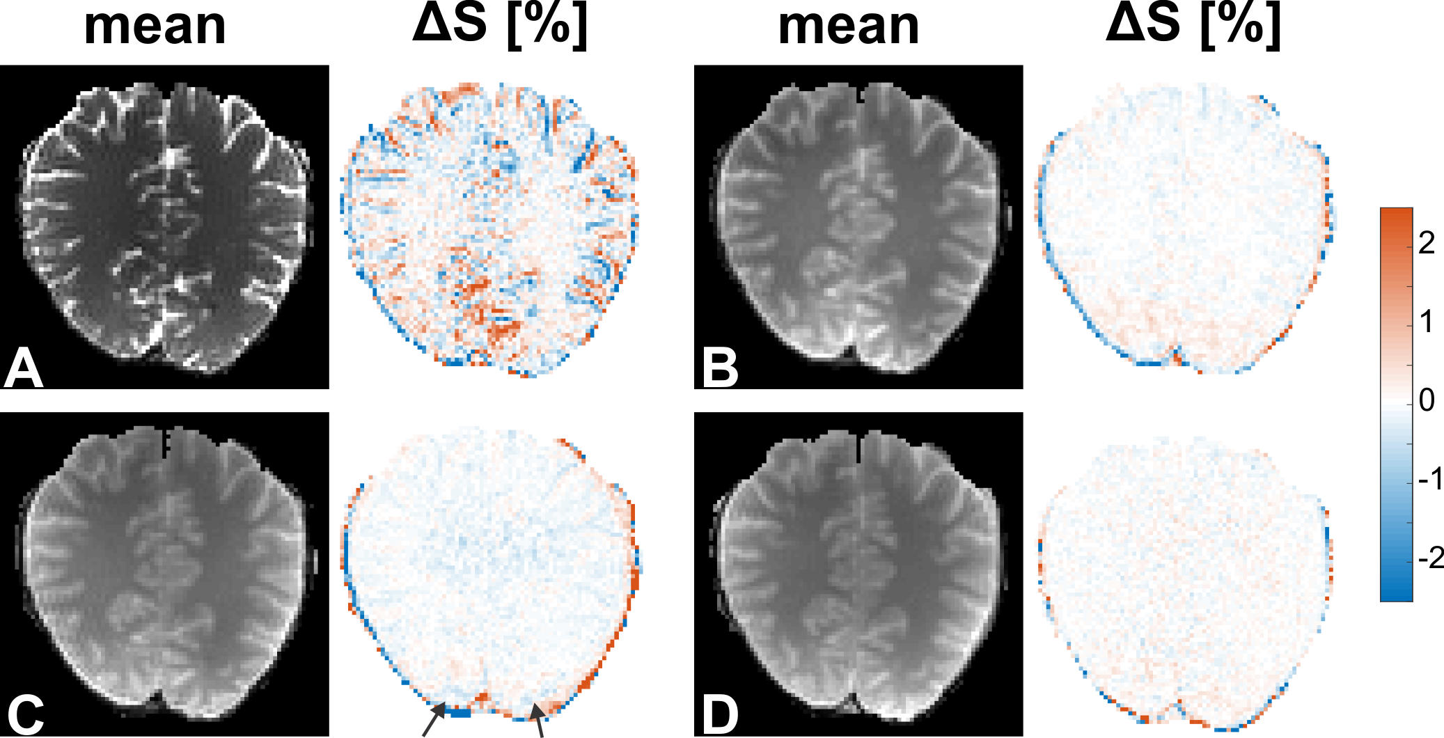
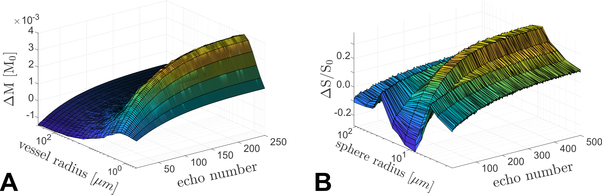
Figure 4. BOLD-related signal changes simulated by a Monte Carlo approach in units of the equilibrium magnetization M0 (A) and measured in microsphere suspensions (B) versus vessel/sphere radius and echo number after a/2-preparation pulse.