3412
Concurrent 1D line-scan fMRI at multiple cortical locations with a novel non-contiguous, non-coplanar multiline acquisition in humans at 7 Tesla1Athinoula A. Martinos Center for Biomedical Imaging, Massachusetts General Hospital, Charlestown, MA, United States, 2Department of Radiology, Harvard Medical School, Boston, MA, United States, 3Department of Radiology, Boston Children's Hospital, Boston, MA, United States, 4Harvard-MIT Division of Health Sciences and Technology, Massachusetts Institute of Technology, Cambridge, MA, United States
Synopsis
Keywords: fMRI Acquisition, fMRI (task based), line-scanning fMRI method
Motivation: Spin-echo-based line-scanning employing inner-volume excitation has been proposed for line-scan fMRI to avoid artifacts stemming from imperfect saturation RF pulses.
Goal(s): To measure non-contiguous and non-coplanar BOLD signals at multiple cerebral cortical areas.
Approach: We developed multi-line T2’- and T2-weighted Gradient-echo sampling of a spin-echo (GESSE) line-scanning fMRI method.
Results: This novel approach enables the detection of fMRI activation at different visual cortical regions, showing pure T2 and TE-dependent BOLD responses. It provides a proof-of-concept to further examine interactions of high-resolution fMRI signals at multiple cortical locations along feedforward and feedback pathways.
Impact: A non-coplanar multi-line gradient-echo sampling of spin-echo (GESSE) line-scanning method was proposed as a novel approach for distinguishing macro- and micro-vascular sensitive fMRI signals by simultaneouslyacquiring T2’- and T2-weighted BOLD fMRI signals at multiple cortical locations.
INTRODUCTION
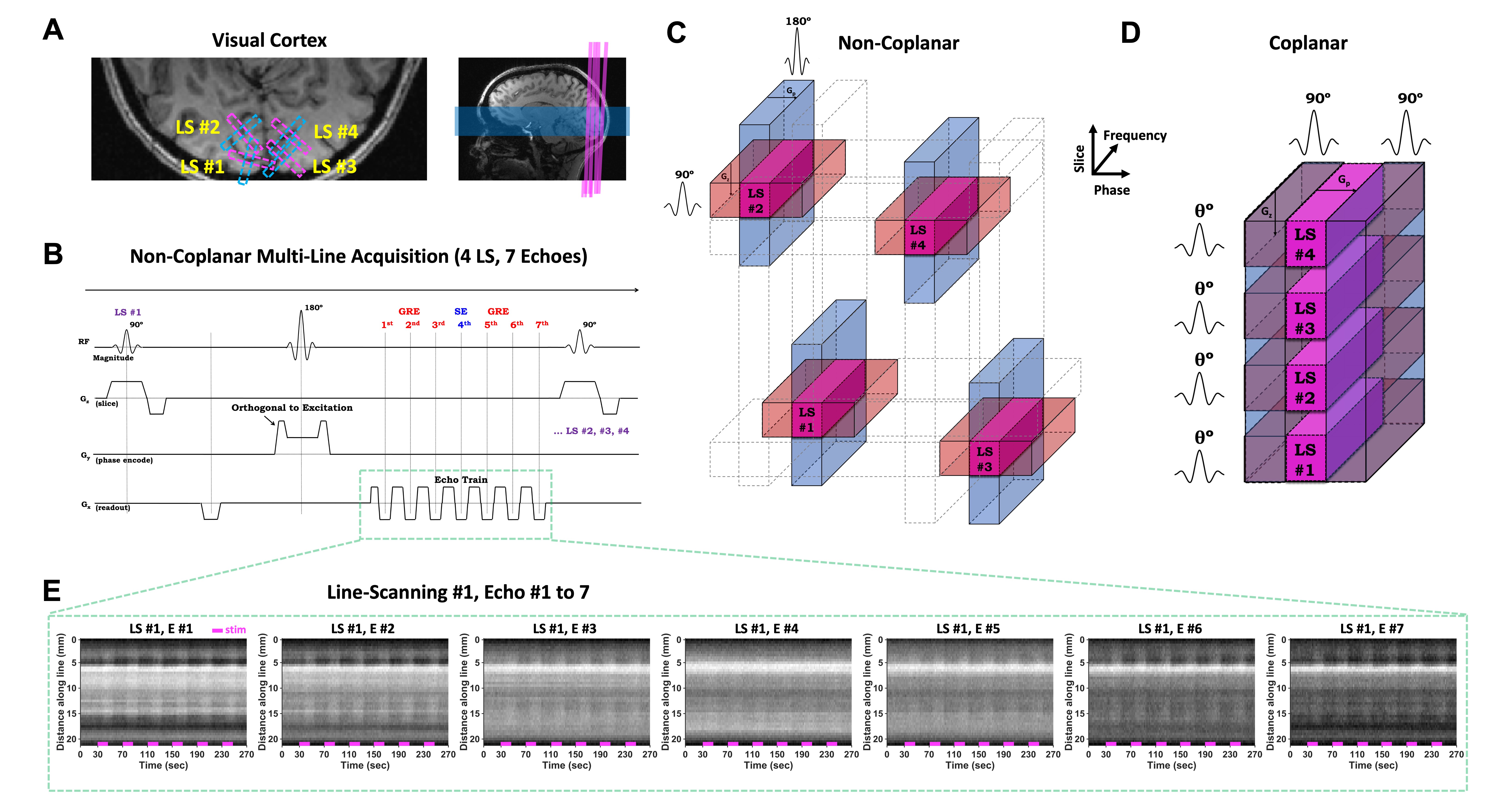
Laminar-specific fMRI provides an opportunity to investigate brain circuity by observing communications between cortical areas along feedforward and feedback pathways. Recently, 1D line-scanning fMRI methods have been successfully applied to elucidate laminar-specific fMRI onset time and dynamic functional connectivity with ultra-high resolution along one direction in animals1–4. Here, we propose a new approach that can perform concurrent imaging at multiple cortical locations in humans to enable measurements of functional interactions between cortical areas. While many line-scan fMRI approaches utilize gradient-echo acquisitions and outer-volume suppression1,5,6, the required saturation bands constrain multi-line acquisitions such that all lines must line in a single plane to avoid substantial signal loss. We employ a spin-echo acquisition utilizing inner-volume excitation7,8 achieved by orthogonal excitation and refocusing slices4, which lifts this constraint and allows for more degrees of freedom in multi-line prescription. Although the excitation and refocusing planes from individual lines may intersect one another, as long as these intersections do not overlap with the chosen cortical regions of interest—which comprise a small subject of the full 1D line—multiple lines can be concurrently measured in an interleaved manner without signal loss. As proof of concept, we present a four-line BOLD fMRI acquisition in human visual cortex at 7 Tesla, demonstrating that activation can be reliably detected at multiple cortical locations.METHODS
FMRI data were acquired in one healthy adult volunteer. Non-coplanar simultaneous T2’ (GRE) and T2 (SE) weighted gradient-echo sampling of spin-echo (GESSE) line-scanning fMRI data were acquired at 4 different cortical regions of visual cortex in both hemispheres (Fig. 1A and 1C) at a whole-body 7T scanner (Magnetom Terra, Siemens Healthineers) with an inhouse-built 64 head coil arrays9. The 180˚ RF pulse was employed on a logical axis perpendicular to the 90˚ excitation RF pulse (Fig. 1B). The acquisition parameters were: TR 2 s, TE 33.68–66.32 ms (7 evenly-spaced echoes), 4 interleaved lines (i.e., one line acquired every 500 ms), line thickness 3 mm, FOV 256 x 192 mm2 (for excitation), readout matrix 512 (in-line resolution 0.5 mm). Visual stimulation consisted of a flickering “black-and-white scaled-noise” stimulus, presented in a block-design paradigm: 30-s baseline, 6 repeated blocks of 16-s stimulation and 24-s inter-stimulus interval (Fig. 1E). Between-run and within-run changes in head position were corrected prospectively (AutoAlign) and retrospectively (a total of 6 runs). An additional run of resting-state fMRI data and phantom data was acquired for 3 m 20 s. All data analyses were implemented in Matlab (Mathworks, Natick, MA).RESULTS
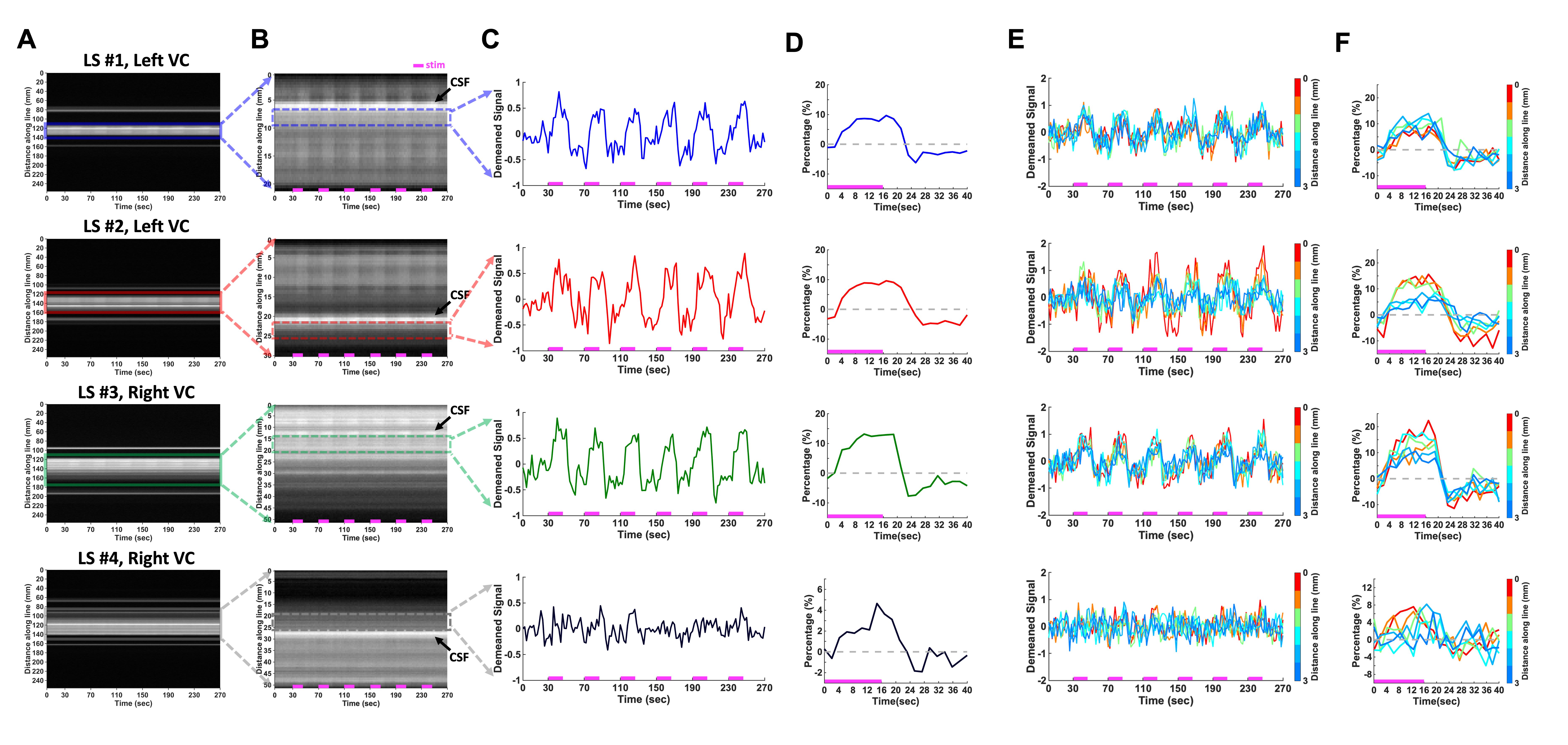
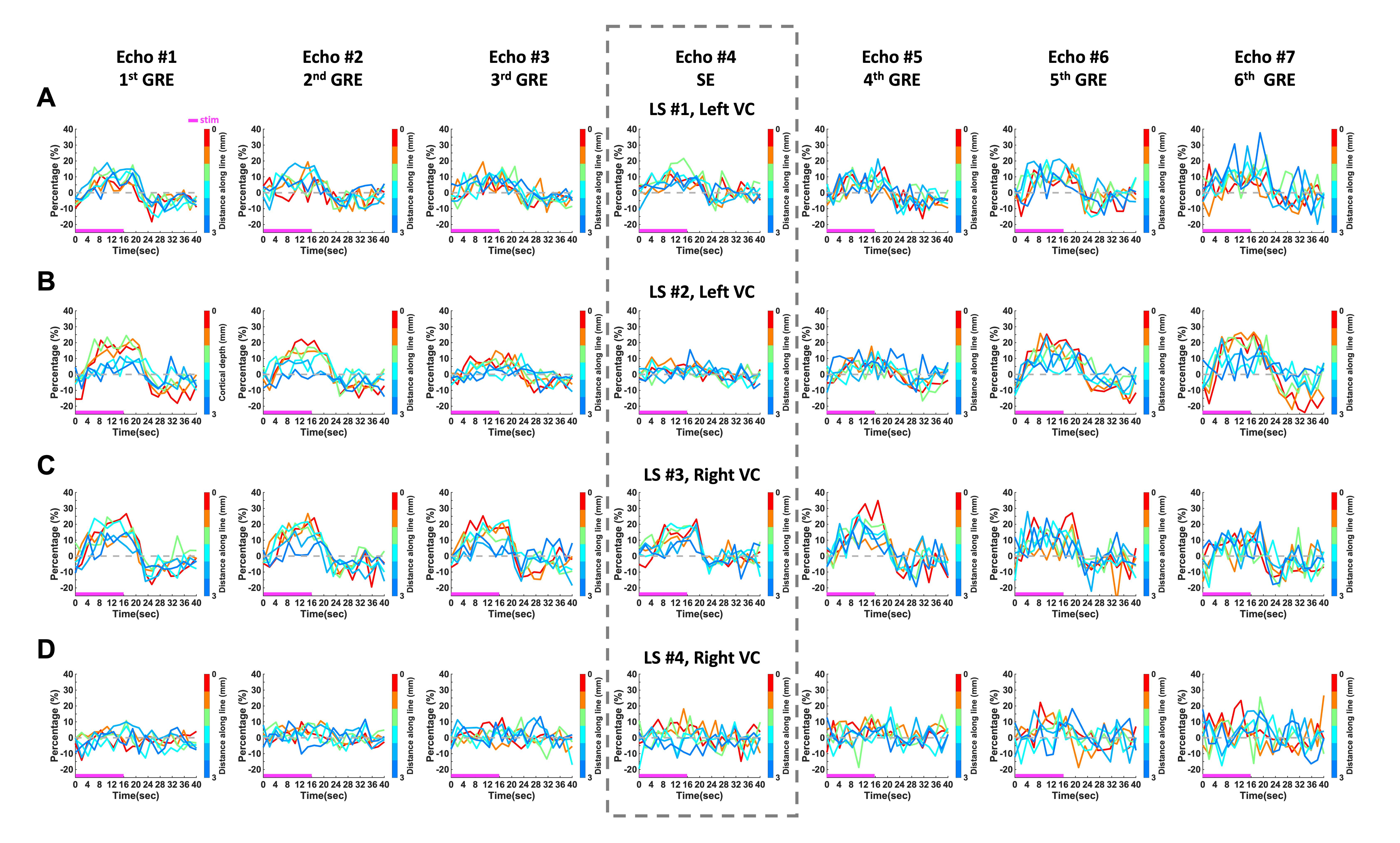
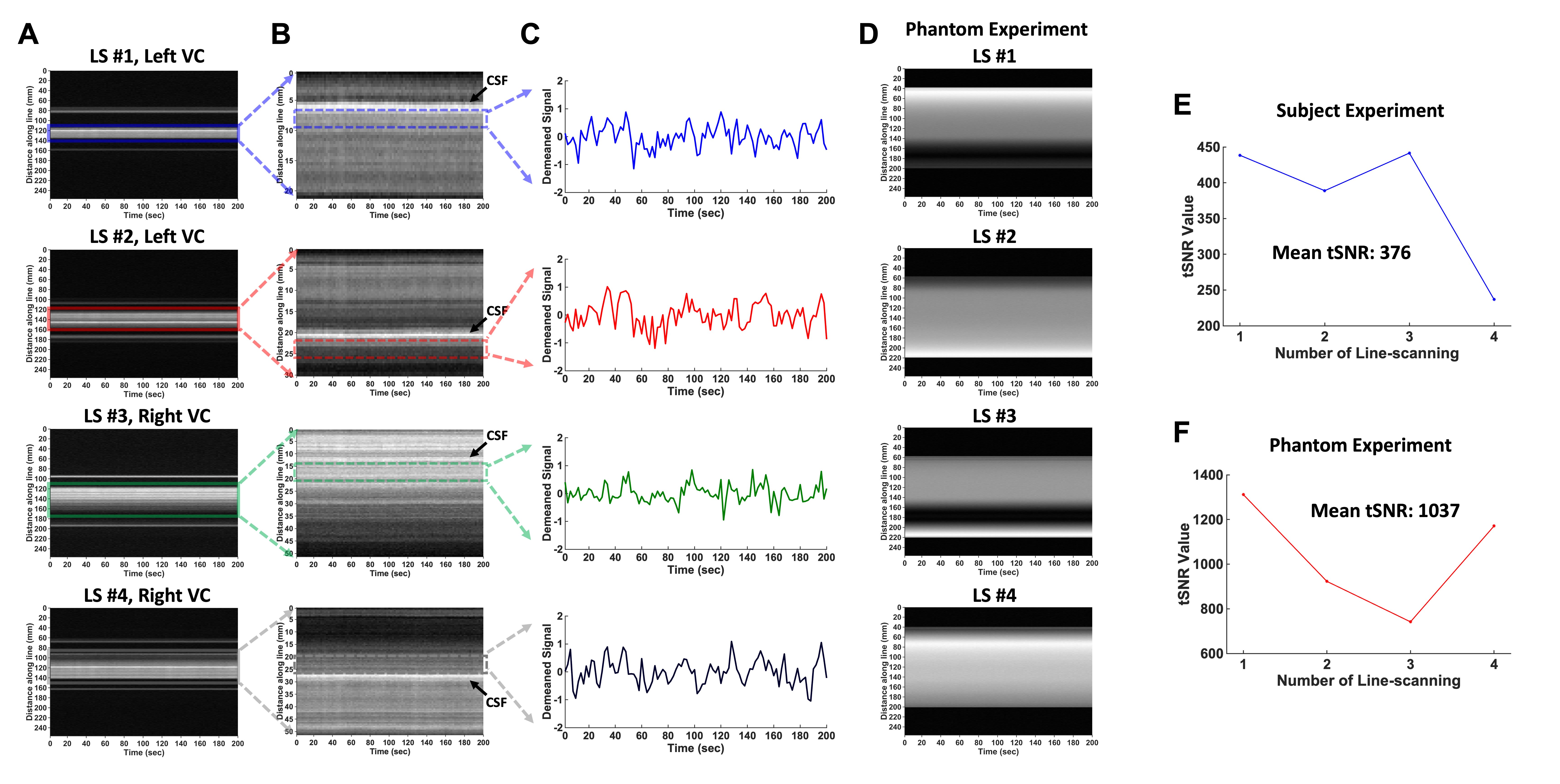
Distinct BOLD-fMRI responses were detected at each location of the visual cortex. To identify cortical ROIs, a line-profile map of the data averaged across echoes was generated (Fig. 2A and 4A) and the cortical ROI was defined by identifying the cerebrospinal fluid (CSF) within the subarachnoid space (Fig. 2B and 4B). An alternating signal intensity matching the stimulus timing could be seen by eye in these line-profile maps, demonstrating the strong functional contrast and sensitivity of the time-series data. Fig. 2C & D presented evoked BOLD responses within the cortical ROI; and Fig. 2E & F presented BOLD responses from individual voxels. The BOLD response amplitude varied systematically across the echoes as expected: the response at the spin-echo exhibited the lowest amplitudes, whereas the responses at the gradient-echoes increased in amplitude with increasing distance from the spin-echo10, indicating increased T2’ weighting and correspondingly higher functional sensitivity (Fig. 3). Finally, temporal SNR (tSNR) was evaluated in each line using the acquired resting-state fMRI and phantom data. The tSNR of the in vivo data from positions #1–3 was distinctly higher than that of position #4 (Fig. 4E); however, the tSNR of the phantom data from position #4 didn’t show lower tSNR (Fig. 4F).DISCUSSION
This work demonstrates the first proof-of-concept of non-contiguous, non-coplanar multi-line GESSE fMRI (ML-GESSE) across different cortical locations in human visual cortex at 7T. While activation could be detected within each line, the sensitivity was not uniform possibly due to functional variability across lines2. While care was taken to position lines to avoid overlap, it is possible that position #4 targeted a lower activated region since line-scanning methods require 1-3 mm line thickness1–5,11–15, which may indicate why some of previous human fMRI works could not report clear BOLD responses15. The 180° RF pulses may contribute more SAR than the 90° saturation pulses, thus for fast fMRI applications SAR may limit the achievable temporal resolution.CONCLUSION
Our ML-GESSE method provides a novel multi-line acquisition that can concurrently sample multiple cortical areas, investigating interactions between brain areas to help decipher information flow and brain circuitry in future studies.Acknowledgements
We thank Sarah Richter and Kyle Droppa for their help with subject recruitment and MRI scanning support and also thank Drs. Klaus Scheffler and Kai Herz for helpful comments on the initial work of the single-line GESSE acquisition. This work was supported in part by the NIH NIBIB (grants P41-EB030006, R01-EB019437 and R01-EB032746), NINDS (RF1-NS113278, R01-NS124778, R01-NS122904), by the BRAIN Initiative (NIH NIMH grant R01-MH111419 and NIH NINDS grants U19-NS123717 and U19-NS128613), and by the MGH/HST Athinoula A. Martinos Center for Biomedical Imaging; and was made possible by the resources provided by NIH Shared Instrumentation Grants S10-OD023637.References
1. Yu X, Qian C, Chen DY, et al. Deciphering laminar-specific neural inputs with line-scanning fMRI. Nat Methods 2014; 11: 55–58.
2. Choi S, Zeng H, Chen Y, et al. Laminar-specific functional connectivity mapping with multi-slice line-scanning fMRI. Cerebral Cortex 2022; 32: 4492–4501.
3. Choi S, Chen Y, Zeng H, et al. Identifying the distinct spectral dynamics of laminar-specific interhemispheric connectivity with bilateral line-scanning fMRI. Journal of Cerebral Blood Flow and Metabolism 2023; 43: 1115–1129.
4. Choi S, Hike D, Pohmann R, et al. Alpha-180 spin-echo based line-scanning method for high resolution laminar-specific fMRI. Biorxiv. DOI: 10.1101/2023.05.09.540065
5. Raimondo L, Knapen T, Oliveira ĺcaro AF, et al. A line through the brain: implementation of human line-scanning at 7T for ultra-high spatiotemporal resolution fMRI. Journal of Cerebral Blood Flow and Metabolism2021; 41: 2831–2843.
6. Morgan AT, Nothnagel N, Petro LS, et al. High-resolution line-scanning reveals distinct visual response properties across human cortical layers. DOI: 10.1101/2020.06.30.179762.
7. Park S, Torrisi S, Townsend JD, et al. Highly accelerated submillimeter resolution 3D GRASE with controlled T2 blurring in T2-weighted functional MRI at 7 Tesla: A feasibility study. Magn Reson Med 2021; 85: 2490–2506.
8. Balasubramanian M, Mulkern R V., Neil JJ, et al. Probing in vivo cortical myeloarchitecture in humans via line-scan diffusion acquisitions at 7 T with 250-500 micron radial resolution. Magn Reson Med 2021; 85: 390–403.
9. Mareyam A, Kirsch J, Chang Y, et al. A 64-Channel 7T array coil for accelerated brain MRI. Proc Intl Soc Mag Reson Med 2020; 0764.
10. Wang F, Dong Z, Wald LL, et al. Simultaneous pure T2 and varying T2′-weighted BOLD fMRI using Echo Planar Time-resolved Imaging for mapping cortical-depth dependent responses. Neuroimage; 245. Epub ahead of print 15 December 2021. DOI: 10.1016/j.neuroimage.2021.118641.
11. Choi S, Yu X, Scheffler K, et al. Simultaneous acquisition of GRE- and SE-type resting-state fMRI signals with GRASE-based line-scanning in the human brain. Proc Intl Soc Mag Reson Med; 30.
12. Choi S, Xie Z, Liu X, et al. Detecting high temporal laminar-specific responses with line-scanning fMRI in awake mice. Proc Intl Soc Mag Reson Med.
13. Albers F, Schmid F, Wachsmuth L, et al. Line scanning fMRI reveals earlier onset of optogenetically evoked BOLD response in rat somatosensory cortex as compared to sensory stimulation. Neuroimage 2018; 164: 144–154.
14. Nunes D, Gil R, Shemesh N. A rapid-onset diffusion functional MRI signal reflects neuromorphological coupling dynamics. Neuroimage 2021; 231: 117862.
15. Raimondo L, Heij J, Knapen T, et al. Towards functional spin-echo BOLD line-scanning in humans at 7T. Magnetic Resonance Materials in Physics, Biology and Medicine. Epub ahead of print 2023. DOI: 10.1007/s10334-022-01059-7.
Figures