3408
SNAKE-fMRI: A modular fMRI simulator from the space-time domain to k-space data and back1CEA/Neurospin, Gif-Sur-Yvette, France, 2Université Paris-Saclay, Gif-sur-Yvette, France, 3Inria Saclay, Palaiseau, France
Synopsis
Keywords: fMRI Acquisition, Simulations
Motivation: fMRI is a powerful tool for neuroimaging, but its optimization in vivo is complicated due to limited reproducibility. Simulation setup can help, but current solutions lack flexibility, control, or efficiency.
Goal(s): Develop a versatile and efficient fMRI simulator generating realistic fMRI data under various conditions: SNR levels, acceleration factors, spatiotemporal resolutions, noise, and artifact sources.
Approach: We propose SNAKE-fMRI, a modular, open-source fMRI simulator that operates from image to k-space and vice-versa. It can generate 3D+time k-space and images, mimicking the entire fMRI acquisition process, with potential k-t accelerations.
Results: We showcase the use of SNAKE-fMRI on a non-Cartesian k-t accelerated scenario.
Impact: With SNAKE-fMRI as an open-source, versatile, and efficient fMRI simulator, researchers can generate realistic fMRI data under a controlled and fully reproducible setup, enabling new advancements in fMRI acquisition and reconstruction methods.
Introduction
Functional Magnetic Resonance Imaging (fMRI) has emerged as a powerful non-invasive tool in neuroscience, enabling scientists to understand human brain function.However, fMRI acquisition and reconstruction techniques are complex to optimize and benchmark due to the lack of ground truth that produces absolute and quantitative metrics. Repeating in-vivo experiments may face the issue of limited reproducibility [1] and is time-consuming and expensive.
fMRI simulators have been developed to generate synthetic fMRI time series, where brain responses are artificially added to existing or synthetic data. However, they often lack flexibility, integration with post-processing, and computational efficiency (summarized in Table 1). Exploring new acceleration schemes in the acquisition setting and innovative reconstruction methods with these tools is not feasible.
To address these unmet needs, we propose SNAKE-fMRI (Simulator from NeuroActivation to Kspace Evaluation for Methodology and Reconstruction of Images). This open-source fMRI simulator operates in the image and k-space domain to yield the data. Its flexibility allows us to investigate various scenarios regarding SNR and acceleration factors to reach higher spatial and temporal resolution and validate reconstruction methods against those scenarios.
Key Features
Simplicity with modularitySNAKE-FMRI has a modular approach for the simulation of fMRI data (Figure 2): A core simulation object will be modified and enriched by so-called handlers, each of them being responsible for a specific part of the model (for instance, defining the anatomical base volume from a phantom or adding noise to a time-series). They are chained together to produce complex behaviors from simple operations. Once the simulation has been done, It is passed down to a reconstruction and statistical analysis pipeline.
Different noise and artifact sources can be modeled and superimposed on one another before or after k-space generation. However, the virtue of SNAKE-fMRI is that it can output both k-space and image-space data, so it can be interfaced with many different image analysis or reconstruction algorithms for validating them.
Efficient K-space data Generation
The acquisition process in SNAKE-fMRI is modeled using a Fourier Transform, which is a simplification that neglects the relaxation aspect of MR physics. In the context of a segmented (or multishot) acquisition, SNAKE-fMRI simulates a new image of the state of the signal at each shot, representing the ideal magnetization after the RF-pulse excitation. The shot is then acquired using the Fourier model, as detailed in Figure 3.
Example Scenario
We illustrate the capabilities of SNAKE-fMRI by simulating fMRI data under a high spatial resolution (e.g., 1mm iso) in a non-Cartesian, k-t accelerated setup consisting of a stack-of-spirals (SoS); the slices are ordered in the “center-out” methods, as described in Figure 3.We simulated a retinotopy mapping by adding brain activation consisting of a 5-minute paradigm of block-on(20s)/block-off(20s) pattern convoluted with canonical HRF that creates a 3% change of contrast in a region of interest, defined as a mask intersecting the gray matter. The high temporal resolution image simulation was then acquired under 3 different acceleration factors 2, 4, and 8 (resulting in volumetric TR of 3.6, 2.2, and 1.5 seconds) and input SNR of 100.
The reconstruction was done using a sequential, per-frame Compressed Sensing-based reconstruction with L1-wavelet regularisation, implemented in PySAP-MRI[10], using density-compensated gpuNUFFT for the data-consistency gradient step. To reduce the number of study parameters, the regularization parameters were estimated using Stein's unbiased Risk Estimate (SURE)[3].
We show an end-to-end process in Figure 4. Using a higher acceleration factor, the effective SNR drops and the sequential reconstruction cannot retrieve the activations in the right region of interest (ROI). This calls for more elaborated methods, with time-aware priors, such as a low-rank and sparsity in time domain one, that fully leverage the k-t aspect of the acquisition.
Conclusion
SNAKE-fMRI is a versatile and efficient fMRI simulator that provides researchers with a valuable tool for generating synthetic fMRI data under various conditions. The simulator can be used to develop and validate new fMRI acquisition and reconstruction methods, including training neural networks for fMRI reconstruction on synthetic data, as well as an educational tool.SNAKE-fMRI will be made available at https://github.com/snake-fmri/snake-fmri
Acknowledgements
No acknowledgement found.References
[1] C. M. Bennett and M. B. Miller, “How reliable are the results from functional magnetic resonance imaging?,” Ann. N. Y. Acad. Sci., vol. 1191, pp. 133–155, Mar. 2010, doi: 10.1111/j.1749-6632.2010.05446.x.
[2] C. Y. Lin and J. A. Fessler, “Efficient Dynamic Parallel MRI Reconstruction for the Low-Rank Plus Sparse Model,” IEEE Trans. Comput. Imaging, vol. 5, no. 1, pp. 17–26, Mar. 2019, doi: 10.1109/TCI.2018.2882089.
[3] D. L. Donoho and I. M. Johnstone, “Threshold selection for wavelet shrinkage of noisy data,” in Proceedings of 16th Annual International Conference of the IEEE Engineering in Medicine and Biology Society, Baltimore, MD, USA: IEEE, 1994, pp. A24–A25. doi: 10.1109/IEMBS.1994.412133.
[4] T. Stöcker, K. Vahedipour, D. Pflugfelder, and N. J. Shah, “High-performance computing MRI simulations,” Magn. Reson. Med., vol. 64, no. 1, pp. 186–193, 2010, doi: 10.1002/mrm.22406.[
5] T. H. Jochimsen, A. Schäfer, R. Bammer, and M. E. Moseley, “Efficient simulation of magnetic resonance imaging with Bloch–Torrey equations using intra-voxel magnetization gradients,” J. Magn. Reson., vol. 180, no. 1, pp. 29–38, May 2006, doi: 10.1016/j.jmr.2006.01.001.[
6] F. Liu, J. V. Velikina, W. F. Block, R. Kijowski, and A. A. Samsonov, “Fast Realistic MRI Simulations Based on Generalized Multi-Pool Exchange Tissue Model,” IEEE Trans. Med. Imaging, vol. 36, no. 2, pp. 527–537, Feb. 2017, doi: 10.1109/TMI.2016.2620961.
[7] E. B. Erhardt, E. A. Allen, Y. Wei, T. Eichele, and V. D. Calhoun, “SimTB, a simulation toolbox for fMRI data under a model of spatiotemporal separability,” NeuroImage, vol. 59, no. 4, pp. 4160–4167, Feb. 2012, doi: 10.1016/j.neuroimage.2011.11.088.
[8] C. T. Ellis, C. Baldassano, A. C. Schapiro, M. B. Cai, and J. D. Cohen, “Facilitating open-science with realistic fMRI simulation: validation and application,” PeerJ, vol. 8, p. e8564, Feb. 2020, doi: 10.7717/peerj.8564.
[9] M. Welvaert, J. Durnez, B. Moerkerke, G. Berdoolaege, and Y. Rosseel, “neuRosim: An R Package for Generating fMRI Data,” J. Stat. Softw., vol. 44, pp. 1–18, Oct. 2011, doi: 10.18637/jss.v044.i10.
[10] S. Farrens et al., “PySAP: Python sparse data analysis package for multidisciplinary image processing,” Astronomy and Computing, vol. 32, p. 100402, 2020, doi: 10/ghw8zm.Figures
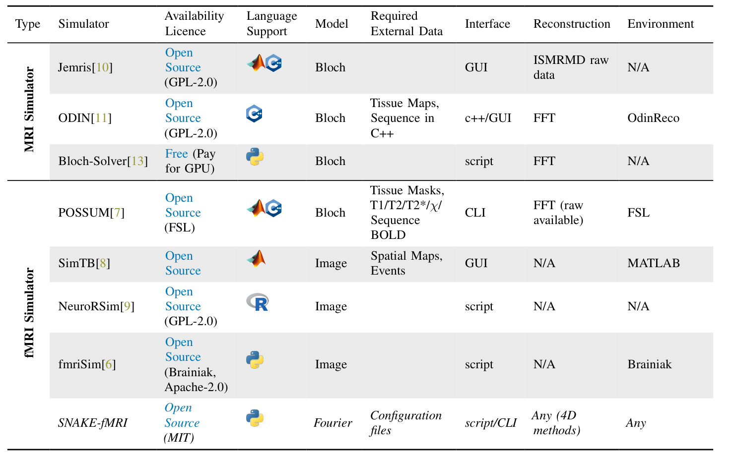
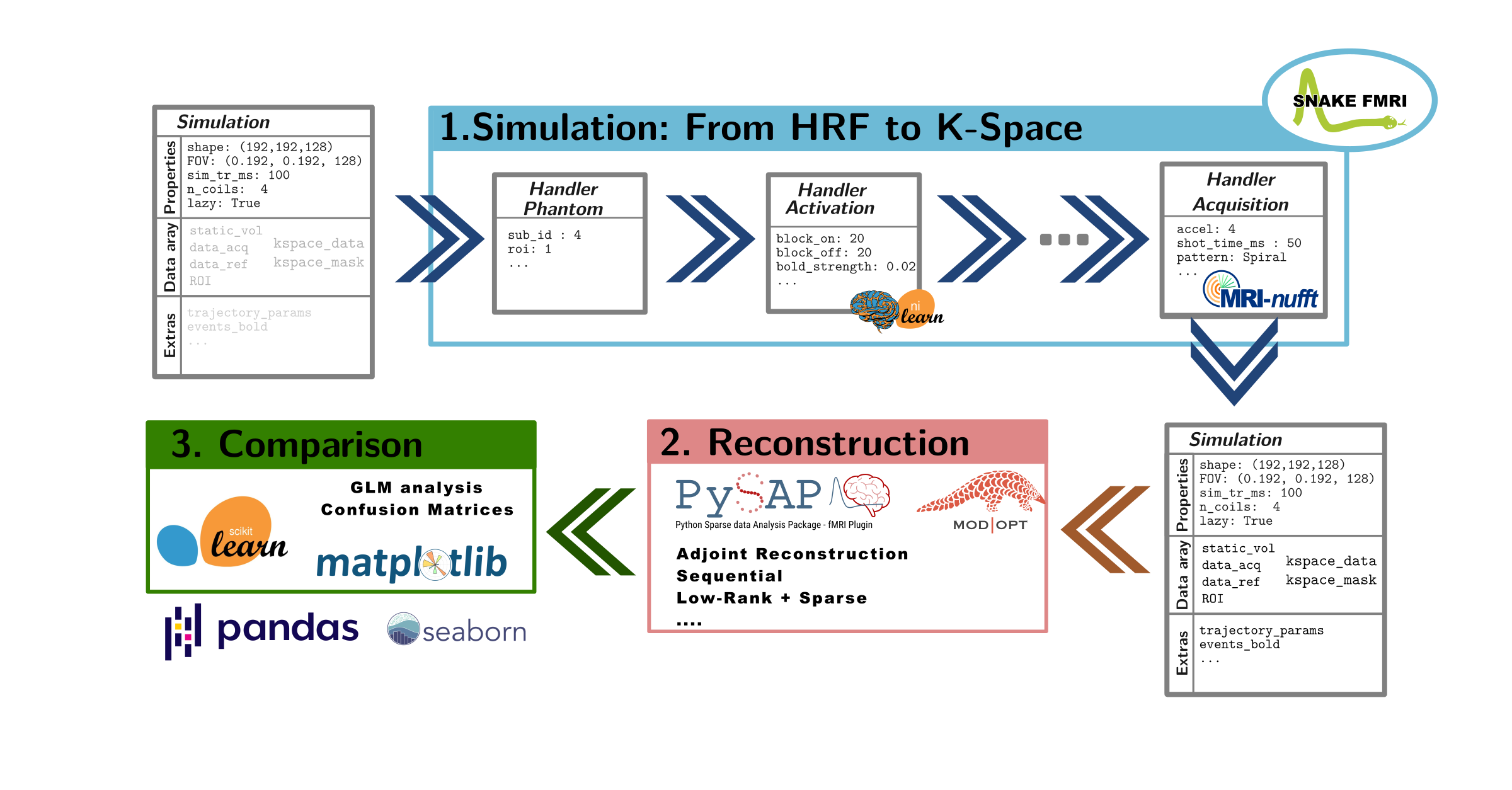
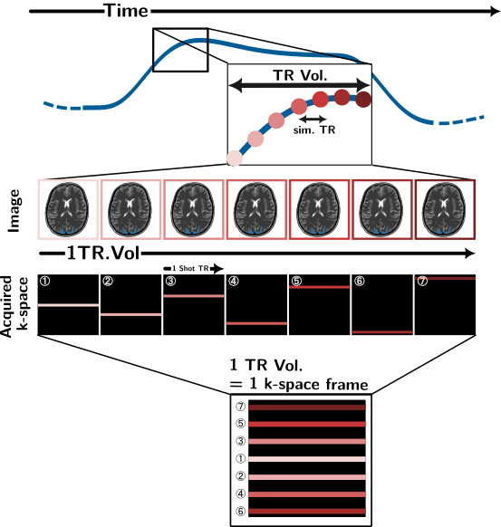
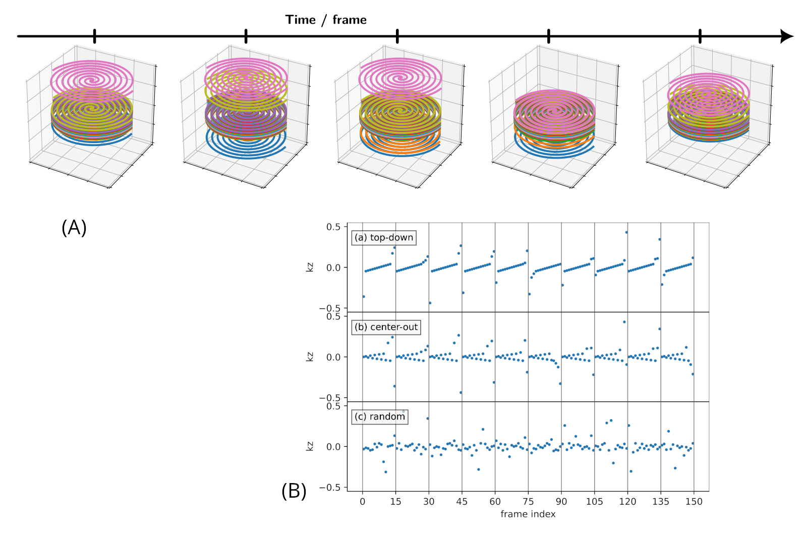
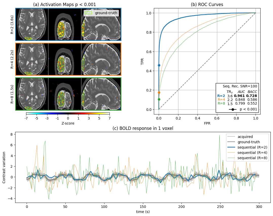
Figure 4: Obtained statistics after simulation, acquisition (center-out SoS) and reconstruction: The downstream analysis and testing against ground truth allow for a clear comparison between different acquisition or reconstruction strategies. Activations (a) are compared with ground-truth ROI; (b) ROC Curve can provide Overall classification metrics such as AUC or Balanced Accuracy (BACC); Finally, time series after the different strategies can be compared (c).
Here, we compared 3 Acceleration factors for the same SNR level and reconstruction method (sequential).