3391
CMR-based estimation of mean pulmonary arterial pressure and pulmonary vascular resistance in CTD-PAH1Department of Radiology, The First Affiliated Hospital of Nanjing Medical University, Nanjing, China, 2Department of Rheumatology, The First Affiliated Hospital of Nanjing Medical University, Nanjing, China, 3Department of Radiology, The Affiliated Cancer Hospital of Nanjing Medical University, Jiangsu Cancer Hospital, Jiangsu Institute of Cancer Research, Nanjing, China
Synopsis
Keywords: Vascular, Cardiovascular
Motivation: This research explores the potential of cardiac magnetic resonance (CMR) as a noninvasive tool for evaluating pulmonary arterial hypertension (PAH) in connective tissue disease-associated PAH (CTD-PAH) patients.
Goal(s): The goal is to estimate mean pulmonary arterial pressure (mPAP) and pulmonary vascular resistance (PVR) accurately using CMR metrics, with right heart catheterization (RHC) as the gold standard.
Approach: Correlations between CMR and RHC parameters were assessed, and predictive models for mPAP and PVR were developed.
Results: Strong correlations were observed between RHC and CMR metrics. Predictive CMR models were established for both mPAP and PVR, offering potential clinical applications.
Impact: CMR parameters can be used to estimate mPAP and PVR in patients with CTD-PAH, potentially providing a noninvasive method for assessing pulmonary arterial hemodynamics during CTD-PAH management.
Background
Cardiac magnetic resonance (CMR) is a promising noninvasive tool for assessing pulmonary arterial hypertension (PAH). This study aimed to estimate mean pulmonary arterial pressure (mPAP) and pulmonary vascular resistance (PVR) based on CMR metrics using right heart catheterization (RHC) as the gold standard in connective tissue disease-associated PAH (CTD-PAH).Methods
This study retrospectively enrolled 70 consecutive patients diagnosed with CTD-PAH who underwent both CMR and RHC within seven days between September 2017 and April 2023. Their ventricular volumes, functions, and morphology; T1 mapping parameters; and main pulmonary artery (MPA) hemodynamics were measured. The correlations between CMR and RHC parameters were assessed using Spearman’s rank correlation coefficient (rs). The thresholds of mPAP changes were identified using segmental linear regression analysis, and predictive models were developed for mPAP and PVR using regression analysis.Results
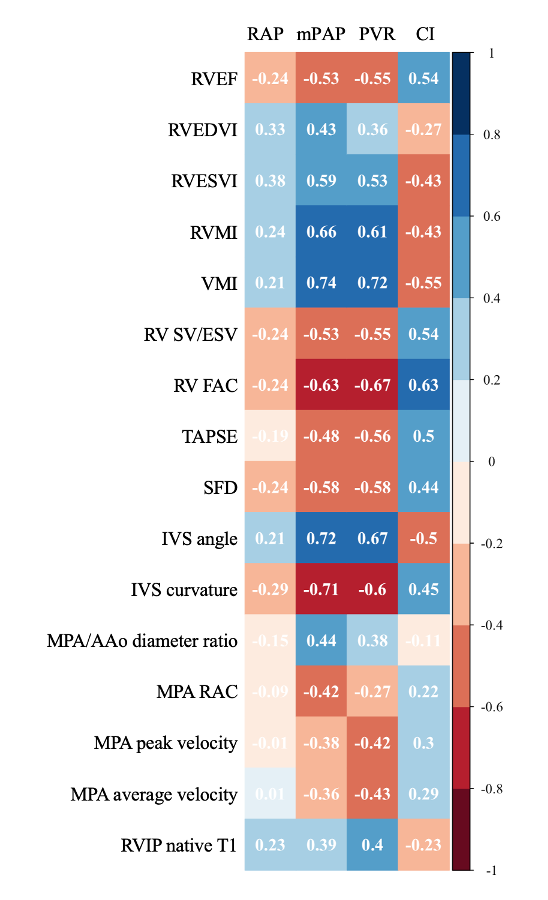
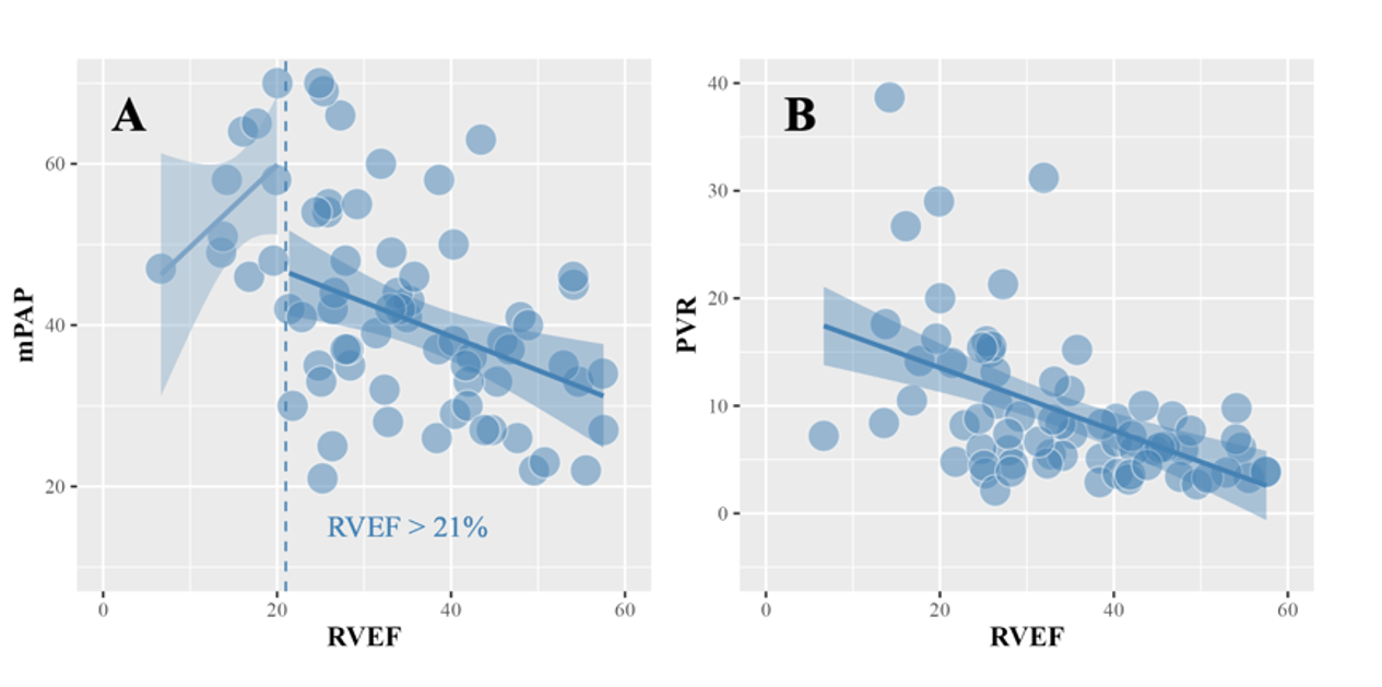
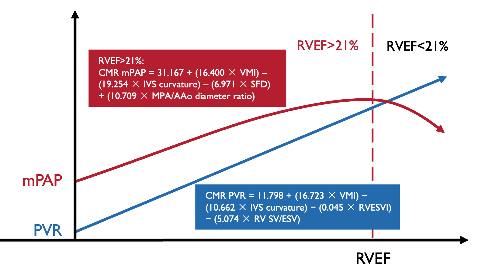
The mPAP was strongly correlated with ventricular mass index (VMI; rs = 0.74), interventricular septal (IVS; rs= 0.72) angle, and IVS curvature (rs = −0.71; all P < 0.01). The PVR was strongly positively correlated with VMI (rs = 0.72, P < 0.01). Segmented linear regression identified a right ventricular ejection fraction (RVEF) threshold of 21%. When the RVEF was > 21%, VMI, septum to free-wall distance change (SFD), IVS curvature, and the MPA/ascending aorta (AAo) diameter ratio were independently correlated with mPAP (P < 0.05). A CMR model for predicting mPAP was established: 31.167 + (16.400 × VMI) – (19.254 × IVS curvature) – (6.971 × SFD) + (10.709 × MPA/AAo diameter ratio). Another CMR model for predicting PVR was developed: 11.798 + (16.723 × VMI) – (10.662 × IVS curvature) – (0.045 × right ventricular end-systolic volume index) – (5.074 × right ventricular stroke volume to end-systolic volume).Conclusions
CMR parameters can be used to estimate mPAP and PVR in patients with CTD-PAH, potentially providing a noninvasive method for assessing pulmonary arterial hemodynamics during CTD-PAH management.Acknowledgements
No acknowledgement found.References
1. Balistrieri A, Makino A, Yuan JX-J. Pathophysiology and pathogenic mechanisms of pulmonary hypertension: role of membrane receptors, ion channels, and Ca2+ signaling. Physiol Rev. 2023;103:1827–1897.2. Mathai SC. Pulmonary Hypertension Associated with Connective Tissue Disease. Cardiol Clin. 2022;40:29–43.
3. Fayed H, Coghlan JG. Pulmonary Hypertension Associated with Connective Tissue Disease. Semin Respir Crit Care Med. 2019;40:173–183.
4. Armstrong I, Billings C, Kiely DG, Yorke J, Harries C, Clayton S, Gin-Sing W. The patient experience of pulmonary hypertension: a large cross-sectional study of UK patients. BMC Pulm Med. 2019;19:67.
5. Hurdman J, Condliffe R, Elliot CA, Davies C, Hill C, Wild JM, Capener D, Sephton P, Hamilton N, Armstrong IJ, Billings C, Lawrie A, Sabroe I, Akil M, O’Toole L, Kiely DG. ASPIRE registry: Assessing the Spectrum of Pulmonary hypertension Identified at a REferral centre. Eur Respir J. 2012;39:945–955.
6. Vonk-Noordegraaf A, Haddad F, Chin KM, Forfia PR, Kawut SM, Lumens J, Naeije R, Newman J, Oudiz RJ, Provencher S, Torbicki A, Voelkel NF, Hassoun PM. Right Heart Adaptation to Pulmonary Arterial Hypertension. J Am Coll Cardiol. 2013;62:D22–D33.
7. Humbert M, Kovacs G, Hoeper MM, Badagliacca R, Berger RMF, Brida M, Carlsen J, Coats AJS, Escribano-Subias P, Ferrari P, Ferreira DS, Ghofrani HA, Giannakoulas G, Kiely DG, Mayer E, Meszaros G, Nagavci B, Olsson KM, Pepke-Zaba J, Quint JK, Rådegran G, Simonneau G, Sitbon O, Tonia T, Toshner M, Vachiery JL, Vonk Noordegraaf A, Delcroix M, Rosenkranz S, Schwerzmann M, Dinh-Xuan AT, Bush A, Abdelhamid M, Aboyans V, Arbustini E, Asteggiano R, Barberà JA, Beghetti M, Čelutkienė J, Cikes M, Condliffe R, de Man F, Falk V, Fauchier L, Gaine S, Galié N, Gin-Sing W, Granton J, Grünig E, Hassoun PM, Hellemons M, Jaarsma T, Kjellström B, Klok FA, Konradi A, Koskinas KC, Kotecha D, Lang I, Lewis BS, Linhart A, Lip GYH, Løchen ML, Mathioudakis AG, Mindham R, Moledina S, Naeije R, Nielsen JC, Olschewski H, Opitz I, Petersen SE, Prescott E, Rakisheva A, Reis A, Ristić AD, Roche N, Rodrigues R, Selton-Suty C, Souza R, Swift AJ, Touyz RM, Ulrich S, Wilkins MR, Wort SJ, Wort SJ. 2022 ESC/ERS Guidelines for the diagnosis and treatment of pulmonary hypertension. Eur Heart J. 2022;43:3618–3731.
8. Simonneau G, Montani D, Celermajer DS, Denton CP, Gatzoulis MA, Krowka M, Williams PG, Souza R. Haemodynamic definitions and updated clinical classification of pulmonary hypertension. Eur Respir J. 2019;53:1801913.
9. D’Alto M, Bossone E, Opotowsky AR, Ghio S, Rudski LG, Naeije R. Strengths and weaknesses of echocardiography for the diagnosis of pulmonary hypertension. Int J Cardiol. 2018;263:177–183.
10. Ferrara F, Zhou X, Gargani L, Wierzbowska-Drabik K, Vriz O, Fadel BM, Stanziola AA, Kasprzak J, Vannan M, Bossone E. Echocardiography in Pulmonary Arterial Hypertension. Curr Cardiol Rep. 2019;21:22.
11. D’Alto M, Romeo E, Argiento P, D’Andrea A, Vanderpool R, Correra A, Bossone E, Sarubbi B, Calabrò R, Russo MG, Naeije R. Accuracy and precision of echocardiography versus right heart catheterization for the assessment of pulmonary hypertension. Int J Cardiol. 2013;168:4058–4062.
12. Broncano J, Bhalla S, Gutierrez FR, Vargas D, Williamson EE, Makan M, Luna A. Cardiac MRI in Pulmonary Hypertension: From Magnet to Bedside. RadioGraphics. 2020;40:982–1002.
13. Simpson CE, Kolb TM, Hsu S, Zimmerman SL, Corona‐Villalobos CP, Mathai SC, Damico RL, Hassoun PM. Ventricular mass discriminates pulmonary arterial hypertension as redefined at the Sixth World Symposium on Pulmonary Hypertension. Pulm Circ. 2022;12:e12005.
14. Saba TS, Foster J, Cockburn M, Cowan M, Peacock AJ. Ventricular mass index using magnetic resonance imaging accurately estimates pulmonary artery pressure. Eur Respir J. 2002;20:1519–1524.
15. Sanz J, Kuschnir P, Rius T, Salguero R, Sulica R, Einstein AJ, Dellegrottaglie S, Fuster V, Rajagopalan S, Poon M. Pulmonary Arterial Hypertension: Noninvasive Detection with Phase-Contrast MR Imaging. Radiology. 2007;243:70–79.
16. Trejo-Velasco B, Cruz-González I, Barreiro-Pérez M, Díaz-Peláez E, García-González P, Martín-García A, Eiros R, Merchán-Gómez S, Pérez del Villar C, Fabregat-Andrés O, Ridocci-Soriano F, Sánchez PL. Mean Velocity of the Pulmonary Artery as a Clinically Relevant Prognostic Indicator in Patients with Heart Failure with Preserved Ejection Fraction. J Clin Med. 2022;11:491.
17. Roller FC, Wiedenroth C, Breithecker A, Liebetrau C, Mayer E, Schneider C, Rolf A, Hamm C, Krombach GA. Native T1 mapping and extracellular volume fraction measurement for assessment of right ventricular insertion point and septal fibrosis in chronic thromboembolic pulmonary hypertension. Eur Radiol. 2017;27:1980–1991.
18. Habert P, Capron T, Hubert S, Bentatou Z, Bartoli A, Tradi F, Renard S, Rapacchi S, Guye M, Bernard M, Habib G, Jacquier A. Quantification of right ventricular extracellular volume in pulmonary hypertension using cardiac magnetic resonance imaging. Diagn Interv Imaging. 2020;101:311–320.
19. Alabed S, Saunders L, Garg P, Shahin Y, Alandejani F, Rolf A, Puntmann VO, Nagel E, Wild JM, Kiely DG, Swift AJ. Myocardial T1-mapping and extracellular volume in pulmonary arterial hypertension: A systematic review and meta-analysis. Magn Reson Imaging. 2021;79:66–75.
20. Johns CS, Kiely DG, Rajaram S, Hill C, Thomas S, Karunasaagarar K, Garg P, Hamilton N, Solanki R, Capener DA, Elliot C, Sabroe I, Charalamopopoulos A, Condliffe R, Wild JM, Swift AJ. Diagnosis of Pulmonary Hypertension with Cardiac MRI: Derivation and Validation of Regression Models. Radiology. 2019;290:61–68.
21. Bhatnagar A, Wiesen J, Dweik R, Chaisson NF. Evaluating suspected pulmonary hypertension: A structured approach. Cleve Clin J Med. 2018;85:468–480.
22. Dong Y, Li Y, Song L. Evaluation of right ventricular function in patients with pulmonary arterial hypertension by different right ventricular-pulmonary artery coupling methods. Medicine (Baltimore). 2022;101:e30873.
23. Sanz J, García-Alvarez A, Fernández-Friera L, Nair A, Mirelis JG, Sawit ST, Pinney S, Fuster V. Right ventriculo-arterial coupling in pulmonary hypertension: a magnetic resonance study. Heart. 2012;98:238–243.
24. Kind T, Mauritz G-J, Marcus JT, van de Veerdonk M, Westerhof N, Vonk-Noordegraaf A. Right ventricular ejection fraction is better reflected by transverse rather than longitudinal wall motion in pulmonary hypertension. J Cardiovasc Magn Reson. 2010;12:35.
25. Swift AJ, Rajaram S, Hurdman J, Hill C, Davies C, Sproson TW, Morton AC, Capener D, Elliot C, Condliffe R, Wild JM, Kiely DG. Noninvasive Estimation of PA Pressure, Flow, and Resistance With CMR Imaging. JACC Cardiovasc Imaging. 2013;6:1036–1047.
26. Roeleveld RJ, Marcus JT, Faes TJC, Gan T-J, Boonstra A, Postmus PE, Vonk-Noordegraaf A. Interventricular Septal Configuration at MR Imaging and Pulmonary Arterial Pressure in Pulmonary Hypertension. Radiology. 2005;234:710–717.
27. Swift AJ, Capener D, Johns C, Hamilton N, Rothman A, Elliot C, Condliffe R, Charalampopoulos A, Rajaram S, Lawrie A, Campbell MJ, Wild JM, Kiely DG. Magnetic Resonance Imaging in the Prognostic Evaluation of Patients with Pulmonary Arterial Hypertension. Am J Respir Crit Care Med. 2017;196:228–239.
28. Rosenkranz S, Preston IR. Right heart catheterisation: best practice and pitfalls in pulmonary hypertension. Eur Respir Rev. 2015;24:642–652.
29. Goh ZM, Balasubramanian N, Alabed S, Dwivedi K, Shahin Y, Rothman AMK, Garg P, Lawrie A, Capener D, Thompson AAR, Alandejani F, Wild JM, Johns CS, Lewis RA, Gosling R, Sharkey M, Condliffe R, Kiely DG, Swift AJ. Right ventricular remodelling in pulmonary arterial hypertension predicts treatment response. Heart. 2022;108:1392–1400.
30. Grignola JC, Domingo E, López-Meseguer M, Trujillo P, Bravo C, Pérez-Hoyos S, Roman A. Pulmonary Arterial Remodeling Is Related to the Risk Stratification and Right Ventricular-Pulmonary Arterial Coupling in Patients With Pulmonary Arterial Hypertension. Front Physiol. 2021;12:631326.
31. Dong Y, Pan Z, Wang D, Lv J, Fang J, Xu R, Ding J, Cui X, Xie X, Wang X, Chen, MD Y, Guo X. Prognostic Value of Cardiac Magnetic Resonance–Derived Right Ventricular Remodeling Parameters in Pulmonary Hypertension. Circ Cardiovasc Imaging. 2020;13:e010568.
32. Swift AJ, Rajaram S, Campbell MJ, Hurdman J, Thomas S, Capener D, Elliot C, Condliffe R, Wild JM, Kiely DG. Prognostic Value of Cardiovascular Magnetic Resonance Imaging Measurements Corrected for Age and Sex in Idiopathic Pulmonary Arterial Hypertension. Circ Cardiovasc Imaging. 2014;7:100–106.
33. Tello K, Dalmer A, Axmann J, Vanderpool R, Ghofrani HA, Naeije R, Roller F, Seeger W, Sommer N, Wilhelm J, Gall H, Richter MJ. Reserve of Right Ventricular-Arterial Coupling in the Setting of Chronic Overload. Circ Heart Fail. 2019;12:e005512.
34. Richter MJ, Peters D, Ghofrani HA, Naeije R, Roller F, Sommer N, Gall H, Grimminger F, Seeger W, Tello K. Evaluation and Prognostic Relevance of Right Ventricular–Arterial Coupling in Pulmonary Hypertension. Am J Respir Crit Care Med. 2020;201:116–119.
35. Hoeper MM, Pausch C, Olsson KM, Huscher D, Pittrow D, Grünig E, Staehler G, Vizza CD, Gall H, Distler O, Opitz C, Gibbs JSR, Delcroix M, Ghofrani HA, Park D-H, Ewert R, Kaemmerer H, Kabitz H-J, Skowasch D, Behr J, Milger K, Halank M, Wilkens H, Seyfarth H-J, Held M, Dumitrescu D, Tsangaris I, Vonk-Noordegraaf A, Ulrich S, Klose H, Claussen M, Lange TJ, Rosenkranz S. COMPERA 2.0: a refined four-stratum risk assessment model for pulmonary arterial hypertension. Eur Respir J. 2022;60:2102311.
36. Boucly A, Weatherald J, Savale L, de Groote P, Cottin V, Prévot G, Chaouat A, Picard F, Horeau-Langlard D, Bourdin A, Jutant E-M, Beurnier A, Jevnikar M, Jaïs X, Simonneau G, Montani D, Sitbon O, Humbert M. External validation of a refined four-stratum risk assessment score from the French pulmonary hypertension registry. Eur Respir J. 2022;59:2102419.
Figures
Graphical Abstracts
Cardiac magnetic resonance (CMR) is a noninvasive tool for assessing connective tissue disease-associated pulmonary arterial hypertension (CTD-PAH). CMR parameters (VMI, IVS curvature, SFD, MPA/AAo ratio) establish a noninvasive model for mPAP. The model's applicability is defined when RVEF > 21%. Additionally, VMI, IVS curvature, RVESVI, and RV SV/ESV can create a PVR model. These simple CMR parameters enable noninvasive mPAP and PVR calculation in CTD-PAH patients.