3388
Automatic Carotid Plaque Segmentation Using Deep Learning Model with Multi-Head Loss Integrating Anatomical Features1Paul C. Lauterbur Research Center for Biomedical Imaging, Shenzhen Institute of Advanced Technology, Chinese Academy of Sciences, Shen Zhen, China, 2Department of Image Advanced Analysis of HSW BU, Shanghai United Imaging Healthcare Co., Ltd., Shanghai, China, 3Department of Radiology, Peking University Shenzhen Hospital, Shenzhen, China, 4Key Laboratory of Biomedical Imaging Science and System, Chinese Academy of Sciences, Shenzhen, China, 5United Imaging Research Institute of Innovative Medical Equipment, Shenzhen, China
Synopsis
Keywords: Vessel Wall, Stroke
Motivation: With the assistance of prior vessel wall mask, segmentation of atherosclerotic plaque can achieve satisfactory performance. However, manual sketching of vessel wall mask is still time-consuming.
Goal(s): To propose a method for fast and accurate plaque segmentation without relying on prior knowledge of vessel walls.
Approach: This study proposes a deep learning model based on a multi-head loss design for automatic segmentation of carotid artery plaques, with the aim of reducing dependence on prior information of vessel walls in plaque segmentation.
Results: In the independent test, the model with the multi-head loss design achieving excellent results similar to using vessel wall prior.
Impact: This study achieved fully automatic and accurate plaque segmentation without manual priors, which will greatly reduce burden of radiologist to segment and quantify plaque, and also contribute to more efficient stroke risk assessment, progress monitoring, and efficacy evaluation for patient.
INTRODUCTION
Atherosclerotic plaque is closely related to ischemic stroke1. Segmentation of plaques is an important step for further risk analysis. However, automatic plaque segmentation requires the manually sketched mask of vessel walls as a prior to achieve good results. This study proposes a deep learning model based on a multi-head loss design for automatic segmentation of carotid artery plaques, with the aim of reducing dependence on prior information of vessel walls in plaque segmentation.METHODS
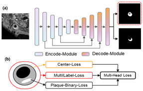
A total of 12,452 2D cross-sectional slices reconstructed from 493 vessel wall MRI scans of patients with carotid artery plaques were used for algorithm development. The annotated data were divided into training, validation, and testing sets at a 7:2:1 ratio. We proposed an improved convolutional neural network model (Fig. 1) based on a multihead loss design for automatic segmentation of carotid artery plaques and vessel walls to avoid dependence on prior information of vessel walls in plaque segmentation. The multihead loss design separated channels in the network's output heatmap to improve segmentation loss and introduced a central loss function for the vessel wall and lumen region. The modified segmentation in the central region of the predicted outcome ultimately achieved optimal plaque segmentation results. The weights of plaque segmentation in the multihead loss are tuned to ensure the performance of the final segmentation target - plaque segmentation. The ground truth (GT) was sketched by five radiologists with more than 10 years of experience. The DSC was used to measure the similarity between the prediction and the ground truth.RESULTS
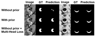
The performance of the comparative experiment is summarized in Figure 2. When only the carotid artery image was input, the DSC between model prediction and ground truth was 0.73. When both the carotid artery image and the vessel wall mask were input, the DSC increased from 0.73 to 0.83. Using the model with the multi-head loss design can also achieve similar improvements. The represented segmentation results of the three models are shown in Figure 3.DISCUSSION AND CONCLUSION
The design of multihead loss improves the vessel wall central loss and weights the central region, achieving excellent results similar to using the vessel wall prior. Therefore, the proposed model can achieve fast and accurate plaque segmentation without relying on prior knowledge of vessel walls.Acknowledgements
The study was partially support by Natural Science Foundation of Guangdong Province-Outstanding Youth Project (2023B1515020002), National Key Technology Research and Development Program of China (2021YFF0501502), Key Laboratory for Magnetic Resonance and Multimodality Imaging of Guangdong Province (2023B1212060052), and Central guidance for local science and technology development project (ZYYD2023D02).References
1. J. A. Madden, Role of the vascular endothelium and plaque in acute ischemic stroke. Neurology 79, S58-S62 (2012).Figures