3377
Paeoniflorin's Cardioprotective Role in High-Altitude MI: A CMR Study1West China Hospital of Sichuan University, Chengdu, China
Synopsis
Keywords: Heart Failure, Cardiovascular
Motivation: To investigate the cardioprotective effects of paeoniflorin on myocardial infarction-induced ventricular remodeling in high-altitude hypobaric hypoxia environments.
Goal(s): To evaluate the potential protective role of paeoniflorin in a rat model of myocardial infarction-induced ventricular remodeling under high-altitude hypobaric hypoxia using cardiovascular magnetic resonance strain analysis.
Approach: Rats were allocated to plain sham operation control, high-altitude sham operation control, high-altitude myocardial infarction operation, and paeoniflorin treatment groups. CMR assessed cardiac function.
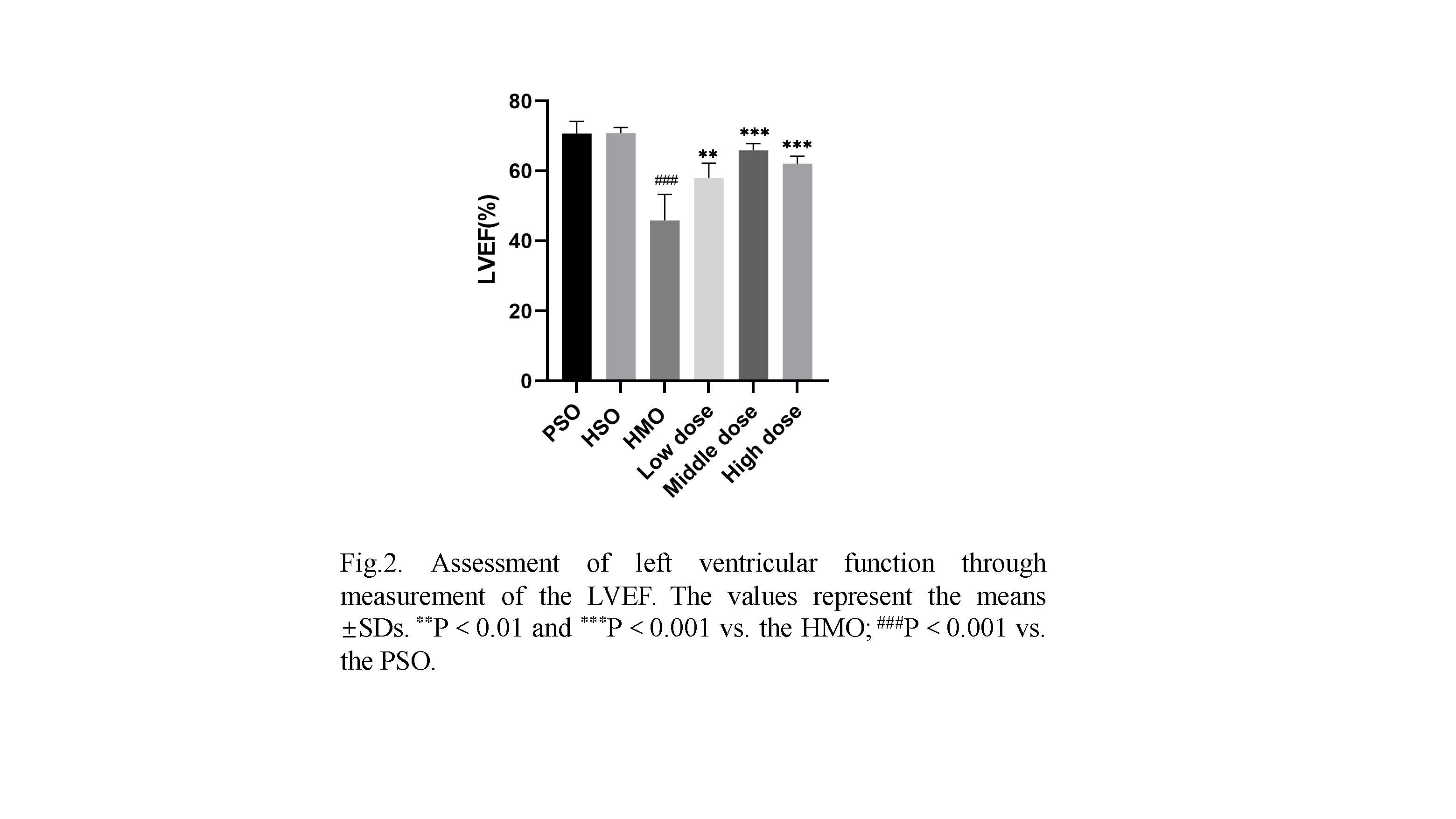
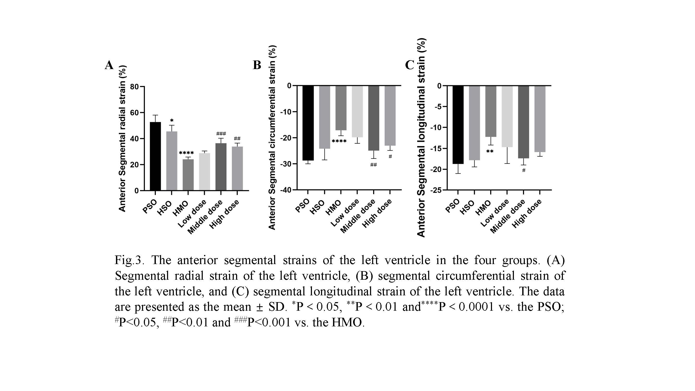
Results: Paeoniflorin treatment groups demonstrated enhanced left ventricular function and regional strains compared to the myocardial infarction group. Histological analyses also showed reduced myocardial injury and fibrosis with paeoniflorin treatment.
Impact: The study's outcomes have implications for managing patients at risk of MI, especially those in high-altitude environments or exposed to hypobaric hypoxia.
Purpose
Investigating the cardioprotective effects of paeoniflorin on myocardial infarction (MI)-induced ventricular remodeling in high-altitude hypobaric hypoxia environments is of great interest in cardiovascular research. This study aimed to evaluate the potential protective role of paeoniflorin in a rat model of MI-induced ventricular remodeling under high-altitude hypobaric hypoxia using 7.0T cardiovascular magnetic resonance (CMR) strain analysis.Materials and Methods
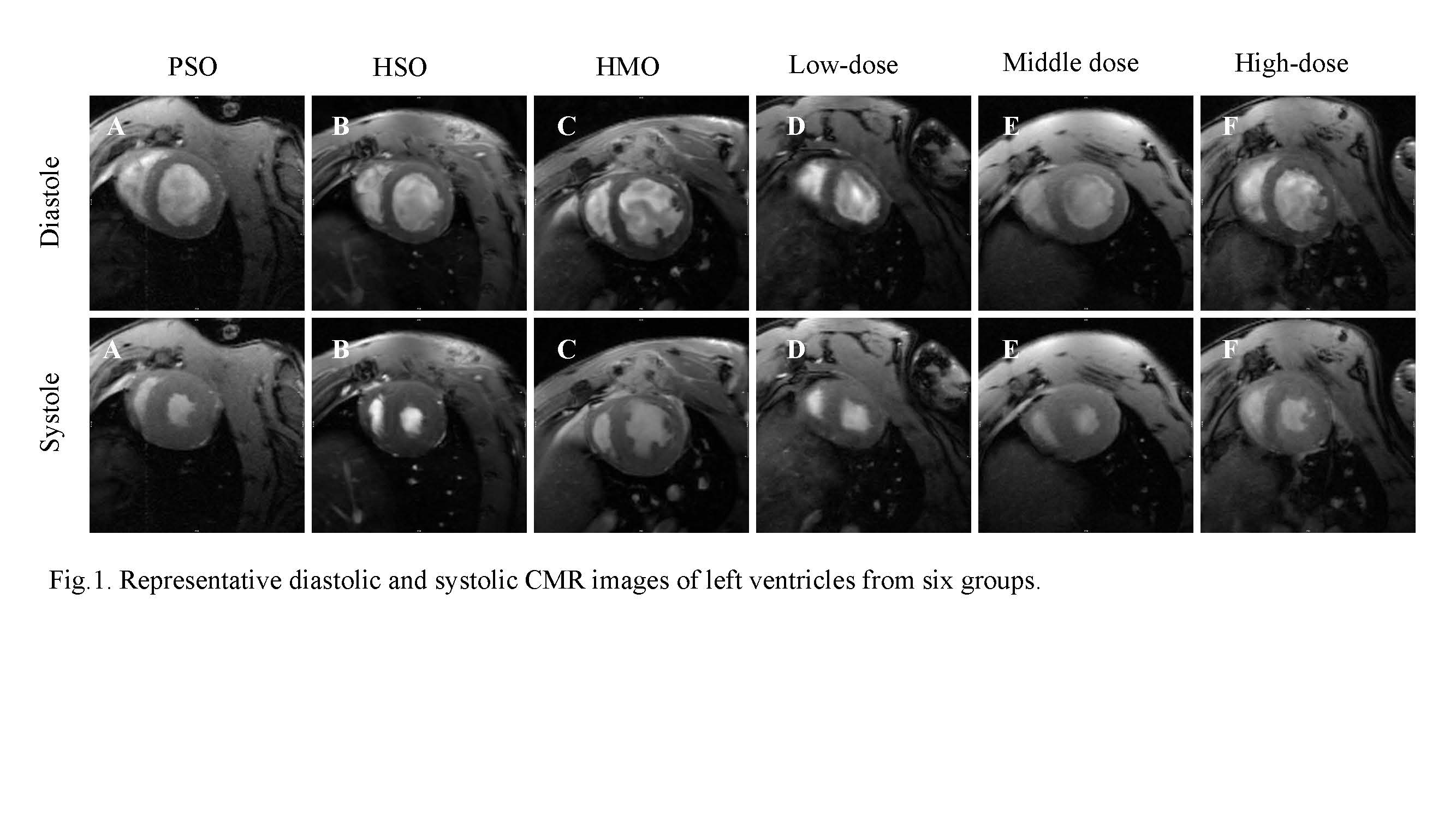
Sixty rats were randomly allocated to six groups (10 rats per group): plain sham operation control (PSO), high-altitude sham operation control (HSO), high-altitude myocardial infarction operation group (HMO), and low-dose, middle-dose, and high-dose paeoniflorin treatment groups. Rats in the high-altitude groups were transported to institution 1 (altitude: 4,250 m), while the plain group rats were transported to institution 2 (altitude: 500 m). The rats acclimatized to their respective environments under standard animal laboratory conditions for three months before undergoing experimental procedures. After four weeks of modeling, CMR was employed to assess cardiac function and left ventricular (LV) structural changes post-interventions. Strain analysis was performed to determine the protective effects of paeoniflorin on MI-induced ventricular remodeling in the high-altitude hypobaric hypoxia rat model.Results
Paeoniflorin treatment groups (low-dose, middle-dose, and high-dose) demonstrated a dose-dependent enhancement in LV function compared to the HMO group. Regional strains, encompassing circumferential, longitudinal, and radial strains, showed significant differences between the paeoniflorin treatment groups and the HMO group. Additionally, histological analyses revealed reduced myocardial injury and fibrosis in the paeoniflorin treatment groups relative to the HMO group.Conclusion
This study's findings indicate that paeoniflorin exerts a protective effect on MI-induced ventricular remodeling in a high-altitude hypobaric hypoxia rat model. These results contribute to understanding the potential therapeutic advantages of paeoniflorin for patients at risk of MI in high-altitude environments, promoting further research into the underlying mechanisms and possible clinical applications.Clinical Relevance
The study's outcomes have implications for managing patients at risk of MI, especially those in high-altitude environments or exposed to hypobaric hypoxia. Recognizing the cardioprotective effects of paeoniflorin could lead to innovative therapeutic approaches, such as preconditioning interventions or pharmacological agents targeting hypoxia-related pathways. Utilizing advanced CMR techniques, including strain analysis, may offer valuable insights into myocardial function and injury, potentially enhancing diagnostic and prognostic precision in clinical settings.Acknowledgements
This study was supported by the National Natural Science Foundation of China (nos. 81930046, 81829003) and The Expert Workstation of Yunnan Province (No. 202105AF150037)References
Liu M, Ai J, Feng J, Zheng J, Tang K, Shuai Z, Yang J. Effect of paeoniflorin on cardiac remodeling in chronic heart failure rats through the transforming growth factor β1/Smad signaling pathway. Cardiovasc Diagn Ther. 2019 Jun;9(3):272-280. doi: 10.21037/cdt.2019.06.01IF: 2.4 Q3 . PMID: 31275817IF: 2.4 Q3 ; PMCID: PMC6603499IF: 2.4 Q3 .Figures