3371
The impact of nonlinear blood behavior on hemodynamic parameters in large vessels: more than expected1School of Electrical Engineering, Pontificia Universidad Católica de Valparaíso, Valparaíso, Chile, 2School of Civil Engineering, Pontificia Universidad Católica de Valparaíso, Valparaíso, Chile, 3Departamento de Informática, Universidad Técnica Federico Santa María, Santiago, Chile, 4Department of Mechanical Engineering, Universidad de Santiago de Chile, Santiago, Chile
Synopsis
Keywords: Flow, Velocity & Flow
Motivation: It has not been exhaustively studied how the nonlinear behavior of the blood impacts the estimation of hemodynamic parameters
Goal(s): To demonstrate the impact of nonlinear viscosities produced by diseases such as anemia and polycythemia on the estimation of hemodynamic parameters
Approach: Hemodynamic parameters were estimated from synthetically generated 4D Flow images obtained from CFD simulations of the aorta using a nonlinear model for the viscosity and different levels of nonlinearity
Results: Important differences were observed in WSS, OSI, and energy loss estimated using a nonlinear model for the viscosity compared to results obtained using constant viscosities. Comparisons were made for several cases
Impact: This investigation could impact how the MR community estimate hemodynamic parameters from 4D MR images.
Introduction
Hemodynamic parameters estimated from 4D flow MRI have been successfully used for the assessment of several cardiovascular diseases including BAV1, coarctations2 and stenosis3. Many of these parameters needs as input a guess of the blood viscosity $$$\eta$$$ which in most of the cases is assumed constant in time and space, and within the range of 0.03 to 0.045 Pa$$$\cdot$$$s4-7. This assumption has been assumed valid in the cardiovascular MRI community for the past decades for large vessels such as the aorta. Moreover, this has been the gold standard for the estimation of hemodynamic parameters from 4D flow MR images. However, the opposite statement, i.e. the shear-thinning non-Newtonian behavior, has not been exhaustively studied nor demonstrated false.The nonlinear behavior of the blood depends on several rheological aspects, with the quantity of red blood cells being one of the most relevant8. Variations in this quantity, characterized by the hematocrit level (HCR), can produce a shear-thinning effect on the blood, making it less or more viscous. The HCR can vary from 10 to 70 in patients with accute anemia and polycythemia8-10, respectively. This variation in the HCR can impact the blood flow dynamics in small and large vessels, generating potential differences in hemodynamic parameters, especially in Wall-Shear Stress (WSS) that depend directly on the apparent viscosity of the fluid.
In this research, we present results that show that the blood nonlinearity, characterized through the HCR, is important for the quantification of hemodynamic parameters from 4D flow MR images. The study was conducted using Computational Fluid Dynamics (CFD) simulations with a nonlinear model of the fluid and realistic synthetic 4D flow MR images.
Methods
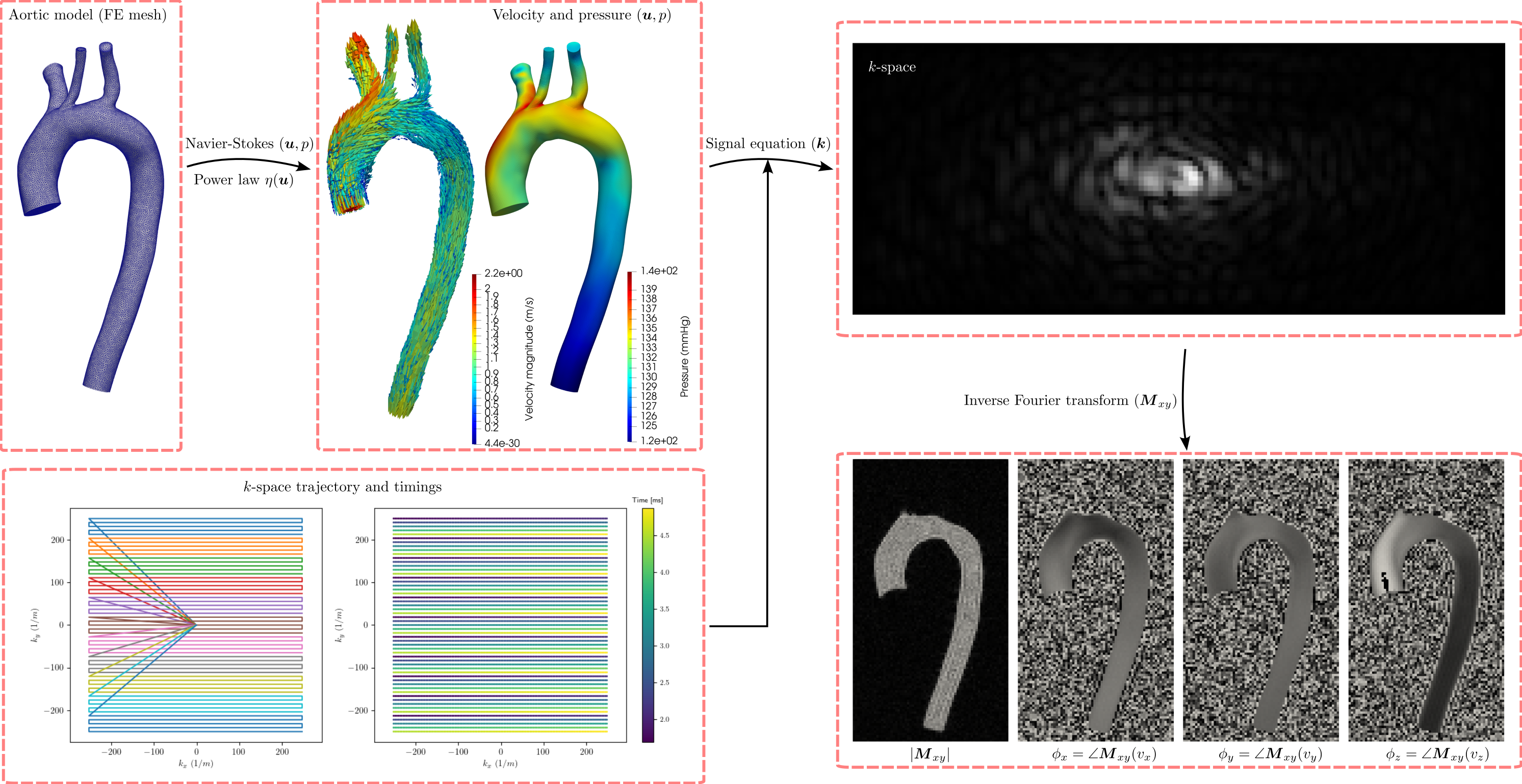
The CFD simulations were obtained solving the Navier-Stokes equations using the Multi-physics simulAtions for engineering and Data assimilation (MAD) library11. The blood nonlinearity was introduced using a power law for the viscosity as follows:$$
(1) \qquad \eta(\boldsymbol{u}) = m\dot{\gamma}(\boldsymbol{u})e^{n-1}, \quad \dot{\gamma} = \sqrt{\frac{1}{2}\dot{\boldsymbol{\gamma}}:\dot{\boldsymbol{\gamma}}}, \quad \dot{\boldsymbol{\gamma}} = \nabla\boldsymbol{u} + \nabla\boldsymbol{u}^T
$$
where the constitutive parameters $$$m$$$ and $$$n$$$ depend on the HCR8.
The aortic model #0012_H_AO_H was obtained from the vascular model repository2,12,13 and the boundary conditions provided in the dataset were used for all the simulations regardless of the HCR.
A 4D flow MR images with parameters taken from the last consensus (VENC of 150 cm/s and voxel size of 2.5$$$\times$$$2.5$$$\times$$$2.5 mm$$$^3$$$), was generated evaluating the signal equation for gradient-echo and multishot EPI (5 lines-per-shot) sequences as:
$$
(2) \qquad s^a(\boldsymbol{k}(t)) = \int_{B} M_0(\boldsymbol{r}_0) e^{-\boldsymbol{i} \pi/\text{VENC} v_a(\boldsymbol{r}_0)}e^{-t/T_2^*(\boldsymbol{r}_0)} e^{-\boldsymbol{i} 2\pi \boldsymbol{k}(t)\cdot \boldsymbol{r}(t)}~d\boldsymbol{r}
$$
with $$$a=\{x,y,z\}$$$ and $$$v_a$$$ the fluid velocity. The $$$k$$$-space locations and timings were generated using realistic magnetic gradients. The motion of flowing spins was considered in Equation (2) using a first order Taylor expansion of $$$\boldsymbol{r}(t)$$$. Gaussian noise with standard-deviation of 2.5% of the maximum amplitude of the image magnitude was added to both real and imaginary channels.
Hemodynamic parameters were estimated using the 4D Flow Matlab Toolbox1 for a constant viscosity (as usual) and the power law given in Equation (1). Wrapping artifacts were corrected using a single-step laplacian method.14
Results
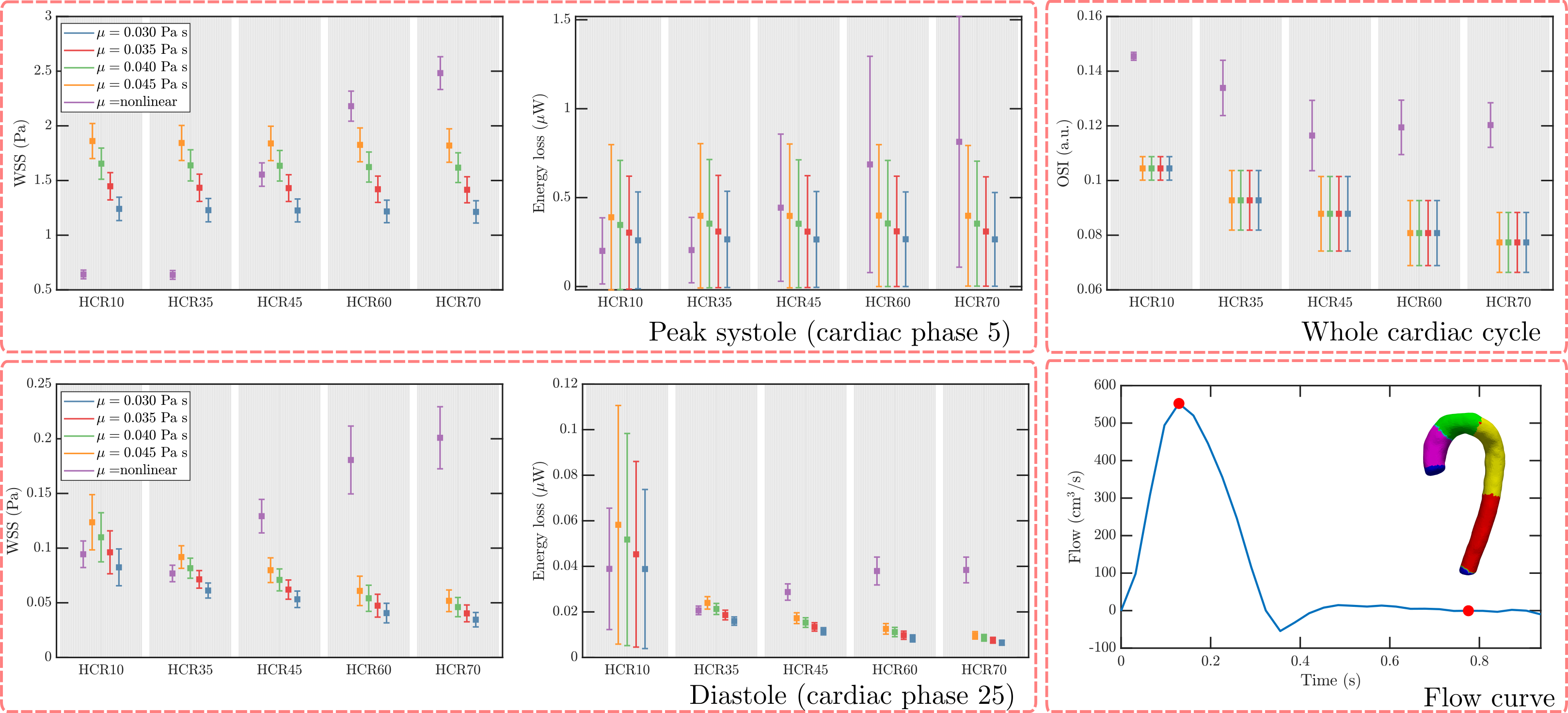
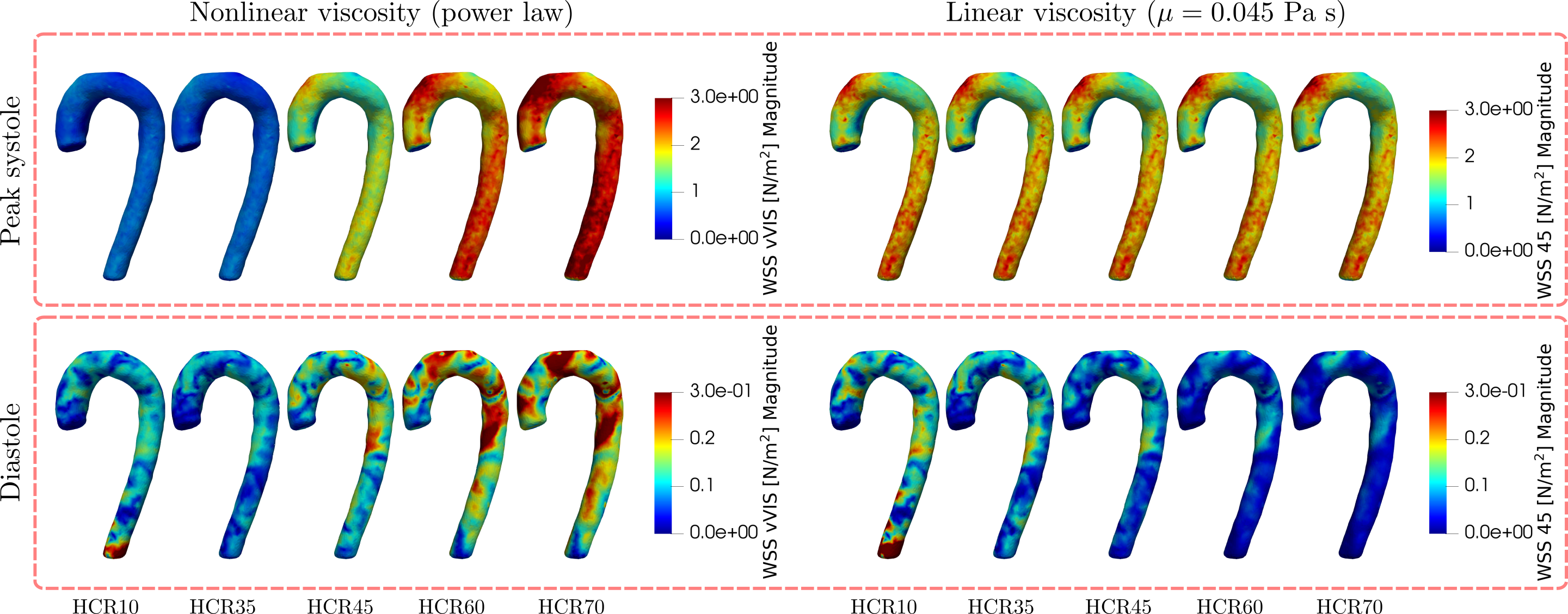
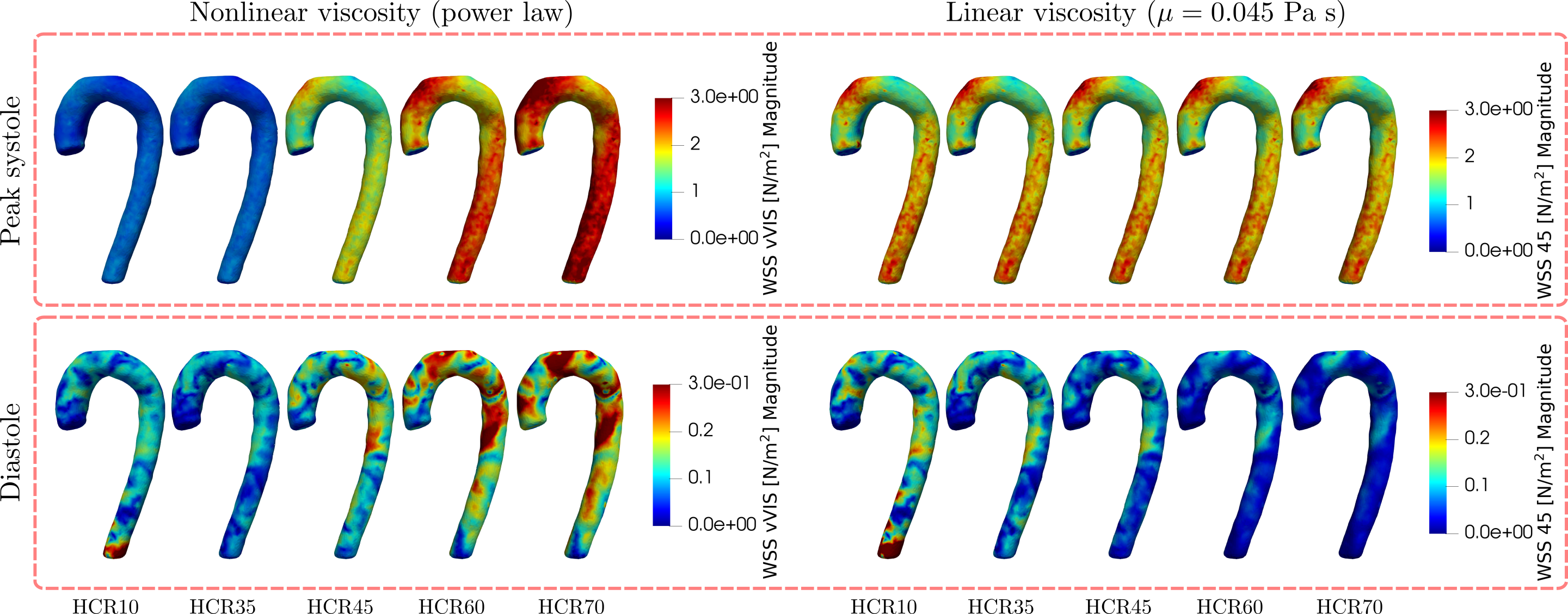
Figure 2 shows the WSS, energy loss, and OSI obtained from synthetic images at peak-systole and diastole the nonlinear viscosity (see Equation 1) and 4 constant viscosities. Important differences were observed for all parameters in any case. Figure 3 shows the WSS maps for the same nonlinear results reported in Figure \ref{fig:fig2} and for a constant viscosity of 0.04 Pa s, where significant variations across HCRs and cardiac phases were observed. Finally, Figure 4 shows the same results depicted in Figure 3 but for an EPI sequence. Almost no differences were observed.Discussion
Results showed that the nonlinear rheology of the blood, characterized using the HCR, can significantly impact the estimation of hemodynamic parameters from 4D flow MR images even at a large vessel such as the aorta. If the HCR is available, the use of the power law for the estimation of hemodynamic parameters could make them more sensitive to different conditions and also give more realistic estimations.Further in-silico and in-vivo experiments are needed to assess the validity of the power law under more demanding imaging and flow conditions.
Conclusion
In this investigation we showed that the nonlinear behavior of the blood produced by conditions affecting the HCR should be considered for the estimation of hemodynamic parameters in large vessels such as the aorta.Acknowledgements
HM and JS acknowledges the financial support given by ANID through the projects ANID-Fondecyt Postdoctorado \#3220266 and ANID-Fondecyt de Iniciación de Investigación \#11200481.References
1. J. Sotelo, P. Franco, A. Guala, L. Dux-Santoy, A. Ruiz-Mu ̃noz, A. Evangelista, H. Mella, J. Mura, D. E. Hurtado, J. F. Rodr ́ıguez-Palomares,S. Uribe, Fully Three-Dimensional Hemodynamic Characterization of Altered Blood Flow in Bicuspid Aortic Valve Patients With Respect toAortic Dilatation: A Finite Element Approach, Frontiers in Cardiovascular Medicine 9 (2022).
2. J. F. Jr. LaDisa, R. J. Dholakia, C. A. Figueroa, I. E. Vignon-Clementel, F. P. Chan, M. M. Samyn, J. R. Cava, C. A. Taylor, J. A. Fe-instein, Computational simulations demonstrate altered wall shear stress in aortic coarctation patients treated by resection with end-to-endanastomosis, Congenital Heart Disease 6 (5) (2011) 432–443. doi:10.1111/j.1747-0803.2011.00553.x.
3. E. S. Farag, P. van Ooij, R. N. Planken, K. C. Dukker, F. de Heer, B. J. Bouma, D. Robbers-Visser, M. Groenink, A. J. Nederveen, B. A.de Mol, J. Kluin, S. M. Boekholdt, Aortic valve stenosis and aortic diameters determine the extent of increased wall shear stress in bicuspidaortic valve disease, Journal of Magnetic Resonance Imaging 48 (2) (2018) 522–530. doi:10.1002/jmri.25956.
4. R. J. van Tuijl, K. M. Timmins, B. K. Velthuis, P. van Ooij, J. J. Zwanenburg, Y. M. Ruigrok, I. C. van der Schaaf, Hemodynamic Parametersin the Parent Arteries of Unruptured Intracranial Aneurysms Depend on Aneurysm Size and Are Different Compared to Contralateral Arteries:A 7 Tesla 4D Flow MRI Study, Journal of Magnetic Resonance Imaging n/a (n/a). doi:10.1002/jmri.28756.
5. E. T. Bieging, A. Frydrychowicz, A. Wentland, B. R. Landgraf, K. M. Johnson, O. Wieben, C. J. Franc ̧ois, In vivo three-dimensionalMR wall shear stress estimation in ascending aortic dilatation, Journal of Magnetic Resonance Imaging 33 (3) (2011) 589–597.doi:10.1002/jmri.22485.
6. P. van Ooij, W. V. Potters, A. Gu ́edon, J. J. Schneiders, H. A. Marquering, C. B. Majoie, E. vanBavel, A. J. Nederveen, Wall shear stressestimated with phase contrast MRI in an in vitro and in vivo intracranial aneurysm, Journal of Magnetic Resonance Imaging 38 (4) (2013)876–884. doi:10.1002/jmri.24051.
7. J. Garay, H. Mella, J. Sotelo, C. C ́arcamo, S. Uribe, C. Bertoglio, J. Mura, Assessment of 4D flow MRI’s quality by verifying its Navier–Stokescompatibility, International Journal for Numerical Methods in Biomedical Engineering 38 (6) (2022) e3603. doi:10.1002/cnm.3603.URL https://onlinelibrary.wiley.com/doi/abs/10.1002/cnm.3603
8. F. J. Walburn, D. J. Schneck, A constitutive equation for whole human blood, Biorheology 13 (3) (1976) 201–210. doi:10.3233/BIR-1976-13307.
9. R. E. Wells, E. W. Merrill, Influence of flow properties upon viscosity-hematocrit relationships, The Journal of Clinical Investigation 41 (8)(1962) 1591–1598. doi:10.1172/JCI104617.
10. R. Mehri, C. Mavriplis, M. Fenech, Red blood cell aggregates and their effect on non-Newtonian blood viscosity at low hematocrit in atwo-fluid low shear rate microfluidic system, PLOS ONE 13 (7) (2018) e0199911. doi:10.1371/journal.pone.0199911.
11. F. Galarce Marin, Inverse problems in hemodynamics. fast estimation of blood flows from medical data.https://gitlab.com/felipe.galarce.m/mad/, Ph.D. thesis, INRIA Paris & Laboratoire Jacques-Louis Lions. Sorbonne Uni-versit ́e (2021).
12. N. M. Wilson, A. K. Ortiz, A. B. Johnson, The vascular model repository: A public resource of medical imaging data and blood flowsimulation results, Journal of Medical Devices 7 (40923) (2013-12-05). doi:10.1115/1.4025983.
13. M. R. Pfaller, J. Pham, A. Verma, L. Pegolotti, N. M. Wilson, D. W. Parker, W. Yang, A. L. Marsden, Automated generation of 0d and 1dreduced-order models of patient-specific blood flow, International Journal for Numerical Methods in Biomedical Engineering 38 (10) (2022)e3639. doi:10.1002/cnm.3639.
14. M. Loecher, E. Schrauben, K. M. Johnson, O. Wieben, Phase unwrapping in 4D MR flow with a 4D single-step laplacian algorithm, Journalof Magnetic Resonance Imaging 43 (4) (2016) 833–842. doi:10.1002/jmri.25045.
Figures