3364
Comparison between 3T non-selective pulse non-enhanced whole-heart bTFE coronary MR angiography and 3T mDixon water-fat separation GRE method
Yong Yuan1, Guangming Lu2, Dongsheng Jin2, Weibo Chen3, Baijun Wang3, Tong Chen4, Qiuju Hu5, Jiajia Zhu5, and Yane Zhao5
1Geriatric Hospital of Nanjing Medical University, Nanjing 210024, China, Nanjing, China, 2Geriatric Hospital of Nanjing Medical University, Nanjing, China, 3Philips Healthcare, Shanghai, China, Shanghai, China, 4Philips Healthcare, Hangzhou, China, Hangzhou, China, 5Geriatric Hospital of Nanjing Medical, Nanjing, China
1Geriatric Hospital of Nanjing Medical University, Nanjing 210024, China, Nanjing, China, 2Geriatric Hospital of Nanjing Medical University, Nanjing, China, 3Philips Healthcare, Shanghai, China, Shanghai, China, 4Philips Healthcare, Hangzhou, China, Hangzhou, China, 5Geriatric Hospital of Nanjing Medical, Nanjing, China
Synopsis
Keywords: Vascular, Cardiovascular
Motivation: To improve the image quality of the non-enhanced whole heart bTFE CMRA sequence at 3T.
Goal(s): To compare the performance of a newly improved bTFE CMRA sequence with the mDixon water-fat separation GRE method at 3T for CMRA.
Approach: Success rate, acquisition time, image quality, and diagnostic performance were compared.
Results: This study demonstrated that the performance of the 3D bTFE CMRA sequence at 3T was brilliant, with a high success rate, appropriate acquisition time, excellent image quality, and good diagnostic performance.
Impact: The non-enhanced bTFE CMRA sequence at 3T may be recommended for more extensive clinical applications.
Introduction
Non-enhanced whole-heart balanced turbo field echo (bTFE) coronary magnetic resonance angiography (CMRA) sequence is conventionally used in 1.5T scanners owing to its inherently high blood signal intensity and blood-myocardial contrast[1]. The increase in magnetic field inhomogeneity at 3T compared with 1.5T and the use of volume-selective pulses in this sequence previously resulted in a greater susceptibility artifact (black banding artifact) at 3T compared with 1.5T, thus limiting the application of this sequence at 3T. Moreover, the application of 3T sequences is limited because their sensitivity to increased field inhomogeneity weakens the effectiveness of frequency-based fat suppression methods.Purpose
To compare the performance of a newly improved three-dimensional (3D) non-selective pulses bTFE sequence with the mDixon water-fat separation GRE method at 3T for CMRA.Methods
From September 15, 2023, patients with suspected coronary artery disease were prospectively recruited who voluntarily underwent CMRA after coronary computed tomography angiography(CCTA)in our hospital. The improved non-enhanced whole-heart CMRA 3D bTFE sequence and the mDixon water-fat separation GRE method were randomly completed on the same 3T MR scanner ( Elition, Philips Healthcare, Best, the Netherlands). Success rate, acquisition time, subjective image quality score, and objective image quality measurement of the above two sequences were compared. The diagnostic performance of bTFE CMRA was compared with CCTA for predicting significant stenosis by dividing stenosis into four levels: no stenosis, mild stenosis, moderate stenosis, and severe stenosis. A compared t-test evaluated the objective image quality measurement. The consistency between bTFE CMRA and CCTA, as well as the consistency between mDixon and CCTA in predicting coronary artery stenosis, were calculated using weighted kappa. With CCTA as the reference standard, the chi-square test also evaluated the diagnostic performance of the bTFE CMRA and the mDixon water-fat separation GRE method for predicting significant stenosis (≥ 50% diameter stenosis) on CCTA.Results
Thirty-eight patients were included (21 males, 17 females; mean age 61 ± 12 years, range 46-81 years). Success rate, acquisition time, subjective image quality score, average signal-to-noise ratio (SNR), and average contrast-to-noise ratio (CNR) of the bTFE sequence and the mDixon water-fat separation GRE method were 92.4% vs 73.6%, 10.7± 3.6 min vs 11.3 ± 4.1min, 4.2± 0.3 vs 3.2± 0.7, 26.7±5.6 vs 20.2±4.2, 13.2±2.6 vs 9.3±2.7 respectively. There was no statistical difference in acquired times, but subjective image quality scores, average SNR, and average CNR differed significantly. Weighted kappa analysis showed the bTFE CMRA and CCTA had consistent predictions for 30 patients, 96 vessels, and 297 segments, but inconsistent for 8 patients, 18 vessels, and 45 segments respectively. Overall, the bTFE CMRA predicted coronary artery stenosis well, with a kappa consistency coefficient of 0.824 per patient, 0.873 per vessel, and 0.894 per segment. With CCTA as the reference standard, the positive percent agreement (PPA), negative percent agreement (NPA), and overall percent agreement (OPA) of the bTFE CMRA were 86.9%, 96.4%, and 90.8% per patient respectively, which were all better than the mDixon water-fat separation GRE method, with PPA 74.1%, NPA 82.3%, and OPA 78.6% respectively.Discussion
This study demonstrated that the performance of the 3D bTFE CMRA sequence at the 3T scanner was brilliant, with a high success rate, appropriate acquisition time, excellent image quality, and good diagnostic accuracy. 3T MRI has provided high SNR and high resolution, and meanwhile, the bTFE CMRA sequence has inherently high blood signal intensity and blood myocardial contrast[2]. High-quality bTFE CMRA at 3T would greatly benefit coronary MRA. The advances in active and passive shimming technology have greatly improved the homogeneity of the magnetic field, making it possible for bTFE CMRA to perform better at 3T, and enhancing the effectiveness of frequency-based fat suppression methods[1, 3]. Based on the above, we applied 3D non-selective pulse technology, which allows for shorter repetition time, echo time, and larger flip angles, and further reduces sensitivity to magnetic field inhomogeneity, enhancing the signal intensity of CMRA[4, 5]. Moreover, compared to the mDixon fat suppression, the use of SPIR fat suppression could reduce cardiac and coronary motion artifacts. While the mDixon sequence uses a dual-echo technique to acquire water and fat images in two separate echoes. This requires a longer TR to accommodate the additional echo time. The longer TR leads to slower imaging speed and increased motion blurring.Conclusions
In 3T CMRA, the improved 3D bTFE sequence performed excellently compared to the mDixon water-fat separation GRE method. The non-enhanced bTFE CMRA sequence at 3T may be recommended for more extensive clinical applications than the mDixon water-fat separation GRE method.Acknowledgements
No acknowledgement found.References
[1] Hajhosseiny R, Bustin A, Munoz C, Rashid I, Cruz G, Manning WJ, et al. Coronary Magnetic Resonance Angiography[J]. JACC: Cardiovascular Imaging. 2020;13(12):2653-72. [2] Tian D, Zhao S-h, Wang Y, Lu H-f, Chen Y-y, Guo J-j, et al. Unenhanced Whole-Heart Coronary MRA: Prospective Intraindividual Comparison of 1.5-T SSFP and 3-T Dixon Water-Fat Separation GRE Methods Using Coronary Angiography as Reference[J]. American Journal of Roentgenology. 2022;219(2):199-211. [3] Lin L, Wang L, Zhang X-N, Li X, Wang J, Shen Z-J, et al. A clinical strategy to improve the diagnostic accuracy of 1.5-T non-contrast MR coronary angiography for detection of coronary artery disease: combination of whole-heart and volume-targeted imaging[J]. European Radiology. 2020;31(4):1894-904. [4] <2009-investigative radiology-Three-Dimensional Imaging of Pulmonary Veins by a Novel Steady-State Free-Precession Magnetic Resonance Angiography Technique Without the Use of Intravenous Contrast Agent.pdf>[J]. [5] <2010-Radiology-Pulmonary vein imaging with unenhanced three-dimensionalFigures
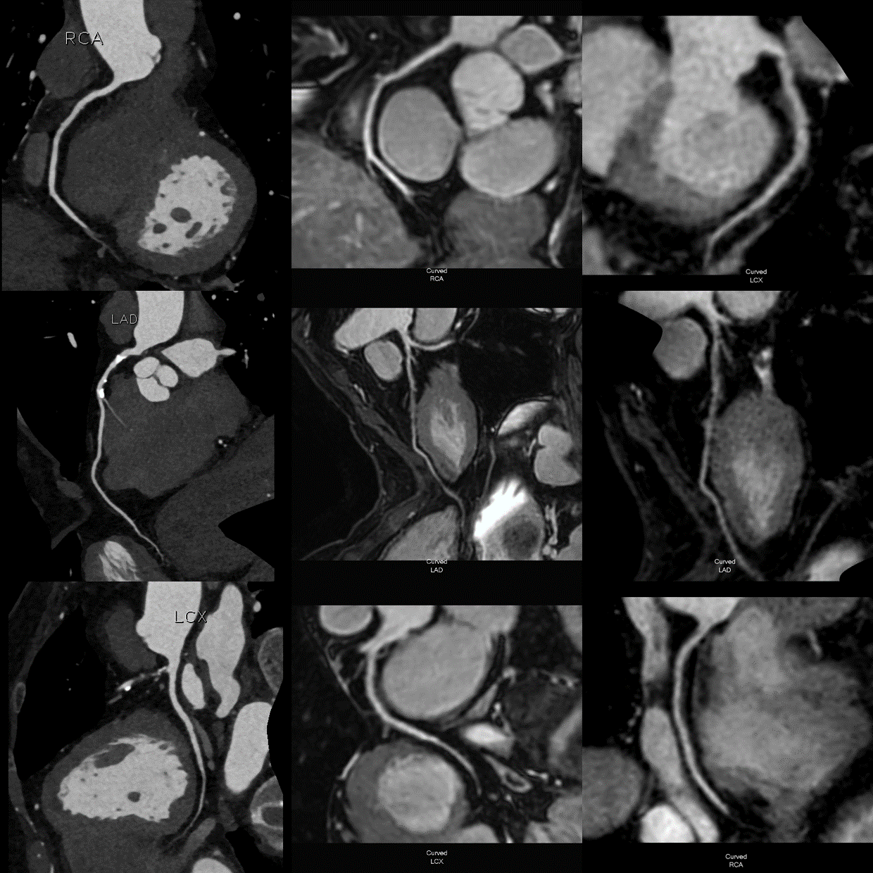
An 82-year-old man, with suspected coronary artery disease, was
confirmed to be normal by CCTA. The upper row was RCA, the middle row LAD, and the
lower row was LCX. CCTA, bTFE CMRA, and
mDxion CMRA images are in sequence from left to right.
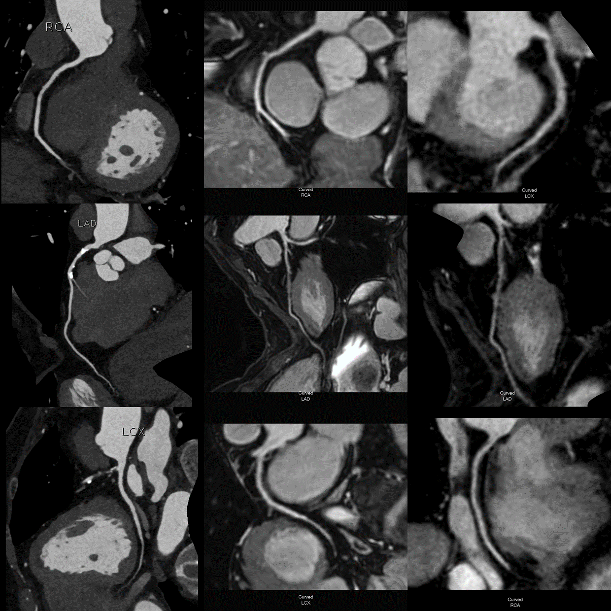
A 69-year-old woman, with suspected coronary artery disease, whose proximal
segment of LAD was confirmed to be moderate stenosis. The upper row was RCA, the
middle row LAD, and the lower row was LCX. CCTA, bTFE CMRA, and mDxion CMRA images are in
sequence from left to right.
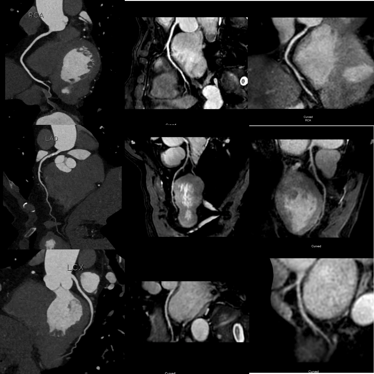
A 68-year-old man, with suspected coronary artery disease, whose
proximal segment of LAD was confirmed to be mild stenosis. The upper row was
RCA, the middle row LAD, and the lower row was LCX. CCTA, bTFE CMRA, and mDxion CMRA images are in
sequence from left to right.
DOI: https://doi.org/10.58530/2024/3364