3362
Single breath hold 3D isotropic whole heart coronary MRA using turbo field echo planar imaging with deep learning constrained Compressed SENSE1Department of Radiological Services, Tokyo Women’s Medical University, Tokyo, Japan, 2Department of Diagnostic imaging & Nuclear Medicine, Tokyo Women’s Medical University, Tokyo, Japan, 3Philips Japan, Tokyo, Japan, 4Philips Healthcare, Best, Netherlands
Synopsis
Keywords: Vascular, Cardiovascular
Motivation: Whole-heart-coronary-MRA (WHC-MRA) is typically performed during free-breathing, and respiratory motion is prospectively compensated by using a 1D-right-diaphragmatic-navigator. However, this method requires long acquisition time and causes a heavy burden on the patient.
Goal(s): Our goal was to obtain high-resolution WHC-MRA with single-breath-hold while ensuring clinically acceptable signal-to-ratio.
Approach: Turbo-field-echo-planar-imaging (TFEPI), a high-speed technique, was combined with deep learning constrained Compressed SENSE (CS-AI) and image quality was evaluated by visual evaluation and contrast-to-ratio.
Results: TFEPI with CS-AI provided high spatial resolution isotoropic single-breath-hold WHC-MRA while maintaining clinically acceptable image quality and scan time compared to Compressed SENSE and SENSE.
Impact: High-resolution single-breath-hold WHC-MRA with clinically acceptable image quality by 3D TFEPI with CS-AI may improve the throughput of cardiac MRI examinations, and in addition reduce patient burden.
Introduction
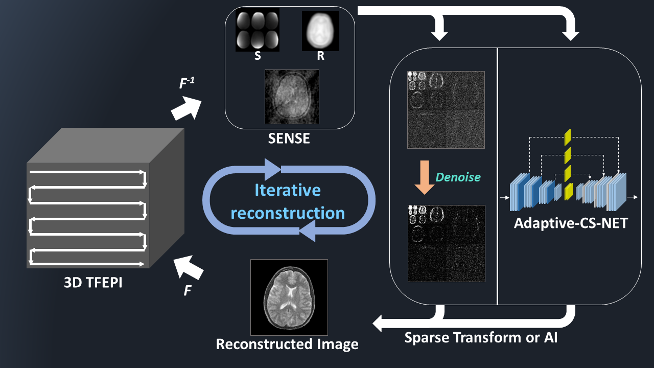
Various sequences have been proposed for whole heart coronary magnetic resonance angiography (WHC-MRA)1-4. In conventional WHC-MRA, it is typically performed during free-breathing, and respiratory motion is prospectively compensated by using a 1D-right-diaphragmatic-navigator (NAV)5. However, an important limitation of the NAV method is its relatively long acquisition time6. Single-breath-hold technique using 3D Turbo-field-echo-planar-imaging (TFEPI) could significantly reduce the acquisition time of WHC-MRA while providing image quality similar to that of conventional free-breathing gradient echo sequence7. TFEPI sequence is a hybrid technique that combines TFE and EPI readouts7. It enables highly accelerated imaging thanks to EPI readout, resulting in shorter scan time by allowing more echoes to be acquired during one heartbeat. Furthermore, EPI with Compressed SENSE (C-SENSE) has been introduced8,9. This approach can be extended to 3D TFEPI without modifying its sampling pattern and ffurther accelerated scan time without degrading the image quality by denoising as previously introduced10. However, TFEPI with C-SENSE is still challenging to obtain high spatial resolution like conventional NAV-WHC-MRA with clinically acceptable signal-to-ratio (SNR). Recently, integrating artificial intelligence (AI) into the Compressed SENSE reconstruction (CS-AI), based on Adaptive-CS-Net11,12 (fig.1), has been introduced and CS-AI dramatically reduced noise artifacts and significantly improved image quality13-15. In addition, it has been shown recently that deep learning-based denoising is an effective tool for image quality improvement in WHC-MRA16. We hypothesize that the combination of TFEPI and CS-AI can provide further high spatial resolution of single-breath-hold WHC-MRA while maintaining clinically acceptable SNR and scan time. In this study, we investigated the feasibility of single-breath-hold TFEPI with CS-AI for WHC-MRA by comparing with single-breath-hold TFEPI with C-SENSE and SENSE.Methods
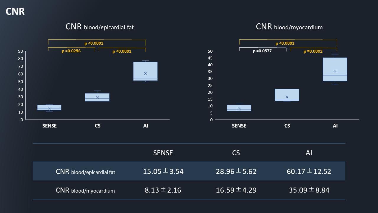
A total of six volunteers (5 males and 1 female; age range: 28~45) were examined on a 3.0T-MRI (Ingenia Elition X, Philips Healthcare). The study was approved by the local IRB, and written informed consent was obtained from all subjects. We compared image quality among WHC-MRA using TFEPI with CS-AI, C-SENSE or SENSE. Three sequences were acquired with cardiac triggering and single-breath-hold for respiratory compensation. Imaging-parameters; TFEPI: FOV=300×280mm2, voxel-size=1.7×1.7×1.7mm3, TR/TE/FA=7.7/3.5/20, TFE-factor=15-22, EPI-factor=7, shot-duration=100~130ms, reduction factor=4.1×2.0, scan time:17~23sec. Curved Planer Reconstructions (CPRs) were performed using Ziostation2 (Ziosoft Co, Tokyo) and image quality was evaluated by visual score at the 10-points (RCA: #1/2/3/4 LAD: #5/6/7/8 CX: #11/13) based on American-Heart-Association classification. For overall image quality, sharpness and noise and artifacts, we evaluated them as 4-point grades (grade “4” was excellent, “1” was severe) by two blinded readers. Visual evaluation was assessed by Steel-Dwass test. For quantitative comparison, contrast-to-noise ratio (CNR) were measured. The CNR was estimated for blood and epicardial fat (CNRblood-epicardial fat) and blood and myocardium (CNRblood-myocardium). To allow quantitative CNR measurements, we used a noise-measurement-method proposed by Zwanenburg et al17. Each sequence was repeated with exactly the same receiver gain, but without any RF and gradient-pulses. The reconstructed images showed only noise, including the noise added due to the C-SENSE/SENSE-reconstruction. The standard-deviation of a region of interest of the corresponding area in the noise image was used as metric for the noise. The CNRblood-epicardial fat and CNRblood-myocardium were calculated by the following equations: CNRA-B = [SI(A) - SI(B)] / 0.5 [SDnoise(A) + SDnoise(B)] Where SI are the mean average signal intensity of the blood, epicardial fat and myocardium respectively, and the corresponding SDnoise is the standard-deviation at the same location on the noise images. The CNR were assessed by one-way repeated measures analysis of variance (ANOVA) and the post-hoc Tukey-Kramer test.Results and Discussion
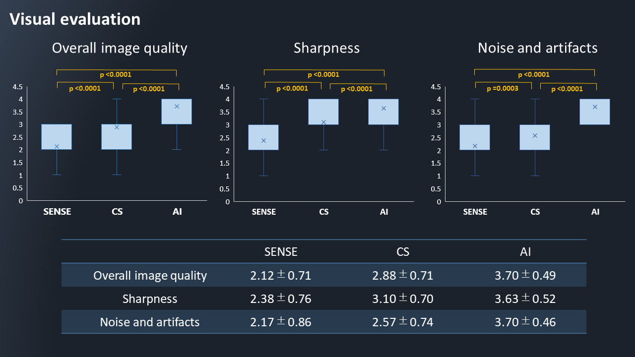
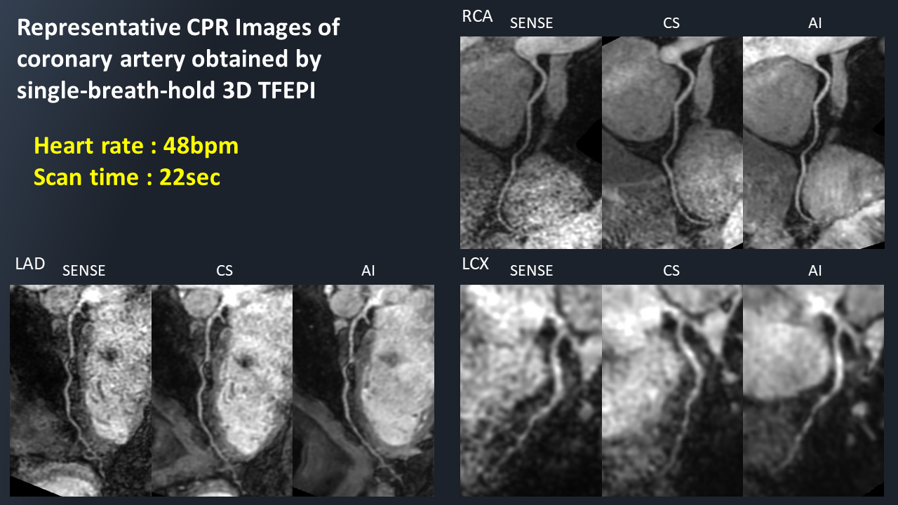
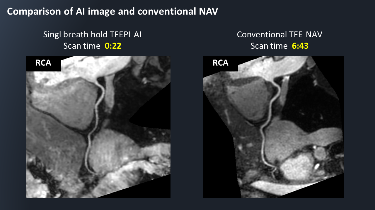
Figure 2 shows the results of visual evaluation. Regarding overall image quality, sharpness and noise and artifacts, TFEPI with CS-AI was significantly higher than TFEPI with C-SNESE and SENSE (p<0.0001). Figure 3 shows CNR comparison among three sequences. For the CNRblood-epicardial fat and CNRblood-myocardium, TFEPI with CS-AI was significantly higher than TFEPI with C-SNESE and SENSE. Figure 4 shows the representative images and actual scan time using TFEPI with CS-AI/C-SENSE/SENSE. Figure 5 shows the representative images and actual scan time of TFEPI with CS-AI and conventional NAV WHC-MRA. TFEPI with CS-AI had high visual score and better image contrast as a result of high SNR and CNR due to the effect of AI-based denoising. Furthermore, the image quality of single-breath-hold TFEPI with CS-AI was comparable to that of conventional TFE with NAV.Conclusion
3D TFEPI with CS-AI has a great potential to obtain 3D high resolution isotropic single-breath-hold WHC-MRA with clinical acceptable image quality with short breath-holding time, around 20 seconds, and it could reduce the burden on the patients.Acknowledgements
No acknowledgements found.References
(1) Kazuo Kodaira, et al. Whole heart coronary MRA using image-based 2D navigator (iNav) and conventional Nav system: comparison of image quality and scan time. Proceedings of the Annual Meeting & Exhibition of ISMRM (2021). (2) Oliver M. Weber, PhD et al. Free-Breathing, Three-Dimensional Coronary Artery Magnetic Resonance Angiography: Comparison of Sequences. Journal of Magenetic Resonance Imaging. 2004; 20: 395-402. (3) Teresa Correia et al. Technical note: Accelerated nonrigid motion-compensated isotropic 3D coronary MR angiography. Med Phys. 2018 Jan; 45(1): 214-222. (4) Yuji Iyama et al. Single-breath-hold whole-heart coronary MRA in healthy volunteers at 3.0-T MRI. Springerplus. 2014; 3: 667. (5) Markus Henningsson et al. Prospaective Respiratory Motion Correction for Coronary MR Angiography Using a 2D Image Navigator. Magn Reson Med. 2013; 69: 486-494. (6) Mehdi H. Monghari et al. Three-dimensional Heart Locator and Compressed Sensing for Whole-heart Magnetic Resonance Angiography. Magn Reson Med. 2016 May; 75(5): 2086–2093. (7) Yuji Iyama et al. Single-Breath-Hold Whole-heart Unenhanced Coronary MRA Using Multi-shot Gradient Echo EPI at 3T: Comparison with Free-breathing Turbo-field-echo Coronary MRA on Healthy Volunteers. Magn Reson Med Sci. 2018; 17: 161-167. (8) Kaga T, et al. Diffusion-weighted imaging of the abdomen using echo planar imaging with compressed SENSE: Feasibility, image quality, and ADC value evaluation. Eur J Radiol. 2021 Sep;142:109889. doi: 10.1016/j.ejrad.2021.109889. (9) Yoneyama M, et al. Noise Reduction in Prostate Single-Shot DW-EPI utilizing Compressed SENSE Framework. Proc Intl Soc Mag Reson Med. 2019:1634. (10) Kazuo Kodaira, et al. Single-breath-hold whole heart coronary MRA using 3D turbo-field-echo-planar-imaging (TFEPI) with Compressed SENSE framework. Proceedings of the Joint Annual Meeting ISMRM-ESMRMB & ISMRT 31st Annual Meeting (2022). (11) Pezzotti N, et al. An Adaptive Intelligence Algorithm for Undersampled Knee MRI Reconstruction. IEEE Access. 2020;8:204825-204838. (12) Pezzotti N, et al. Adaptive-CS-Net: FastMRI with Adaptive Intelligence. arxiv. 2019;(NeurIPS). (13) Yoneyama M, et al. Rapid submillimeter high-resolution prostate T2 mapping with a deep learning constrained Compressed SENSE reconstruction. Proc Intl Soc Mag Reson Med. 2021:4117. (14) Yoneyama M, et al. SNR boost in whole-body DWIBS utilizing deep learning constrained Compressed SENSE reconstruction. Proceedings of the Joint Annual Meeting ISMRM-ESMRMB & ISMRT 31st Annual Meeting (2022). (15) Yoneyama M, et al. SNR enhancement in rapid high b-value prostate single-shot DW-EPI utilizing deep learning constrained Compressed SENSE reconstruction. Proceedings of the Joint Annual Meeting ISMRM-ESMRMB & ISMRT 31st Annual Meeting (2022). (16) Xi Wu, et al. Feasibility of accelerated non-contrast-enhanced whole-heart bSSFP coronary MR angiography by deep learning-constrained compressed sensing. Euro Radiol 2023, 33:8180-8190. (17) Zwanenburg et al. MR Angiography of the cerebral perforating arteries with magnetization prepared anatomical reference at 7T: comparison with time-of-flight. J Magn Reson Imaging. 2008; 28: 1519–1526.Figures