3357
Investigating Brain Health in Coronary Artery Disease: Vascular and Metabolic Biomarkers, Cognition, and Implications1Physics, Concordia University, Montreal, QC, Canada, 2Montreal Heart Institute, Centre Epic and Research center, Montreal, QC, Canada, 3Hurvitz Brain Sciences Program, Sunnybrook Research Institute, Toronto, ON, Canada, 4Department of Radiology, Université de Sherbrooke, Sherbrooke, QC, Canada, 5Computer Science Department, Université de Sherbrooke, Sherbrooke, QC, Canada, 6Department of Psychology, Concordia University, Montreal, QC, Canada, 7Department of Chemistry and Biochemistry, Concordia University, Montreal, QC, Canada, 8Department of Medicine, Université de Montreal, Montreal, QC, Canada, 9Research Center, Institut Universitaire de Gériatrie de Montréal, Montreal, QC, Canada
Synopsis
Keywords: Vascular, Brain
Motivation: There is limited understanding regarding the physiological mechanisms underlying the impact of coronary artery disease (CAD) on brain health.
Goal(s): In this study, we sought to explore the influence of CAD on vascular and metabolic biomarkers.
Approach: Specifically examining Cerebrovascular Reactivity (CVR), Cerebral Blood Flow (CBF), and Oxygen Extraction Fraction (OEF) in the brain, and how these factors relate to cognitive performance.
Results: Our initial findings, based on a preliminary dataset, indicate that individuals with CAD exhibit reduced CBF and CVR, with increased OEF across several brain regions. Low CVR and high OEF are also associated with poorer executive function in these regions.
Impact: The influence of coronary artery disease (CAD) on brain health remains unclear. We show that CAD is associated with elevated OEF and reduced CVR and CBF. High OEF and low CVR are linked to poorer cognition, especially executive functions.
Introduction
The World Health Organization estimates that 30% of deaths in high-income countries result from chronic vascular diseases, which are associated with cerebrovascular damage [1]. Vascular impairments lead to brain lesions and cognitive decline, impacting quality of life for decades [2, 3]. To understand this process, we need sensitive biomarkers of brain health. While previous studies focused on vascular lesions and atrophy, we lack knowledge about early changes that precede lesions. Recent advances in MRI enable us to quantify brain vascular, metabolic, and tissue health, allowing us to study brain health, cognition, and the brain-heart connection comprehensively. This project uses calibrated functional Magnetic resonance Imaging (fMRI) to quantify Cerebral Blood Flow (CBF), Cerebrovascular Reactivity (CVR), Oxygen Extraction Fraction (OEF), and Cerebral Metabolic Rate of Oxygen Consumption (CMRO2) as metabolic and vascular biomarkers to understand the physiological changes associated with coronary artery disease (CAD).Methods
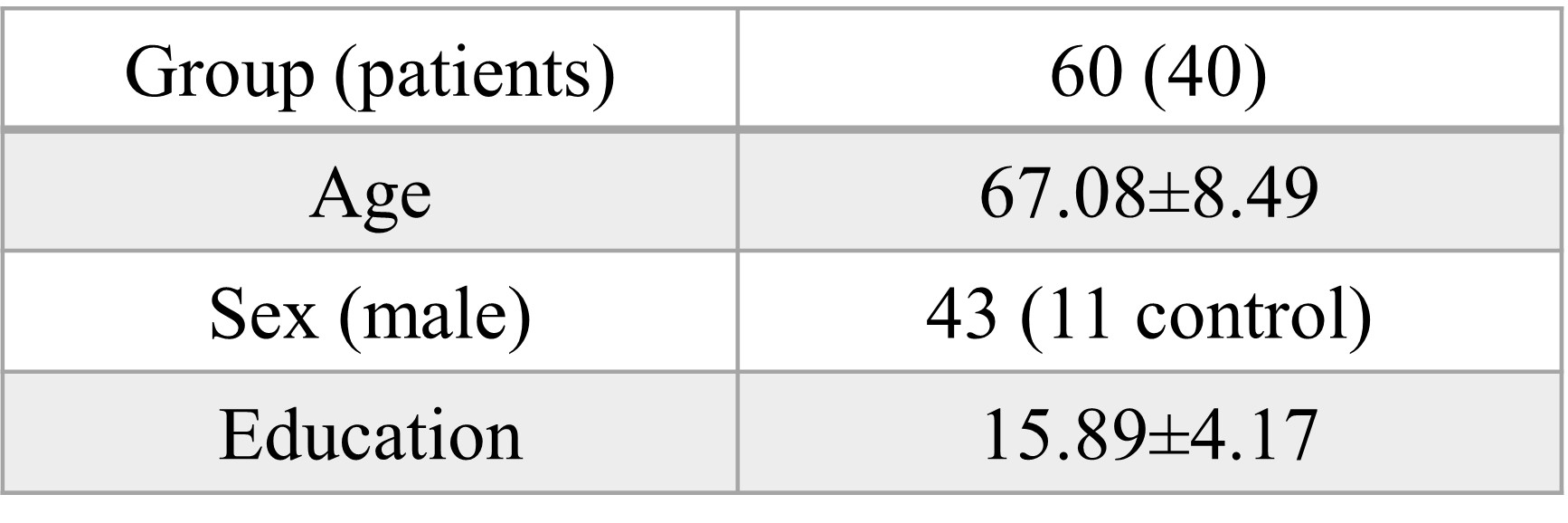
Data was acquired from 60 participants (Table 1 for demographics), on a 3T Skyra MRI. Data acquisition includes structural MRI and a dual echo pseudo-continuous Arterial Spin Labeling (pCASL) with TR/TE1/TE2: 4000/10/30ms conducted during a 14-minute period of breathing manipulation. During this manipulation, participants breathed three mixed gases. EtO2 and EtCO2 were controlled using a computer-driven system (RespirAct, Thornhill Research Inc., Toronto, Canada). The respiratory pattern involved baseline breathing, one 2-minute hypercapnic block (+5 mmHg EtCO2), one 2-minute hyperoxic block (300 mmHg EtO2), and a 2-minute carbogen block (hyperoxia and hypercapnia). Participants used a re-breathing face mask connected to the Respiract gas delivery system. Preprocessing and quantification of CBF were performed using MATLAB and FSL [4,5]. BOLD images were estimated using the surround addition method and then utilized for CVR map estimation. CVR was calculated by incorporating the CO2 gas trace as a regressor in a General Linear Model [6]. To quantify OEF and CMRO2 maps, end-tidal PO2 during each gas manipulation, along with CBF and BOLD signal percent changes for each gas, were utilized in a General Calibration Model (GCM) [6]. Participants also underwent a neuropsychological battery to assess verbal reasoning, short-term memory, working memory, psychomotor speed, attention, and executive functions [7,8]. To compare all parameters in each region of interest (ROI), extracted from lpba40 atlas [9], between the CAD and healthy control (HC) groups, a one-way ANCOVA test was conducted in R, with sex, GM volume, and age as covariates. All p-values were corrected using the Family Wise Error method. Linear regression was subsequently employed to identify associations between each biomarker and cognitive performance.Results
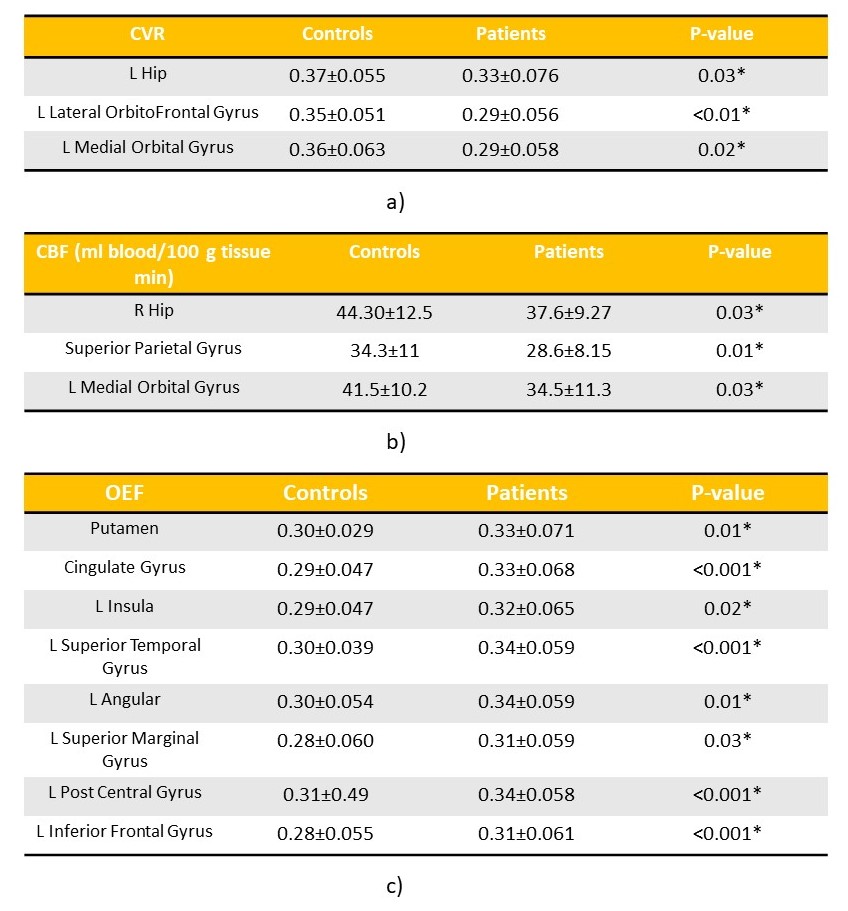
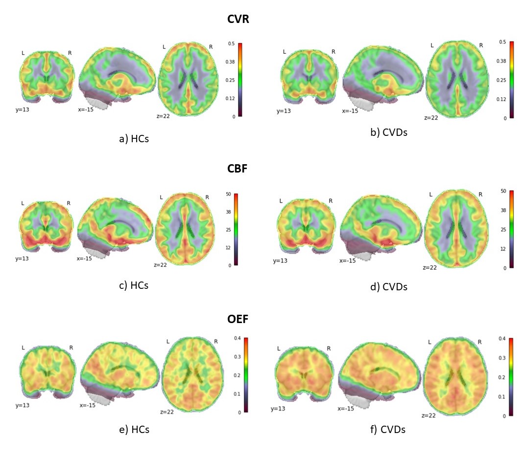
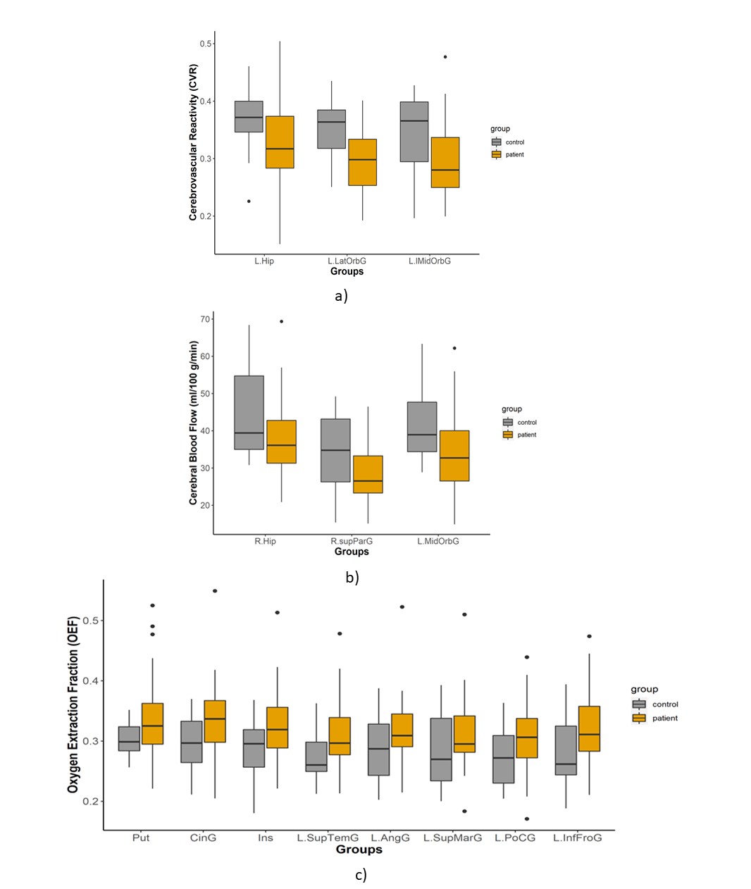
Results reveal significant differences in various brain regions between HCs and CADs:1. CVR is significantly higher in HCs in the Left (L) Hippocampus, L Medial Orbital Gyrus, and L Lateral OrbitoFrontal Gyrus (Table 2, Fig. 1, Fig. 2).
2. CBF is significantly higher in HCs in the Right (R) Hippocampus, L Medial Orbital Gyrus, and the superior parietal gyrus (Table 2, Fig. 1, Fig. 2).
3. OEF was found to be significantly higher in CADs compared to HCs in several cortical and sub-cortical areas (Table 2, Fig. 1, Fig. 2).
4. No significant changes were identified in CMRO2.
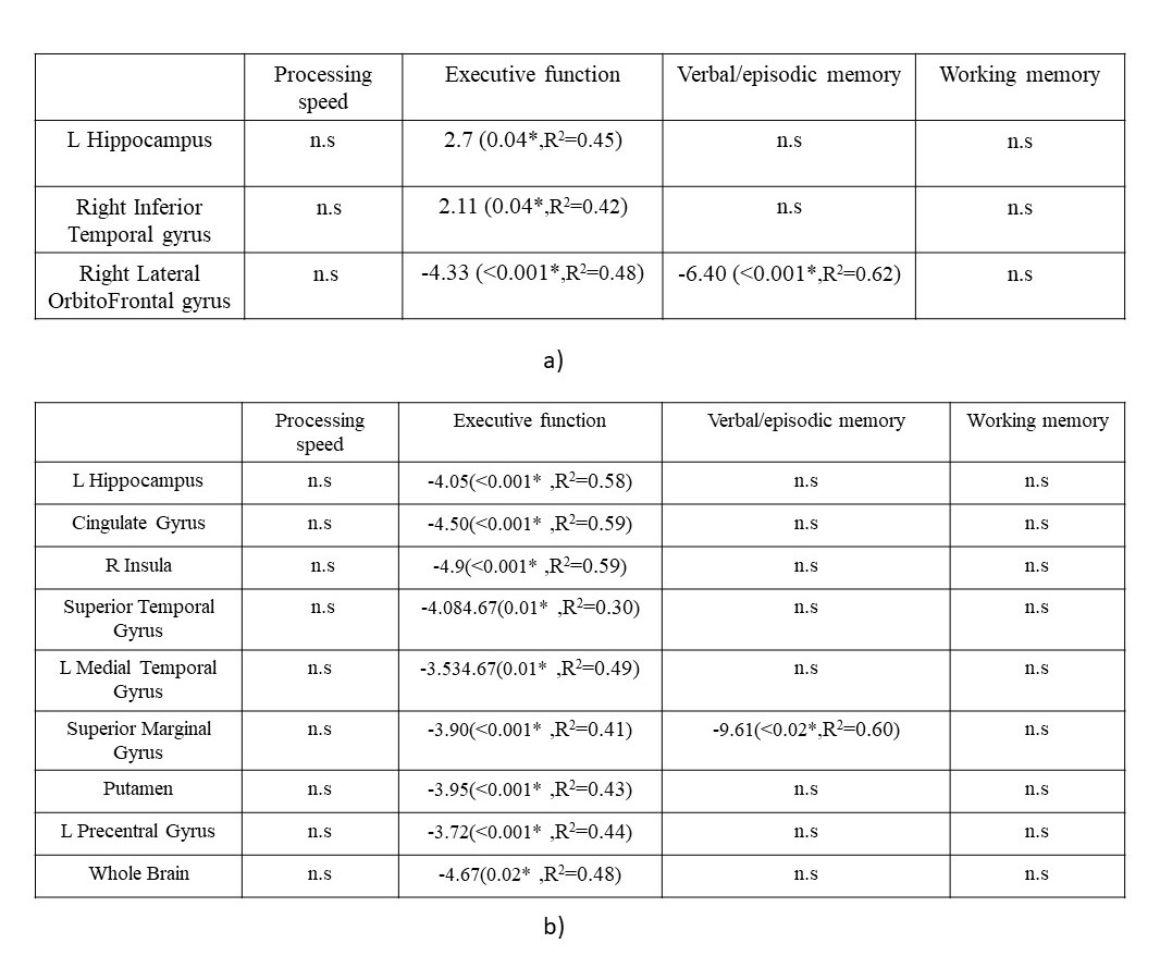
Moreover, a significant positive relationship was found between executive function and CVR in the regions where CVR was higher in CAD, except for the L Lateral OrbitoFrontal Gyrus (Table. 3). Additionally, there was a negative significant relationship between OEF and Executive functions in many of the regions where differences between CAD and HC were identified (Table. 3).
Discussion
We revealed several noteworthy findings in relation to CAD: Firstly, CAD patients exhibit lower CBF and CVR, but higher OEF in numerous brain regions, showing a predominantly vascular deficit and preserved metabolism. These observations are consistent with previous studies that demonstrated vascular changes in CAD [10], and vascular structure changes resulting in cerebral hypoperfusion [11,12]. These vascular alterations not only impair endothelial cells [13] but also disrupt neurovascular coupling, leading to damage to endothelial cells that hampers the production of vasodilators and decreases vessel elasticity, ultimately reducing CVR [14]. Metabolism seems to be maintained in our patients however, likely showing an early misery perfusion stage that may develop into atrophy in the future. Furthermore, the positive association between CVR and cognition aligns with existing literature, indicating that better cerebrovascular reactivity, and therefore vascular reserve, is necessary to maintain cognitive function [15]. Conversely, the negative association between OEF and cognition suggests that an elevated OEF may indicate inadequate perfusion that affects cognition. These early relationships between brain physiological components and cognition in treated CAD patients could underlie the increased incidence of late life dementia in this population.Acknowledgements
We would like to acknowledge the contribution of Paule Samson, MR technologist, Julie Lalongé, research assistant and medical electrophysiology technologist,and of all the staff of the EPIC center that has contributed to this project.
We also want to thank all students and research assistants who have helped in data acquisitions: Roni Zaks, Robert Hovey, Stephanie Beram, Alexandre bailey, Agathe Godet and Kathia Saillant and the research participants who took part in this study.
SAT was supported by the Canadian Institutes of Health Research (CIHR: FBD 175862).
CJG was supported by the Heart and Stroke Foundation New Investigator Award and J.M.Barnett fellowship, the Michal and Renata Hornstein Chair inCardiovascular Imaging, and the Heart and Stroke Foundation Grant-inAid G-17-0018336.
LB was supported by Mirella and Lino Saputo Research Chair in Cardiovascular Health and the Prevention of Cognitive Decline from the Universite de Montrealat the Montreal Heart Institute.
References
[1] Mendis, Shanthi, et al. "World Health Organization (WHO) and International Society of Hypertension (ISH) risk prediction charts: assessment of cardiovascular risk for prevention and control of cardiovascular disease in low and middle-income countries." Journal of hypertension 25.8 (2007): 1578-1582.
[2] Van Der Flier, Wiesje M., et al. "Vascular cognitive impairment." Nature Reviews Disease Primers 4.1 (2018): 1-16.
[3] Eisenmenger, Laura B., et al. "Vascular contributions to Alzheimer's disease." Translational Research (2022).
[4] https://www.mathworks.com/products/matlab.htm
[5] Alsop, David C., et al. "Recommended implementation of arterial spin‐labeled perfusion MRI for clinical applications: a consensus of the ISMRM perfusion study group and the European consortium for ASL in dementia." Magnetic resonance in medicine 73.1 (2015): 102-116.
[6] Gauthier, Claudine Joëlle, and Richard D. Hoge. "Magnetic resonance imaging of resting OEF and CMRO2 using a generalized calibration model for hypercapnia and hyperoxia." Neuroimage 60.2 (2012): 1212-1225.
[7] Homack, Susan, Donghyung Lee, and Cynthia A. Riccio. "Test review: Delis-Kaplan executive function system." Journal of clinical and experimental neuropsychology 27.5 (2005): 599-609.
[8] Reitan, Ralph M. "Validity of the Trail Making Test as an indicator of organic brain damage." Perceptual and motor skills 8.3 (1958): 271-276.
[9] https://www.loni.usc.edu/research/atlases
[10] Anazodo, Udunna C., et al. "Impaired cerebrovascular function in coronary artery disease patients and recovery following cardiac rehabilitation." Frontiers in aging neuroscience 7 (2016): 224.
[11] Brown, William R., and Clara R. Thore. "Cerebral microvascular pathology in ageing and neurodegeneration." Neuropathology and applied neurobiology 37.1 (2011): 56-74.
[12] Girouard, Helene, and Costantino Iadecola. "Neurovascular coupling in the normal brain and in hypertension, stroke, and Alzheimer disease." Journal of applied physiology 100.1 (2006): 328-335.
[13] Bai, Tao, Shijia Yu, and Juan Feng. "Advances in the role of endothelial cells in cerebral small vessel disease." Frontiers in Neurology 13 (2022): 861714.
[14] Lattanzi, Simona, Artur Słomka, and Afshin A. Divani. "Blood pressure variability and cerebrovascular reactivity." American Journal of Hypertension 36.1 (2023): 19-20.
[15] Kim, Donghoon, et al. "Relationship between cerebrovascular reactivity and cognition among people with risk of cognitive decline." Frontiers in Physiology 12 (2021): 645342.
Figures