3356
A Hybrid Deep Learning and Graphics method for Vessel Centerline Extraction and Vessel Segment Labeling to Assist Plaque Detection and Evaluation1Paul C. Lauterbur Research Center for Biomedical Imaging, Shenzhen Institute of Advanced Technology, Chinese Academy of Sciences, Shen Zhen, China, 2Department of Image Advanced Analysis of HSW BU, Shanghai United Imaging Healthcare Co., Ltd., Shanghai, China, 3Department of Radiology, Peking University Shenzhen Hospital, Shenzhen, China, 4Key Laboratory of Biomedical Imaging Science and System, Chinese Academy of Sciences, Shenzhen, China, 5United Imaging Research Institute of Innovative Medical Equipment, Shenzhen, China
Synopsis
Keywords: Vessel Wall, Blood vessels
Motivation: Manual centerline Extraction based on black-blood Magnetic resonance vessel wall imaging is a difficult and time-consuming but important step for further analysis of plaques.
Goal(s): To propose a method for quickly, automatically and accurately extracting the centerline and label the segments of the target arteries.
Approach: This study proposes a method that combines deep learning and traditional graphics method for the automatic and accurate centerline extraction and even segments labeling, which is applicable to flexible MR sequences (only black-blood images, only bright-blood images, or both).
Results: Compared with the ground truth, the proposed method achieved excellent completeness and accuracy in a short time.
Impact: This study introduces a flexible (MRVWI or/and TOF-MRI), swift, and precise approach for extracting and labeling the centerline of target vessel. With this method, radiologists can more conveniently and efficiently observe potential abnormal areas around vessel and diagnose vessel-related diseases.
INTRODUCTION
Magnetic resonance vessel wall imaging (MRVWI) can directly visualize vessel walls, allowing for the characterization and quantitative assessment of vessel walls and plaques1. Vessel centerline extraction is the first and most important step for further analysis of plaques2. However, centerline extraction based on MRVWI is challenging because it is susceptible to interference from nearby areas with similar signal strengths. Using the centerline extracted from bright-blood images directly in black-blood images may result in errors due to image registration deviation. This study proposes a method that combines deep learning and traditional graphics methods for automatic and accurate centerline extraction and even segment labeling, which is applicable to flexible MR sequences (only black-blood images, only bright-blood images, or both).METHODS
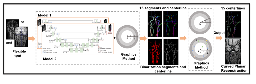
All images were obtained from 552 patients who underwent black-blood T1 MRVWI and bright-blood TOF/CE MRA or just bright-blood TOF/CE MRA from the neck to the whole brain in four different hospitals. Among them, images of 236 T1 MRVWI scans and 510 TOF/CE MRA scans were used for model development. The images of the remaining 42 patients who simultaneously scanned two sequences were used for independent testing.The automatic method can be conceptualized in three steps. As depicted in Figure 1, first, the main vessel segments are segmented, and the centerline of the vessels is extracted. The improved convolutional neural network (CNN)-based models were developed to segment 15 main vessel segments based on the flexible MR sequence input, which were fused to extract the 15 segment centerlines of the vessels based on the boundary distance field (BDF), finite difference method (FDM) and steepest descent method (SDM). Then, the centerlines are connected and extended. Based on the selected sequence, another CNN-based model was utilized to predict the binarization vessel segments. The same BDF, FDM and SDM were utilized to extract the binary centerline. This binary centerline was used to connect and extend the 15 segment centerlines based on the vessel tracking algorithm. In this way, the performance of potential interruption areas and end of centerlines has been improved. Finally, the neighborhood localization algorithm is used to check and correct the potential errors in vessel centerline bifurcation.
Completeness (correct extraction length to ground truth ratio) and average centerline distance (ACD, distance between extraction and ground truth) were used to evaluate the performance. This study recorded the performance of the automatic method and the performance after the automatic method was manually corrected by junior doctors.
RESULTS
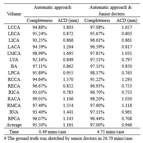
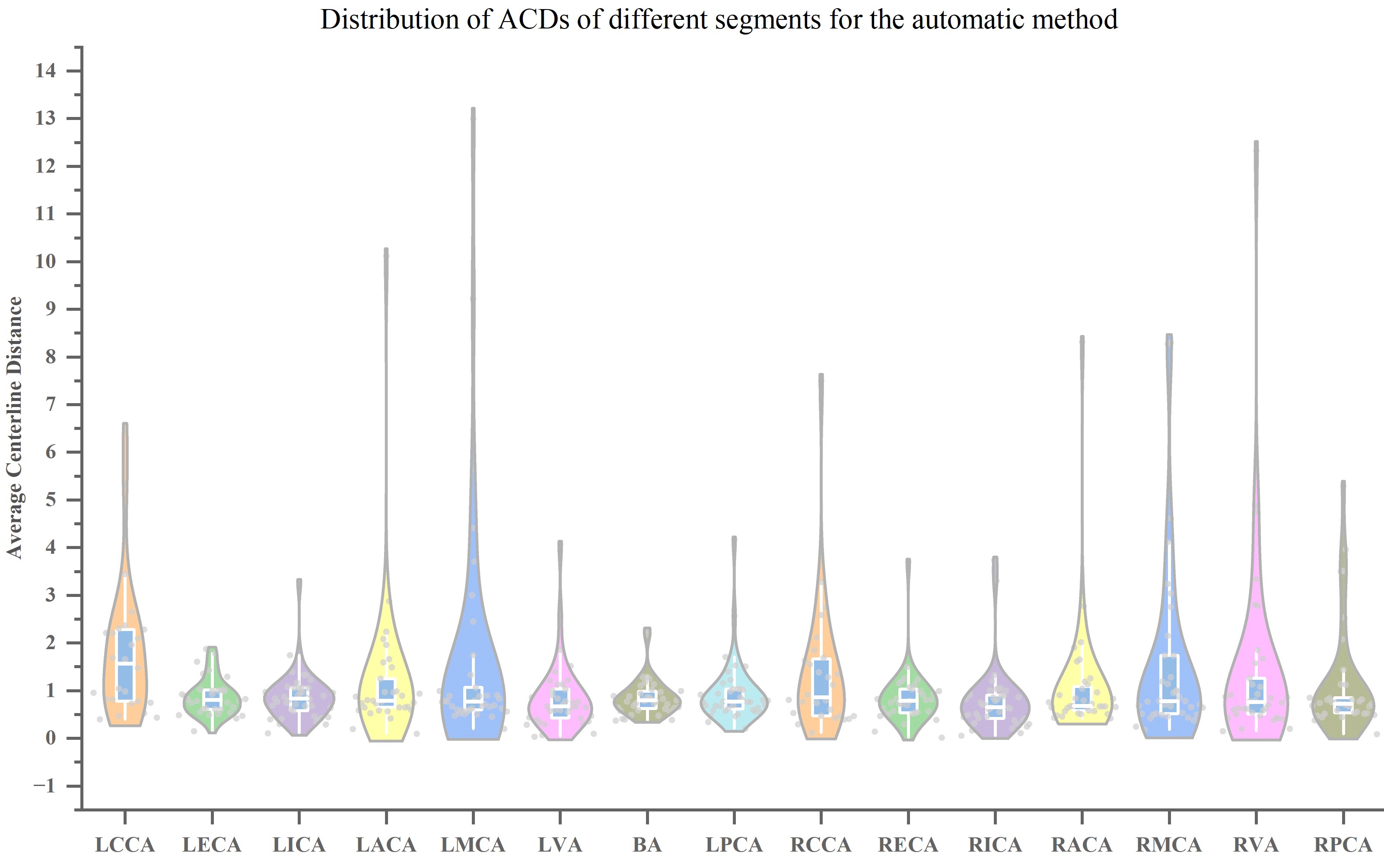
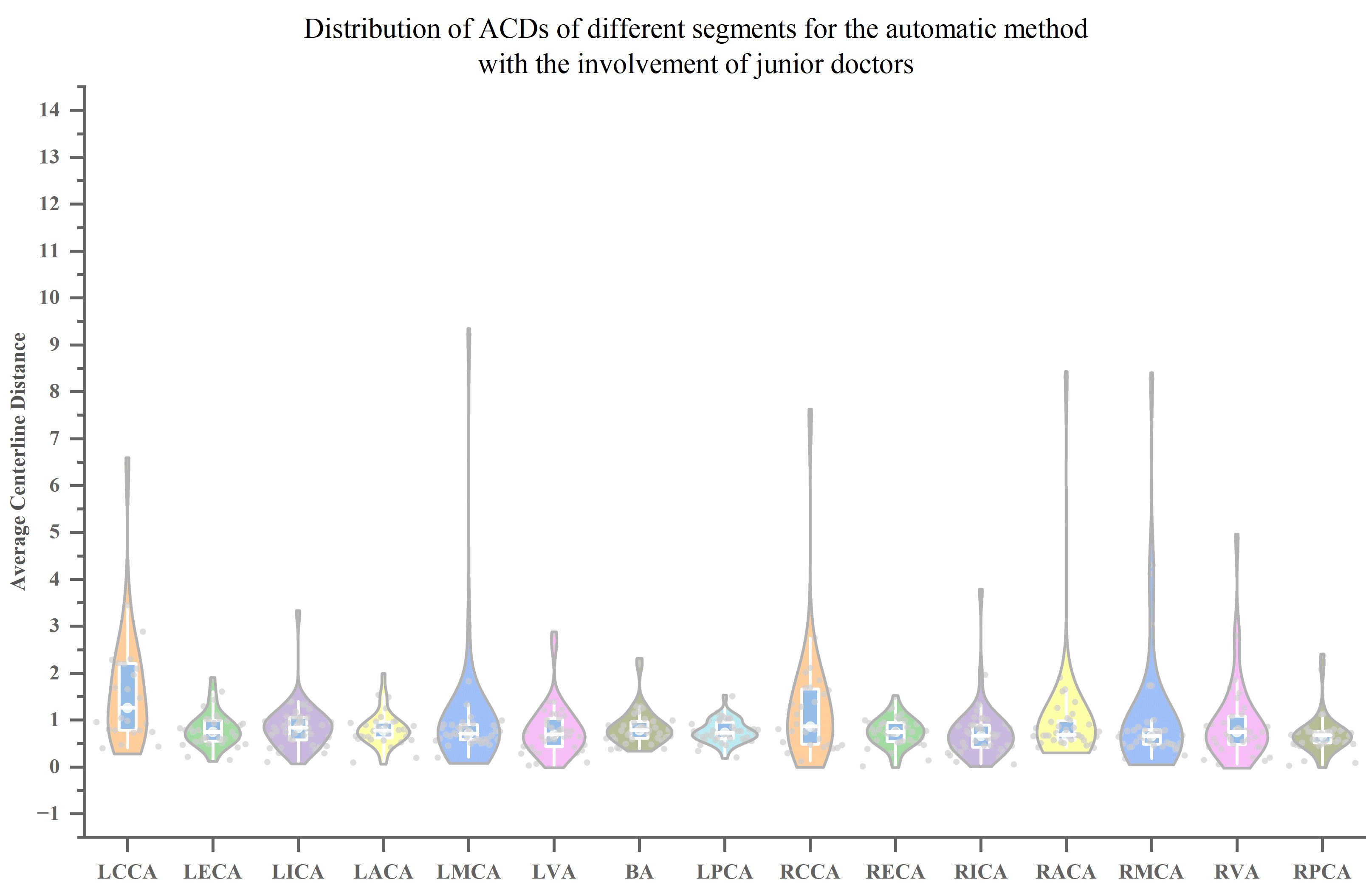
The independent test results are shown in Figure 2. The proposed method achieves an average completeness of 95.5% for centerline extraction and labeling of 15 vessel segments in an average time of just 0.49 minutes, with an average surface distance of 1.161 mm, which is very similar to the ground truth. After the involvement of junior doctors, the average completeness increased from 95.5% to 97%, the ACD decreased from 1.161 to 0.946, and the time increased only increased from 0.49 minutes to 4.75 minutes per case. Figure 3 and Figure 4 utilize violin plots with box and scatter to illustrate the distribution of ACDs of different segments for the automatic method and the automatic method with the involvement of junior doctors. As shown in Figure 3, the ACDs of most vessel segments are approximately 1 mm, but there are a handful of outliers in small vessels. Comparing Figures 3 and 4, it can be observed that the outliers have been effectively mitigated after the involvement of junior doctors.DISCUSSION AND CONCLUSION
This study combines the improved CNN model with traditional graphics methods to extract centerlines and label segments from the neck to brain vessels. The improved CNN model was used to segment the intact vessel, and the graphics methods were used to extract and correct the centerlines and labels. Compared with the ground truth, the proposed method achieved excellent completeness and accuracy in a short time. Therefore, the proposed method can quickly, automatically and accurately extract the centerline and label the segments of the whole carotid and intracranial arteries. Fully automated methods have achieved excellent performance, furthermore, after the involvement of junior doctors, with only a slight increase in time, the ACD of the centerline was reduced to within 1 mm. This demonstrates that the method possesses substantial potential for clinical application.Acknowledgements
The study was partially support by Natural Science Foundation of Guangdong Province-Outstanding Youth Project (2023B1515020002), National Key Technology Research and Development Program of China (2021YFF0501502), Key Laboratory for Magnetic Resonance and Multimodality Imaging of Guangdong Province (2023B1212060052), and Central guidance for local science and technology development project (ZYYD2023D02).References
1.Choi, Young Jun, Seung Chai Jung, and Deok Hee Lee. "Vessel wall imaging of the intracranial and cervical carotid arteries." Journal of stroke 17, no. 3 (2015): 238.
2.Huang, Xianjue, Jun Wang, and Zhiyong Li. "3D carotid artery segmentation using shape-constrained active contours." Computers in Biology and Medicine 153 (2023): 106530.
Figures