3355
wall characteristics and clinical indicators correlate with acute ischemic stroke events in patients with middle cerebral atherosclerosis1Baoji Central Hospital, Bao ji, China, 2Jincheng General Hospital, Jin cheng, China, 3GE HealthCare MR Research, Bei Jing, China, 4Baoji Central Hospital, Bei Jing, China
Synopsis
Keywords: Atherosclerosis, Atherosclerosis, High resolution magnetic resonance vascular wall imaging, Middle cerebral artery, Acute ischemic stroke
Motivation: The occurrence of acute ischemic stroke (AIS) in patients with middle cerebral artery (MCA) atherosclerosis is related to the status of atherosclerotic plaque itself and level of homocysteine (Hcy) and glycated hemoglobin (HbA1c).
Goal(s): To analyze wall characteristics and clinical indicators in patients with MCA atherosclerosis with a view to finding predictors of AIS events.
Approach: We gathered data from 38 participants, segregated them into the AIS and transient ischemic attack (TIA) groups, and subjected the data from both groups to statistical analysis.
Results: Combined stenosis rate and Hcy have higher value for predicting AIS events.
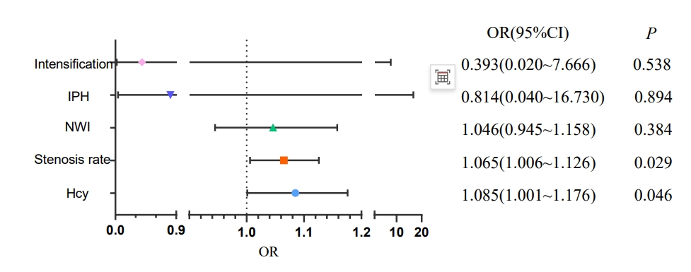
Impact: Hcy and stenosis rate were independent risk factors for AIS events in patients with MCA atherosclerosis; combined Hcy and stenosis rate had a higher predictive value of AIS events.
Introduction
Intracranial atherosclerotic stenosis (ICAS) is a primary cause of acute ischemic stroke (AIS) in China, with the middle cerebral artery (MCA) being the most susceptible intracranial artery. AIS is associated with high mortality and recurrence rates. It has been established that the occurrence of AIS is linked not only to the condition of atherosclerotic plaque but also to the levels of Homocysteine (Hcy) and Glycated hemoglobin (HbA1c). However, limited research has focused on the combination of these two factors. Therefore, this study aims to assess the wall characteristics of atherosclerotic MCA using High-Resolution Magnetic Resonance Vascular Wall Imaging (HR-MRVWI). Additionally, it seeks to explore the relationship between the arterial wall, patients' clinical indicators, their combination, and AIS. The study aims to identify the risk factors that predict AIS events.Methods
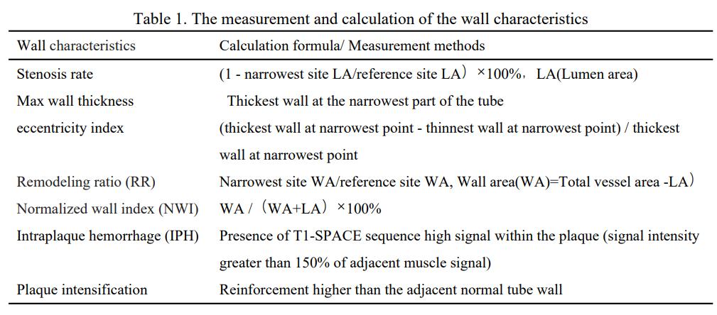
This study received approval from our institutional review board, and informed consent was waived due to its retrospective nature. The study included 38 patients meeting the criteria between March 2022 to February 2023. All participants underwent MR examinations using a 3.0T MR scanner equipped with a 20-channel head coil. The scan sequences comprised routine brain sequences (T1-weighted, T2-weighted, T2 FLAIR, TOF MR angiography, and DWI) as well as pre- and post-contrast HR-MRVWI. The specific parameters for HR-MRVWI were as follows: TR=900ms, TE=16ms, resolution=0.5×0.5×0.6mm³. Based on whether DWI showed hyperintensity in the middle cerebral artery supply territory, subjects were divided into two groups: AIS and TIA. Following the scans, HR-MRVWI images were transferred to a workstation, and vessel wall characteristics listed in Table 1 were measured and calculated. Clinical indices, including Hcy and HbA1c, were also obtained. The SPSS 26.0 software was used for intergroup comparisons of vessel wall characteristics and clinical indices. Logistic one-way regression and logistic multifactorial regression analyses were performed for factors that exhibited significant differences between the groups. Additionally, an ROC curve test was conducted to determine the diagnostic efficacy of factors with a P-value < 0.05.Result
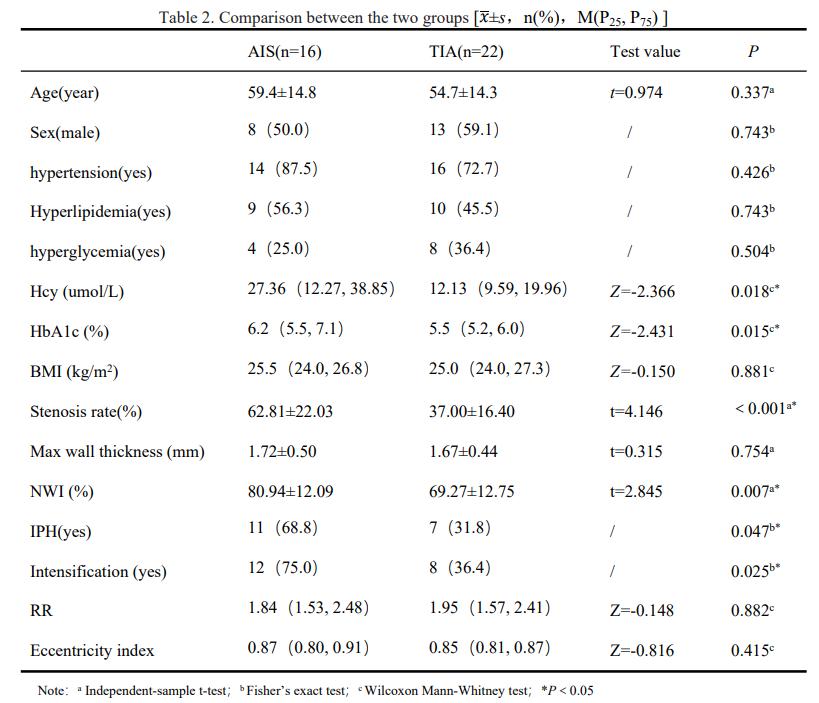
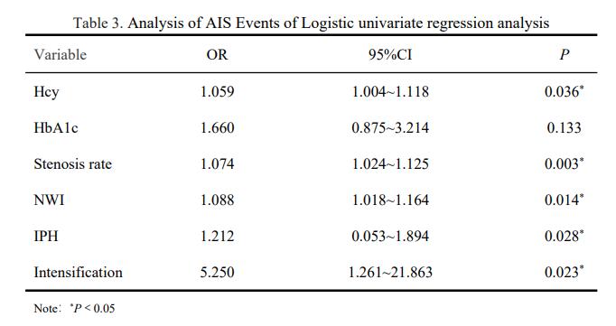
(1) Hcy, HbA1c, stenosis rate, NWI, IPH, and intensification exhibited higher levels in the acute stroke group compared to the non-acute stroke group (Table 2);(2) Binary logistic univariate regression analyses indicated that the primary risk factors for AIS events in patients with MCA atherosclerosis included Hcy, stenosis, NWI, IPH, and intensification (Table 3);
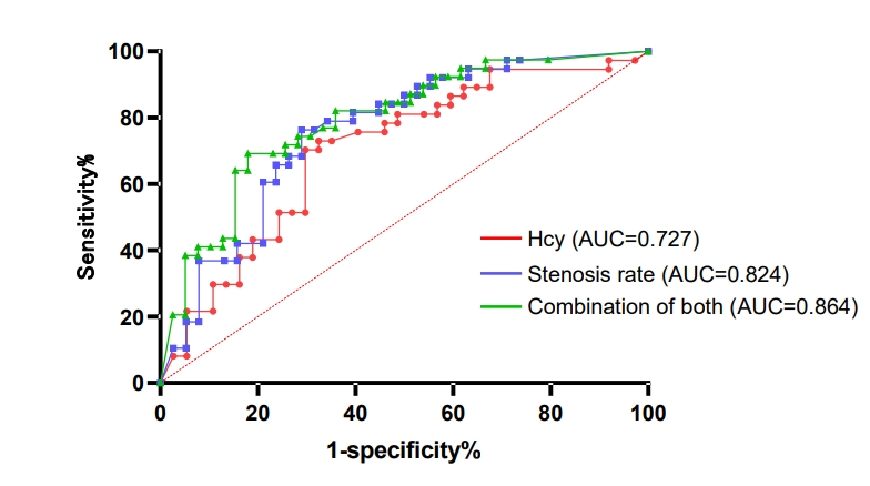
(3) Binary logistic multifactorial regression analyses revealed that Hcy and stenosis independently served as risk factors for AIS events in patients with MCA atherosclerosis. Furthermore, the combination of these two factors demonstrated an even larger area under the curve, as illustrated in Figure 1-2
Discussion
In this study, we explored the association between vascular wall characteristics, clinical parameters, and the predictive value of acute ischemic stroke (AIS) events in individuals with middle cerebral atherosclerosis. The findings revealed that patients in the AIS group exhibited elevated levels of Homocysteine (Hcy), Glycated hemoglobin (HbA1c), stenosis rate, normalized wall index (NWI), intraplaque hemorrhage (IPH), and plaque intensification. These results align with those reported in several previous studies[5-9]. Hcy and stenosis rate were identified as independent predictors of AIS events in patients with middle cerebral atherosclerosis. Notably, the combination of these two factors demonstrated enhanced predictive value for AIS events. The mechanisms underlying these findings are as follows: Hcy may contribute to stroke by promoting thrombosis and exerting pro-oxidant effects[10]. In the prothrombotic pathway, Hcy interacts with chemolipoproteins and microorganisms, leading to aggregate formation. These aggregates can obstruct blood vessels when vulnerable arterial plaques are present, consequently increasing the risk of AIS events[11]. In the prooxidative pathway, Hcy, either independently or in conjunction with other factors, can induce endothelial damage through its conversion to Hcy-thiolactone, thereby causing or exacerbating atherosclerosis[12]. Concomitantly, luminal stenosis results in restricted blood flow. A narrow lumen suggests the presence of thick plaque on the vessel wall. The combined effect of these factors promotes plaque rupture, hemorrhage, and detachment, subsequently elevating the risk of AIS events[13]. Hence, it is imperative to closely monitor Hcy levels and stenosis rates in patients with middle cerebral atherosclerosis. Appropriate interventions should be considered when Hcy levels approach 25.675 umol/L and stenosis rates exceed 49.5%.Conclusions
Wall characteristics include, stenosis rate, NWI, IPH, and plaque intensification, and the level of Hcy demonstrated positive correlations with acute AIS events. Further analysis revealed that both Hcy and stenosis rate independently served as risk factors for AIS events in individuals with MCA atherosclerosis. Notably, the combination of Hcy and stenosis rate displayed even greater predictive value for AIS events.Acknowledgements
No acknowledgement found.References
[1] Zhang D F, Chen Y C, Chen H, et al. A High-Resolution MRI Study of Relationship between Remodeling Patterns and Ischemic Stroke in Patients with Atherosclerotic Middle Cerebral Artery Stenosis [J]. Front Aging Neurosci, 2017, 9: 140.
[2] Fukuda K, Iihara K, Maruyama D, et al. Relationship between carotid artery remodeling and plaque vulnerability with T1-weighted magnetic resonance imaging [J]. J Stroke Cerebrovasc Dis, 2014, 23(6): 1462-1470.
[3] Leng X, Wong K S, Liebeskind D S. Evaluating intracranial atherosclerosis rather than intracranial stenosis [J]. Stroke, 2014, 45(2): 645-651.
[4] Wang B. Correlation between homocysteine glycosylated hemoglobin and neurological function and long-term prognosis in acute ischemic stroke [J]. Medical Review, 2016, 22(22): 4500-4503.
[5] Li DX, Zhang Y, Zhang HW, et al. Expert understanding of diagnosis, treatment and prevention of hyperhomocysteinemia[J]. Journal of Rare Diseases, 2022, 29(6): 1-4.
[6] Selvin E, Coresh J, Shahar E, et al. Glycaemia (haemoglobin A1c) and incident ischaemic stroke: the Atherosclerosis Risk in Communities (ARIC) Study [J]. Lancet Neurol, 2005, 4(12): 821-826.
[7] Chung G H, Kwak H S, Hwang S B, et al. High resolution MR imaging in patients with symptomatic middle cerebral artery stenosis [J]. Eur J Radiol, 2012, 81(12): 4069-4074.
[8] Lee J, Kil J, Kim D W, et al. Usefulness of Plaque Magnetic Resonance Imaging in Identifying High-Risk Carotid Plaques Irrespective of the Degree of Stenosis [J]. J Cerebrovasc Endovasc Neurosurg, 2017, 19(4): 291-300.
[9] Huang J, Jiao S, Zhao X, et al. Characteristics of patients with enhancing intracranial atherosclerosis and association between plaque enhancement and recent cerebrovascular ischemic events: a high-resolution magnetic resonance imaging study [J]. Acta Radiol, 2019, 60(10): 1301-1307.
[10] Kim Y, Lee J H, Lee S H, et al. Serum Homocysteine Is Associated With HDL Only in Stroke Patients With Small Vessel Occlusion [J]. Front Neurol, 2020, 11: 565506.
[11] Bone Health and Nutrition Committee of the Chinese Nutrition Society, Parenteral and Enteral Nutrition Branch of the Chinese Medical Association, Chinese Society of Geriatrics, Northern Chronic Disease Control Branch. Chinese Society of Geriatrics, Northern Chronic Disease Prevention and Control Branch. Expert consensus on the diagnosis and treatment of hyperhomocysteinemia [J]. Electronic Journal of Oncometabolism and Nutrition, 2020, 7(3): 283-288.
[12] Harris S, Rasyid A, Kurniawan M, et al. Association of High Blood Homocysteine and Risk of Increased Severity of Ischemic Stroke Events [J]. Int J Angiol, 2019, 28(1): 34-38.
[13] Wang XY, Li XH, Qian YF, et al. Study on the relationship between middle cerebral artery plaque and stroke type by high resolution MRI [J]. Journal of Practical Radiology, 2021, 37(3): 356-359, 364.
Figures