3353
Hemodynamics Contrast Investigation of Intracranial Aneurysms before and after Flow Diverter Stent Treatment1Center for Biomedical Imaging Research, Department of Biomedical Engineering, Tsinghua University, Beijing, China, 2Changhai Hospital, Shanghai, China
Synopsis
Keywords: Flow, Velocity & Flow, aneurysm
Motivation: The research aims to investigate the changes in hemodynamics in intracranial aneurysms before and after treatment with blood flow shunt stents treatment.
Goal(s): The technical goal is to quantify common hemodynamic parameters, and use statistical comparison to analyze the variation patterns.
Approach: A set of program was utilized to separate aneurysms from complex environments, calculate hemodynamic values within the region, and analyze the trend of changes in preoperative and postoperative data.
Results: The results of simple statistical analysis showed that the average flow velocity, average WSS and OSI at the aneurysm presented a decreasing trend in different degrees after the shunt stent treatment.
Impact: In this study, the number of subjects and hemodynamic parameters can be increased to obtain more comprehensive experimental results. Researchers can also focus on the relationship between aneurysm stability and hemodynamics, and establish corresponding prediction and evaluation models.
Introduction
Aneurysms are localized diffuse dilation or bulging of the vessel wall due to lesions or injuries in the arterial wall. With high morbidity, disability and mortality, aneurysms pose a great threat to people's health, so it has been paid great attention in clinic1. In the clinical diagnosis and treatment of intracranial aneurysms, hemodynamics is an important evaluation reference index. In recent years, with a low complication rate, flow diverter has become the more promising treatment method2. The basic principle of the flow diverter is to reduce the blood flow rate and pressure, prevent the rupture of intracranial aneurysms, and provide conditions for intimal growth to facilitate endothelialization and reconstruct the parent artery. In this study, 4D Phase-Contrast MRI was used to calculate the hemodynamics of intracranial aneurysms before and after the implantation of shunt stents, which is of great significance to evaluate the treatment effect and further optimize the treatment method.Methods
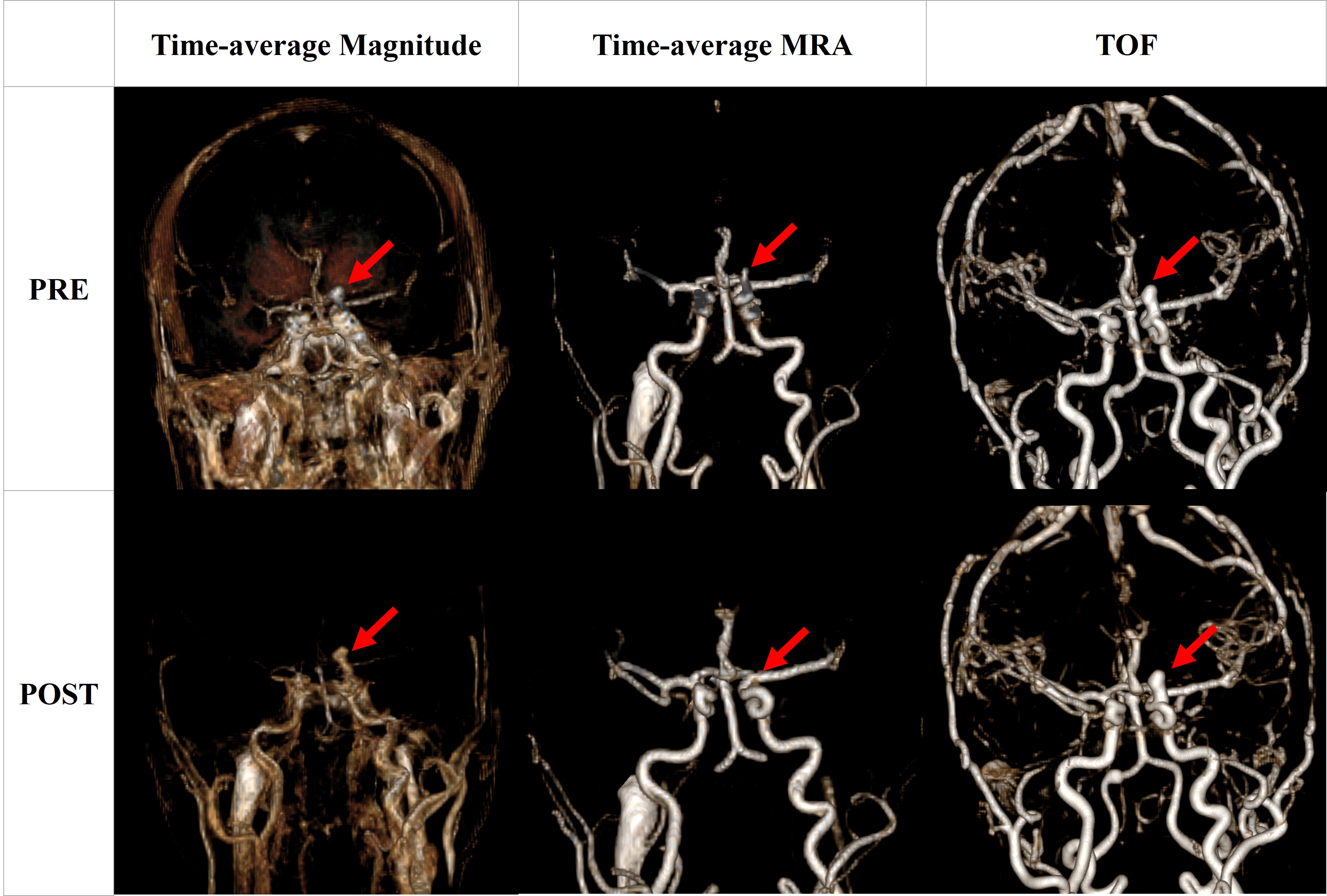
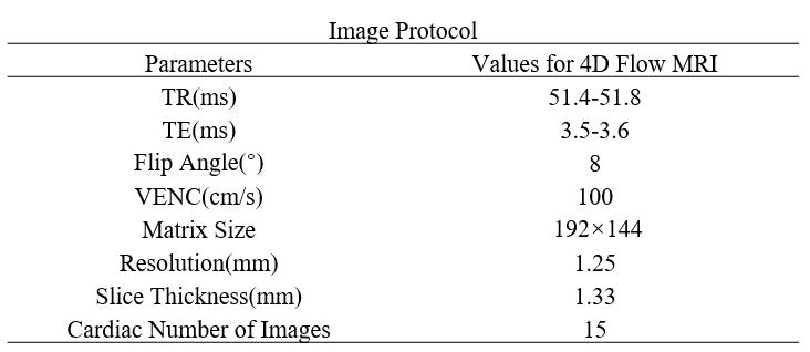
Patient and Data AcquisitionData from twenty patients with intracranial aneurysms following flow diverter stent treatment provided by Changhai Hospital (Shanghai, China) was used in this study. Each set of patient data included intracranial 4D flow images and MRA images before and after flow diverter stent implantation. As a reference, preoperative or postoperative TOF MRA images were also utilized in aneurysm recognition, selection and segmentation. All scans were performed at 3T Siemens Skyra. The dataset example was displayed in Figure 1 and the image protocol was shown in Table 1.
Due to the individual differences of aneurysm cases, the limitations of current algorithms and the quality problems of data sets, not all data can get accurate calculation results. In the data of twenty patients, fourteen cases of preoperative and postoperative aneurysms data could be measured accurately, and six cases of postoperative data could not be utilized due to different degrees of blood flow chaos and low signal value at the aneurysm.
Aneurysm Extraction and Hemodynamic Calculation
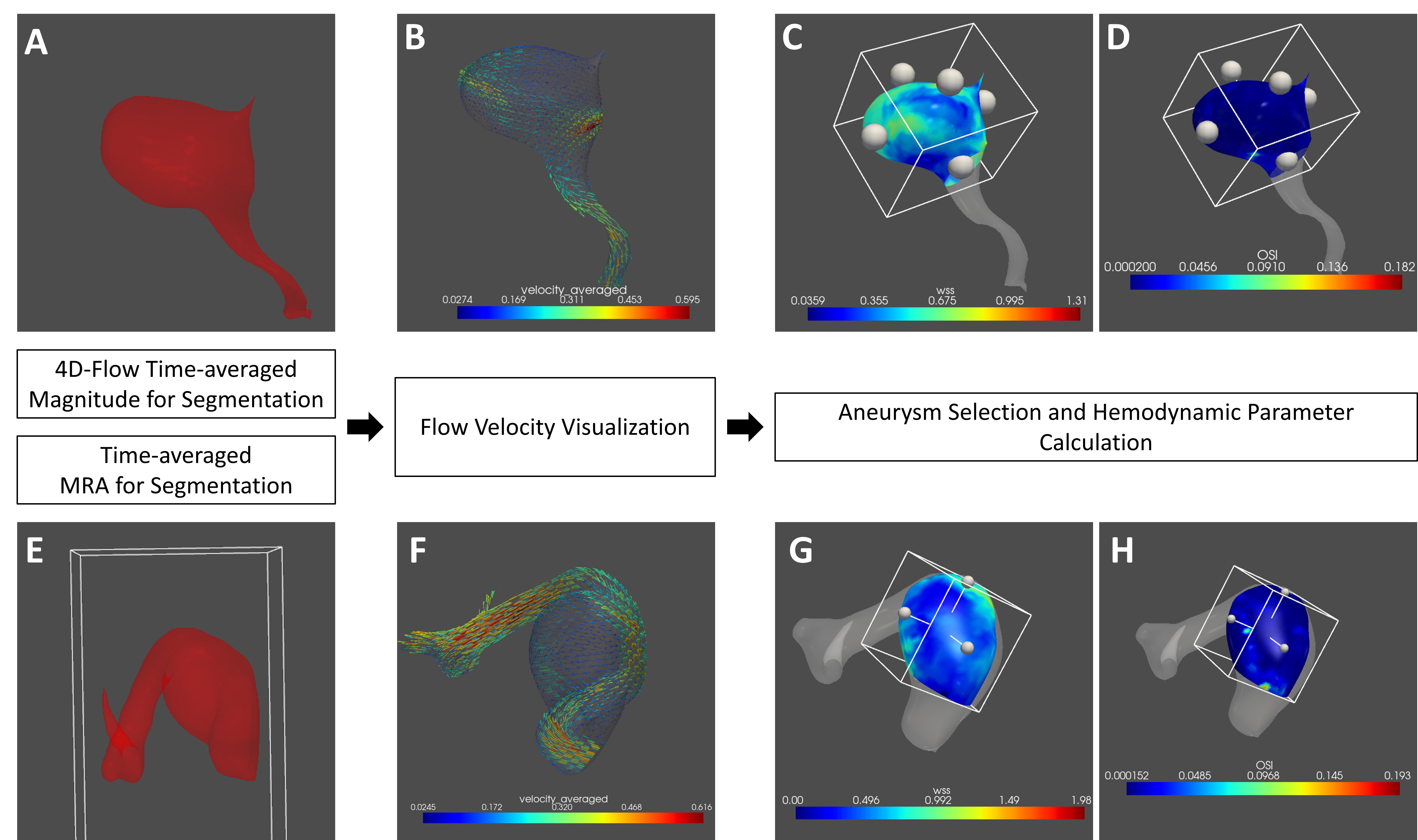
In this study, we used Fu's workflow3 to visualize aneurysm and calculate hemodynamics. First, we adopted simple and effective threshold segmentation to extract aneurysms from complex surroundings. There are two specific segmentation principles. One is based on the difference between the high signal of the aneurysm and the surrounding environment in the 4D flow time-average magnitude image, and the other is based on time-average MRA signal difference. The two methods have their own advantages and disadvantages. We chose the method with better effect according to the specific situation of each patient's data in order to find the location of the aneurysm more accurately, and then ensured that the calculated hemodynamic parameters are accurate. The workflow was shown in Figure 2.
Results
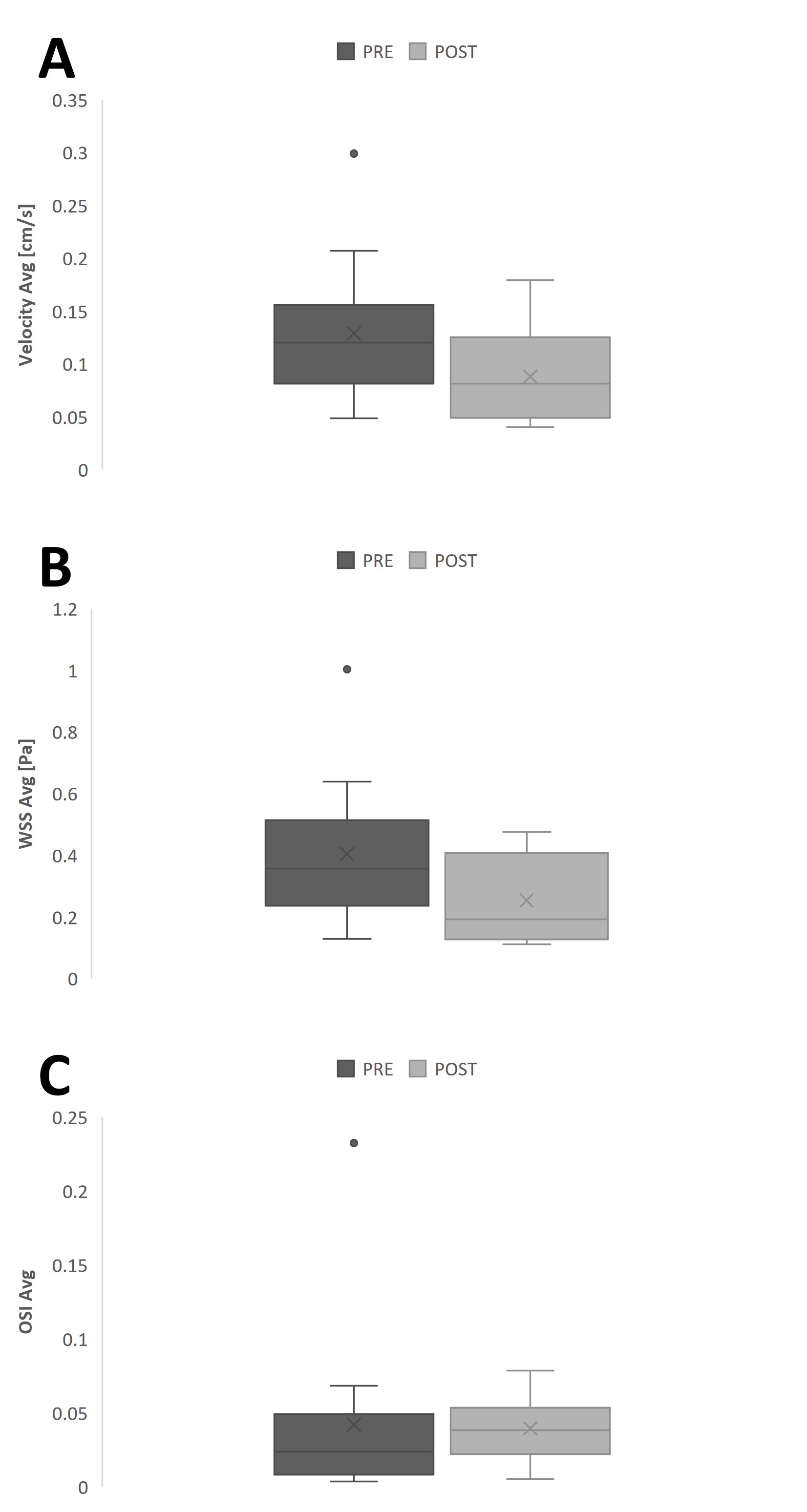
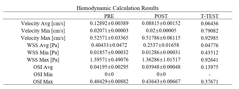
The hemodynamic calculation results of fourteen intracranial aneurysm data before and after shunt stent implantation are shown in Table 2 and Figure 3. The results of simple statistical analysis showed that the average flow velocity, average WSS and average OSI at the aneurysm presented a decreasing trend in different degrees after the shunt stent treatment. Statistical t-test was performed on the pre-operative and post-operative data of the two groups, and the results showed that the average wall shear stress was significantly different between pre-operative and post-operative.Discussion and Conclusion
The decreasing trend of blood flow velocity at the aneurysm indicates that the implantation of shunt stent led to the effect of guiding blood flow shunt and reducing blood flow in the aneurysm. The decrease of OSI indicates that the change of blood flow direction and intensity was slowed down after surgery. It is worth noting that although the current study believes that low WSS and high OSI are related to the rupture of aneurysms4, this study reflected the downward trend of postoperative WSS, rather than a certain low value. This trend may be a normal phenomenon after the blood flow velocity slows down.Due to the limitation of amount of study subjects, this study failed to obtain statistically significant results. In the future, more aneurysm data should be included, and the research content should be further expanded to evaluate the trend of aneurysm growth, healing and rupture in terms of hemodynamic changes. The post-operative data will always produce chaotic, low and inconspicuous signals near the aneurysm due to the stent implantation, resulting in difficulty in determining the aneurysm location. Subsequent studies can also focus on localization algorithms that independent of postoperative blood flow data at the aneurysm.
Acknowledgements
No acknowledgement found.References
[1] Etminan, N., & Rinkel, G. J. (2016). Unruptured intracranial aneurysms: development, rupture and preventive management. Nature reviews. Neurology, 12(12), 699–713.
[2] Chong, W., Zhang, Y., Qian, Y., Lai, L., & Parker, G. Computational Hemodynamics Analysis of Intracranial Aneurysms Treated with Flow Diverters: Correlation with Clinical Outcomes.
[3] Fu M, Peng F, Zhang M, Chen S, Niu H, He X, Xu B, Liu A, Li R. Aneurysmal wall enhancement and hemodynamics: pixel-level correlation between spatial distribution. Quant Imaging Med Surg 2022.
[4] Yu, C. , Chang, L. , Qi, Z. , Yue, S. , & Bin, G. . (2017). Hemodynamic study of the risk factors of intracranial aneurysm rupture. Journal of Beijing University of Technology.
Figures