3351
Improving background suppression of intracranial spiral time-of-flight MRA with phase-sensitive water-fat separation1Department of Radiology, Mayo Clinic, Rochester, MN, United States, 2Department of Radiology, University of Wisconsin-Madison, Madison, WI, United States
Synopsis
Keywords: Flow, Fat, time-of-flight, phase-sensitive, water-fat separation, fat suppression
Motivation: Efficient fat suppression can improve flow visualization in time-of-flight (TOF) MRA. We hypothesize that the phase difference of water and fat may be used for efficient fat suppression for out-of-phase (OP) TOF MRA.
Goal(s): To demonstrate the feasibility of fat suppression for spiral TOF with sliding-slice localized quadratic (ssLQ) encoding using the phase-sensitive approach.
Approach: After the general reconstruction of ssLQ OP TOF, the global slowly varying phase was estimated and removed. Water and fat voxels were then identified according to their phase.
Results: Background suppression was substantially improved with the proposed method, resulting in enhanced visualization of small vessels.
Impact: The proposed phase-sensitive approach requires no changes of pulse sequence and negligible computational cost. It might be implemented with a wide range of OP TOF MRA for fat suppression.
Introduction
Localized quadratic (LQ) encoding is a hybrid 2D/3D imaging technique, in which a ‘chirp’ radiofrequency (RF) pulse with a linearly swept frequency is used to excite a slab and thus the position along the slice direction is encoded by a quadratic phase1. Spiral sliding-slice LQ (ssLQ) time-of-flight (TOF)2 combines the through-plane LQ encoding and sliding-slice in-plane spiral readout for a fast, flexible and SNR efficient acquisition. Spiral Dixon water-fat separation3 can be combined with spiral ssLQ TOF for fat suppression to improve the visualization over the conventional out-of-phase (OP) method at a trade-off of doubling the scan time. The goal of this study is to achieve the fat suppression with spiral ssLQ OP TOF by an approach of phase-sensitive water-fat separation4.Method
a. Data AcquisitionVolunteer data of spiral ssLQ OP TOF of the intracranial vasculature were collected on a 3T scanner (Ingenia Elition, Philips Healthcare, Best, The Netherlands) and a 1.5T scanner (Ambition X, Philips Healthcare, Best, The Netherlands) with 15-channel head coils around the circle of Willis. Informed consent was obtained from each volunteer before scanning, per the guidelines of our institutional review board. Scan parameters included: field of view (FOV) = 200x200x91 mm3, resolution = 0.63x0.63x1.4 mm3 (3T) and 0.73x0.73x1.4 mm3 (1.5 T), TR=23ms, TE= 3.45 ms (second OP at 3T) and 2.4 ms (close to first OP at 1.5 T), flip angle = 21o, slab-to-slice ratio M = 16, readout length = 6.0 ms (3T) and 9.7 ms (1.5T). A venous saturation pulse was applied in each acquisition. Flow compensation gradients were applied in the slice direction. A Cartesian pre-scan was also performed to obtain a low-resolution off-resonance field map for deblurring. Scan time was 2 min 13 sec for 3T and 2 min 52 sec for 3T and 1.5T respectively.
b. Reconstruction
The reconstruction steps are illustrated in figure 1. After the general reconstruction for spiral ssLQ, including phase corrections for LQ and sliding slice, gridding and deblurring at off-resonance frequency for water, an initial slowly varying global phase was estimated from the OP images by low-pass filtering. This background phase was then removed from the OP images at the phase correction stage. Subsequently, water and fat components were assigned according to the phase of each voxel as shown in figure 1, exploiting the fact that the phase of water and fat is close to 0o and 180o, respectively. The slowly varying background phase was then re-estimated using the water-fat in-phase images obtained by complex subtraction of water and fat. This procedure was iterated before output of the final water and fat images.
Results and Discussion
The results at 3T are demonstrated in figures 2-3. An overall flat background phase close to 0o can be achieved after several iterations, as shown in figure 2. The proposed method demonstrated better background suppression in images of maximum intensity projection (MIP), enabling increased visualization of small vessels (figure 3), although the difference is subtle. At 1.5T, the fat signal is relatively brighter since the spiral ssLQ sequence can acquire data at the first water/fat OP echo point rather than the second one. Therefore, the overall improvement of background suppression and visualization of vessels is much more substantial (figures 4-5).There are several limitations of this approach. First, this approach essentially identifies a voxel as water or fat, it does not calculate the water/fat compositions within one voxel. Therefore, partial volume effect needs to be further investigated. Second, since fat is not deblurred, the water-fat separation with the proposed approach is not as accurate as the spiral Dixon approach3 in which water and fat are jointly deblurred. Finally, like the Dixon approach, misclassification of flow as fat could happen due to the flow accumulated phase (figure 5). Nevertheless, the water images obtained by the proposed approach may be used as complementary information in addition to the OP images at negligible computational cost.
Acknowledgements
This work is supported in part by Philips Health.References
1. Pipe JG. Analysis of localized quadratic encoding and reconstruction. Magn Reson Med. 1996;36(1):137-146. doi:10.1002/mrm.1910360122.
2. Wang D, Krishnamoorthy G, Ooi M, Pipe JG. Spiral inflow MRA with sliding-slice localized quadratic encoding. Magn Reson Med. 2023 Nov;90(5):1818-1829. DOI: 10.1002/mrm.29770.
3. Wang D, Zwart NR, Pipe JG. Joint water-fat separation and deblurring for spiral imaging: Joint Water-Fat Deblurring. Magn Reson Med. 2018;79(6):3218-3228. doi:10.1002/mrm.26950.
4. Hargreaves BA, Vasanawala SS, Nayak KS, Hu BS, Nishimura DG. Fat-suppressed steady-state free precession imaging using phase detection. Magn Reson Med 2003;50:210–213.
Figures
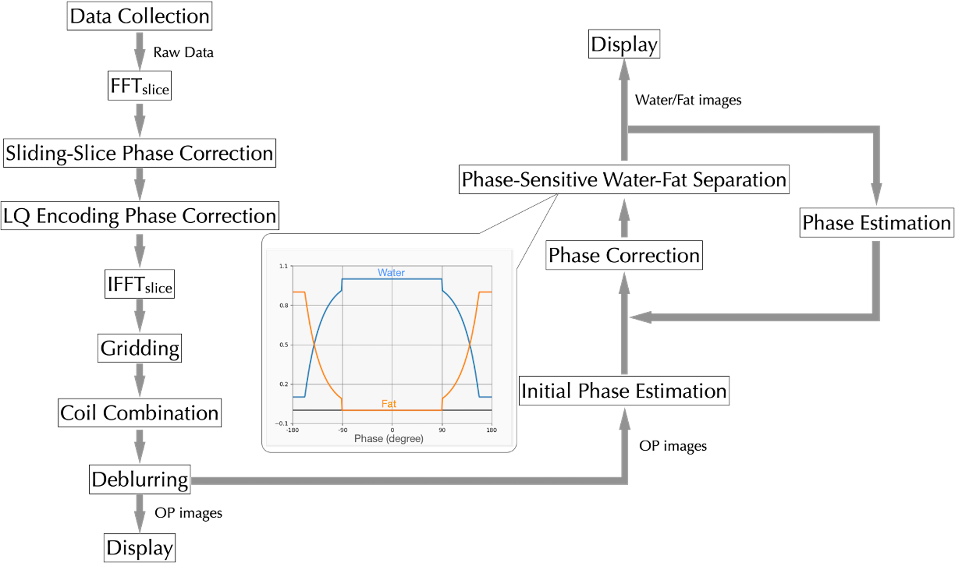
Figure 1 Flow chart of reconstruction. After the general ssLQ spiral reconstruction (left column), the proposed approach of phase-sensitive water-fat separation (right column) was applied to the OP images to obtain the water/fat images. ssLQ: sliding-slice localized quadratic; OP: out-of-phase.
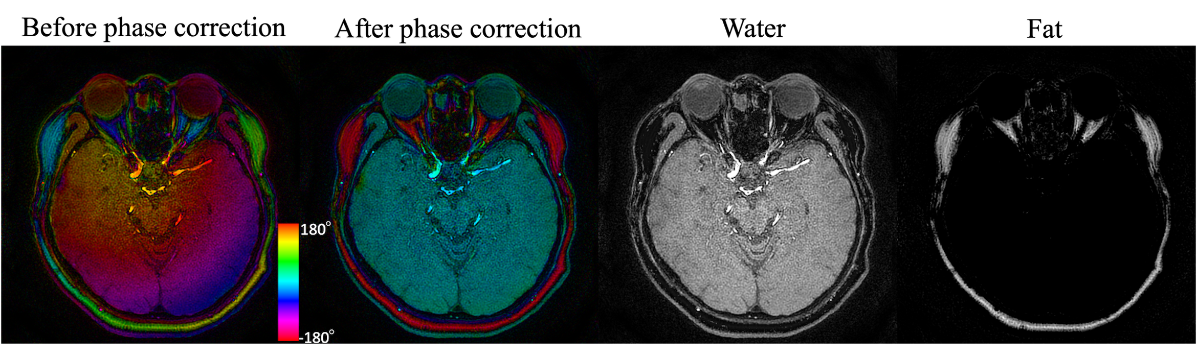
Figure 2 Demonstration of phase-sensitive water-fat separation in a healthy volunteer at 3T. The phase of the coil-combined OP images was estimated and corrected. After the phase correction, the phase of water and fat is close to 0o and 180o respectively, which can be used to obtain the water and fat images. Note that the color of the left two images represents phase. OP: out-of-phase.
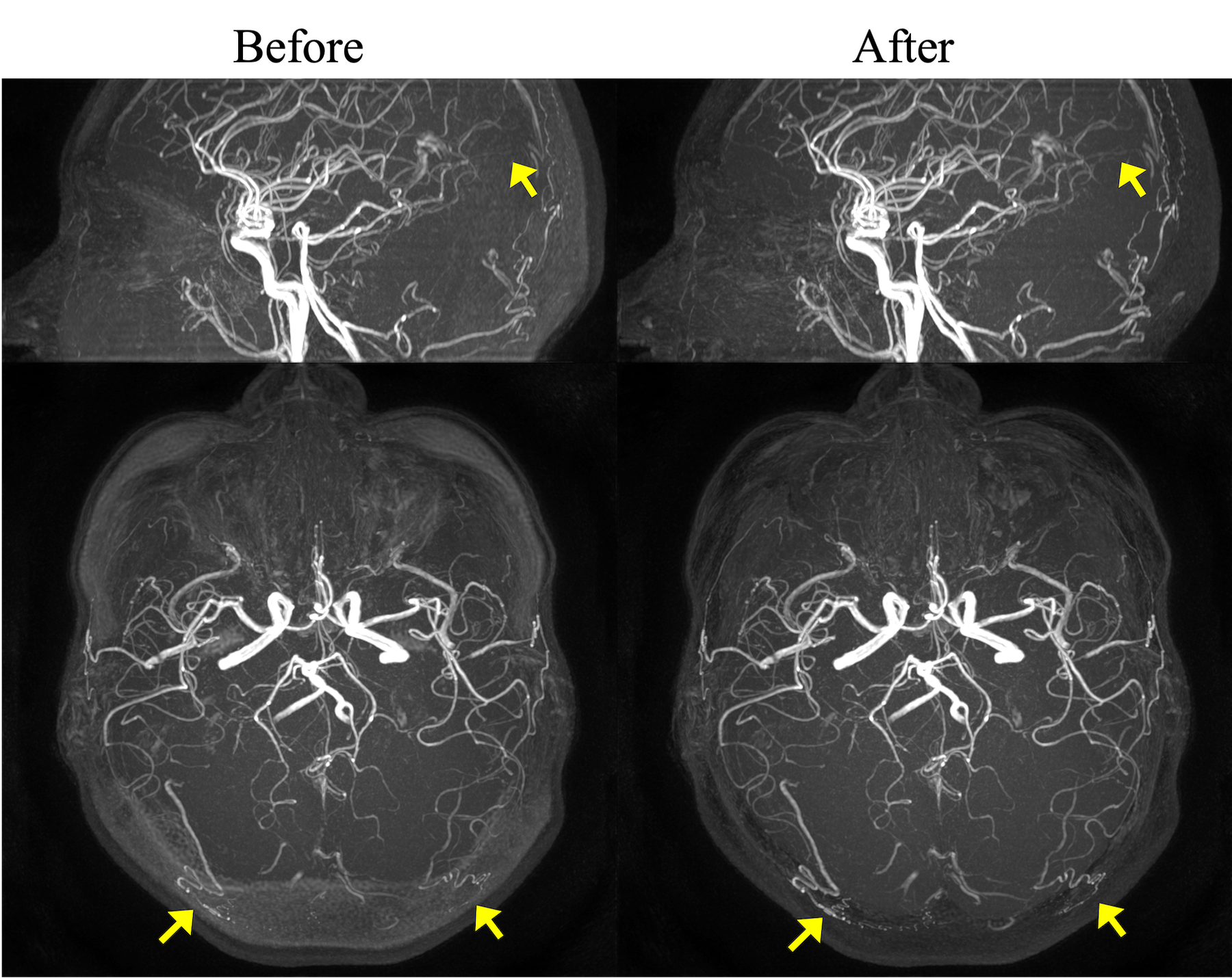
Figure 3 MIPs of the OP images and water images at 3T before and after the phase-sensitive water-fat separation. The proposed method reveals small vessels (arrows) that are otherwise not visible in OP MIPs. MIP: maximum intensity projection; OP: out-of-phase.
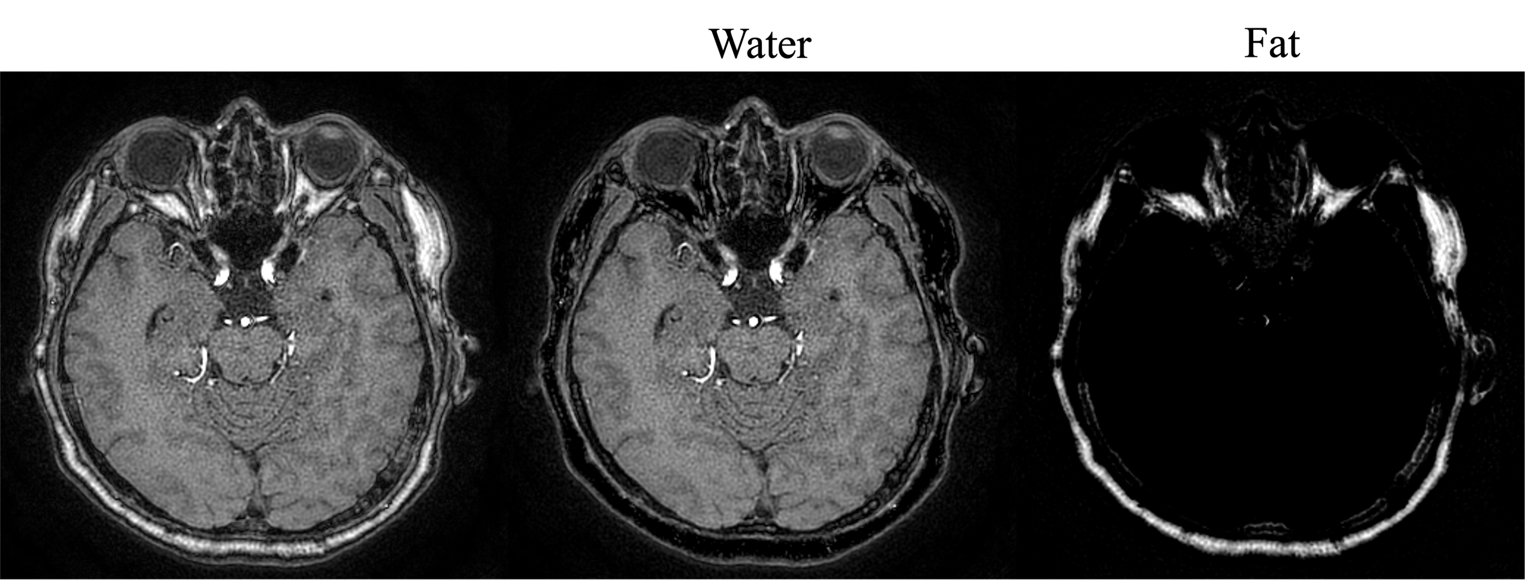
Figure 4 Results of images of another volunteer data at 1.5 T. OP: out-of-phase.
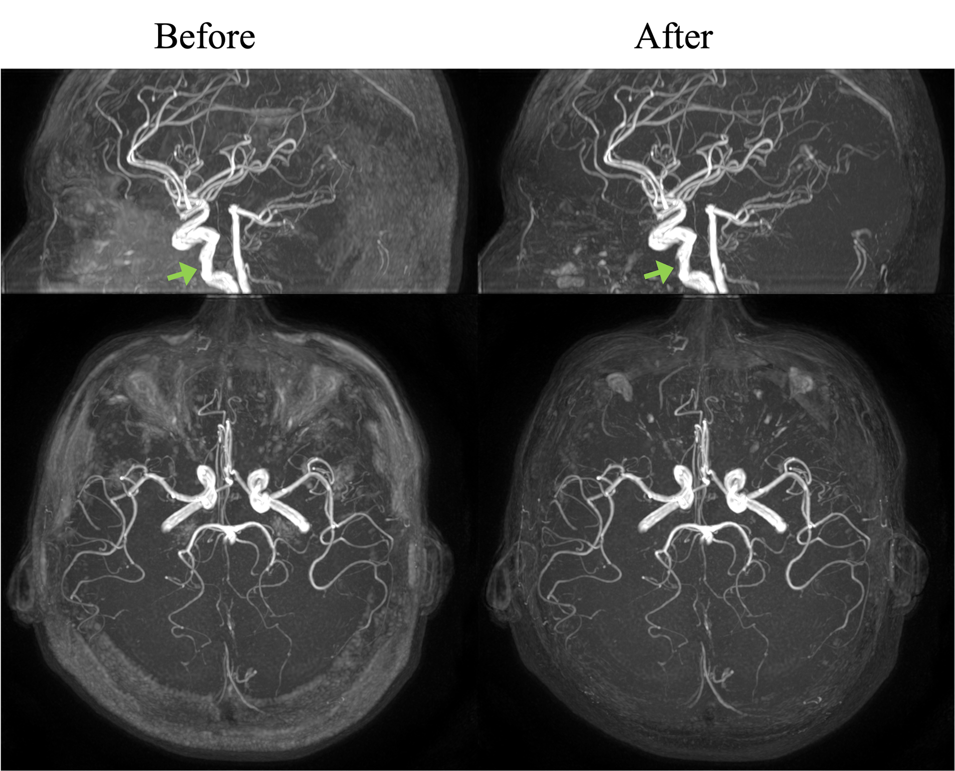
Figure 5 MIPs of the OP images and water images at 1.5 T before and after the phase-sensitive water-fat separation. Reduced flow signal (green arrows) is observed with the proposed method. MIP: maximum intensity projection. OP: out-of-phase.