3349
Enhancing reliability in TOF-MRA: minimizing spin-dephasing effects and improving fat suppression1Neusoft Medical Systems, Shanghai, China
Synopsis
Keywords: Vascular, Blood vessels, vessels; TOF; Angiography
Motivation: Clinicians express concerns about false-positive findings, leading to uncertainties in the reliable interpretation of TOF-MRA.
Goal(s): This study focuses on improving the reliability of TOF-MRA for cerebrovascular imaging.
Approach: By incorporating a small flip-angle spatially-selective fat-suppression and utilizing the shortest echo-time, signal loss is minimized, and uniform fat-suppression is achieved.
Results: The results, compared to conventional protocols, demonstrate superior quality and reliability by reducing spin-dephasing and achieving uniform fat-suppression without compromising blood signal quality or requiring extensive post-processing. This approach effectively mitigates the risk of false-positive findings and overestimation of stenosis, potentially establishing it as a more effective routine clinical examination for MRA.
Impact: By improving the reliability of TOF-MRA through optimized fat-suppression and echo-time reduction, it enhances the accuracy of evaluating cerebrovascular abnormalities. This advancement enables clinicians to have greater confidence in interpreting TOF-MRA images and reduces the risk of false-positive findings.
Introduction
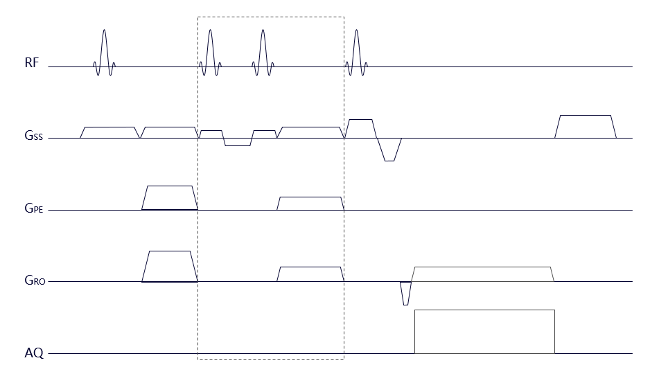
Time-of-flight MR angiography (TOF-MRA) has long been the most widely utilized noninvasive tool for cerebrovascular imaging. Nonetheless, clinicians occasionally raise concerns about reliable interpretation, particularly the false positive findings due to the intra-vascular signal loss, which can undermine their confidence in excluding anatomical abnormalities or hemodynamic issues effectively[1]. Current understanding suggests these signal losses are commonly attributed to spin-dephasing caused by factors such as long echo time, hemodynamic complexities and local field distortions. Additionally, incomplete and non-uniform fat suppression can further hinder vessel visibility, especially in maximum intensity projection (MIP) angiograms.In our pursuit to enhance the reliability and performance of TOF-MRA, we explored the implementation of a spatial selective fat suppression module positioned after the tracking vein saturation pulse and immediately preceding the spoiled GRE imaging (Figure 1). This strategic placement inherently leads to a reduction in echo time, effectively minimizing spin-dephasing effects and facilitating a more uniform fat suppression. To assess the effectiveness of the new strategy, we compared the results with several conventional TOF-MRA protocols utilizing different fat saturation techniques, including out-phase echo time, water excitation [2], spectral-selective fat suppression and shortest TE with manual skull and fat scraping in post-processing.
Methods
In our new strategy, the spatial selective fat suppression module utilizes the simplest binomial pulse (1-1) with inter-pulse delay (tau=2.2ms) chosen to allow 180°phase evolution between water and fat spins. The first RF pulse (α°) rotates both fat and water magnetization toward the transverse plane, while the second inverted RF pulse (-α°) further tips fat protons down towards the transverse plane and water protons back up to the longitudinal axis. The value for the flip angle α° was initially optimized as 15° (data not shown).Twenty healthy subjects were scanned on a 1.5T MRI scanner using a 24-channel head and neck coil. The scan parameters were set as follows: TR 23ms; FOV 200X180X180 mm3, voxel size 0.8X0.8X1.2 mm3, flip angle 21°, and the scan time of 3min54s. A TE of 2.59/6.9/4.21/2.59/2.59 was employed in our new methods, out-phase echo time, water excitation, spectral-selective fat suppression and direct shortest TE without fat suppression, respectively. The TOF angiograms were reconstructed using the maximum intensity projection for all methods, except for the last method, which required additional post-processing involving manually scraping of the background tissue due to the absence of fat suppression.
Results
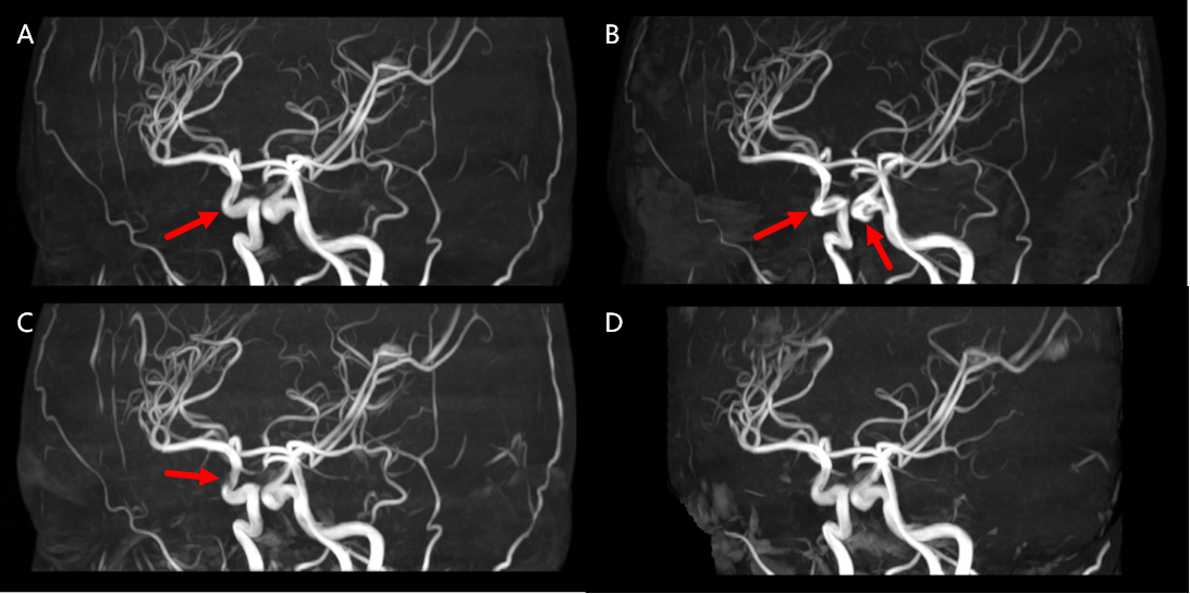
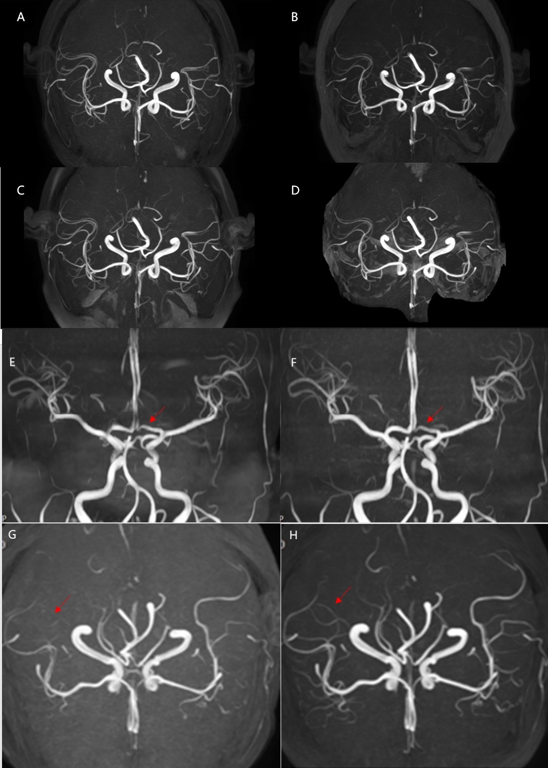
After conducting a thorough comparison of all the images, we found the described method outperformed the conventional TOF-MRA in various aspects. Figure 2 highlights the improvement in uniformity and continuity of the cerebrovasculature achieved through the implementation of the new strategy, when compared to TOF-MRA using out-phase TE and water excitation module. Furthermore, the vascular image quality obtained with our new method is comparable to that of TOF-MRA with the shortest TE and without fat suppression. Figure 3 depicts the notable reduction in false positive findings (signal loss in the anterior cerebral artery) and the enhanced uniformity of fat suppression when compared to TOF-MRA utilizing water excitation and spectral-selective fat suppression techniques.Discussion and Conclusion
The reduction of echo time has proven to be highly effective in minimizing the spin-dephasing effects in TOF-MRA. Although scans utilizing direct shortest TE without fat suppression successfully provided clear arterial depiction, the manual removal of background signals by operators can be time-consuming and impractical for consistent clinical workflow. The signal loss observed in TOF MRA with water excitation can be attributed to flow dephasing between binomial pulses. Furthermore, the non-spatial spectral-selective fat suppression technique may lead to suppression of vessel signals due to imperfect frequency tuning caused by field inhomogeneity, as well as the saturation of blood flow in the feeding arteries located outside the homogenous magnetic field region. In conclusion, the incorporation of the optimized small flip-angle spatial-selective fat suppression module with shortest echo time TOF MRA allows us to benefit from minimum spin dephasing and uniform fat suppression without compromising the flowing blood signal or requiring additional post-processing efforts. The results of this project establish the superiority of the new strategy in enhancing the quality and reliability of cerebrovascular imaging compared to conventional TOF-MRA techniques. The investigated method holds significant potential for implementation as a routine clinical examination for MRA, effectively mitigating the risk of overestimation of stenosis.Acknowledgements
No acknowledgement found.References
[1] Okahara M, Kiyosue H, Yamashita M, Nagatomi H, Hata H, Saginoya T, Sagara Y, Mori H. Diagnostic accuracy of magnetic resonance angiography for cerebral aneurysms in correlation with 3D-digital subtraction angiographic images: a study of 133 aneurysms. Stroke. 2002 Jul;33(7):1803-8
[2] Gizewski ER, Ladd ME, Paul A, Wanke I, Goricke S, Forsting M. Water excitation: a possible pitfall in cerebral time-of-flight angiography. AJNR Am J Neuroradiol 2005;26:152–155
Figures