3347
Feasibility of Local Stiffness Measurement for Intracranial Arteries Using Time-resolved 3D Black-blood Cine MRI1Department of Radiology and Imaging Sciences, University of Utah, Salt Lake City, UT, United States, 2Computer Vision, Mohamed bin Zayed University of Artificial Intelligence, Abu Dhabi, United Arab Emirates, 3University of Washington, Seattle, WA, United States, 4Department of Computer Science, Utah Valley University, Orem, UT, United States, 5Cardiovascular Medicine Division, Internal Medicine Department, University of Utah, Salt Lake City, UT, United States
Synopsis
Keywords: Flow, Vessels
Motivation: Local stiffness of intracranial arteries may provide regional assessment of vessel changes and information about vessel pathologies, but so far there is no reliable method to measure it.
Goal(s): To propose a black-blood MRI technique with submillimeter isotropic resolution and multiple cardiac phases, and to explore its feasibility of measuring local stiffness of intracranial arteries.
Approach: A novel time-resolved 3D black-blood cine MRI was proposed combining MERGE, golden-angle radial, retrospective gating, and GRASP reconstruction. MOCHA pipeline was used to measure cardiac-driven lumen changes.
Results: Images obtained with our proposed technique can capture cardiac-driven lumen area changes that are essential for local stiffness measurement.
Impact: The proposed method, after validation, can serve as a unique local stiffness measurement tool for intracranial arteries that will highly benefit vascular imaging studies.
Background
Arterial stiffness is a key indicator of vascular aging, and it was recently shown to be an independent risk factor for cognitive impairment and dementia1,2. Traditionally, stiffness is evaluated in large arteries such as the aorta and carotid artery by measuring pulse wave velocity (PWV) using applanation tonometry or ultrasound devices. However, evaluating the large artery stiffness alone does not provide insights into the local stiffness of intracranial arteries.Recently, several methods for measuring regional pulse wave velocity (PWV) in intracranial arteries have been proposed, using transcranial doppler (TCD)3 or 4D flow MRI4, but they cannot measure local stiffness in each particular artery location. In this study, we aim to propose a novel time-resolved 3D black-blood cine MRI, and explore the feasibility of using it to measure local stiffness in intracranial arteries.
Methods
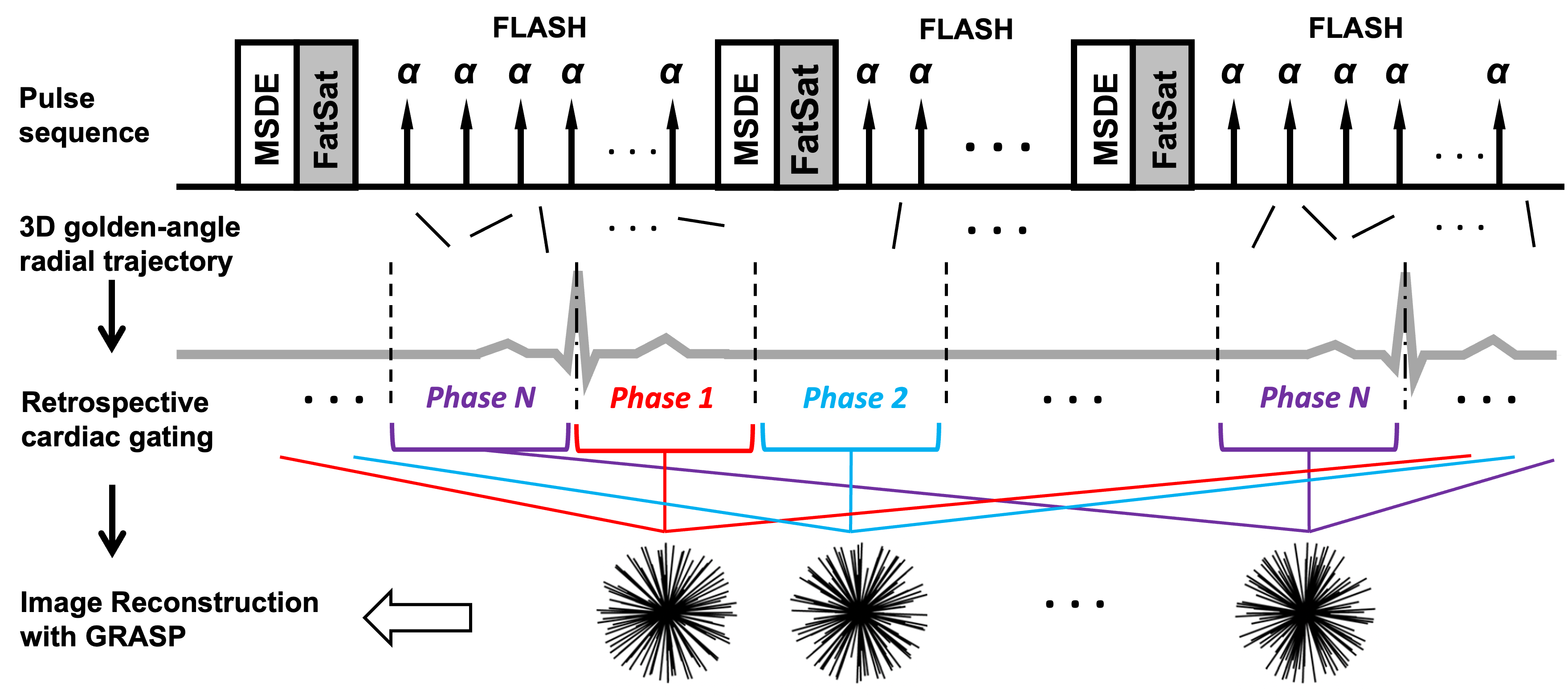
Pulse sequence and image reconstruction: In this study, we propose a time-resolved 3D black-blood cine MRI technique (Fig. 1). The pulse sequence combines 3D motion-sensitizing driven equilibrium (MSDE) prepared FLASH, a.k.a., MERGE5, and 3D golden-angle radial acquisition. Retrospective cardiac gating is achieved by re-organizing the k-space data into several cardiac phases. Images are reconstructed offline using GRASP6 by solving the following equation exploiting the sparsity along different phases, i.e., temporal total variation (TTV):$$f\left(x \right)=||F\cdot S\cdot x||^2_2+\lambda||TTV\cdot x||_1$$This time-resolved 3D black-blood cine MRI technique is similar to the one proposed in a previous study7, except that a 3D golden-angle radial trajectory is used instead of 3D Cartesian. Different from the 4D MERGE using prospective gating proposed in another study8, our method uses retrospective cardiac gating so it is more suitable for cine MRI.
Image processing pipeline: Data were processed following the MOCHA (Multi-planar, multi-contrast and multi-time point analysis tOol for intracranial vessel wall CHAracterization) pipeline9 using 3D Slicer (https://www.slicer.org/), including 1) artery tracing and labeling on bright-blood TOF images, 2) image registration between TOF and black-blood cine images, and 3) generation of MPR views for different arteries and cardiac phases. Then the lumen contour for each 2D cross-sectional image was obtained using an AI-based automatic segmentation algorithm10.
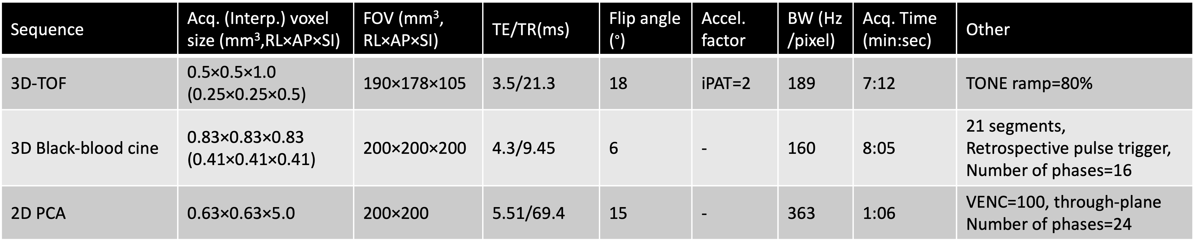
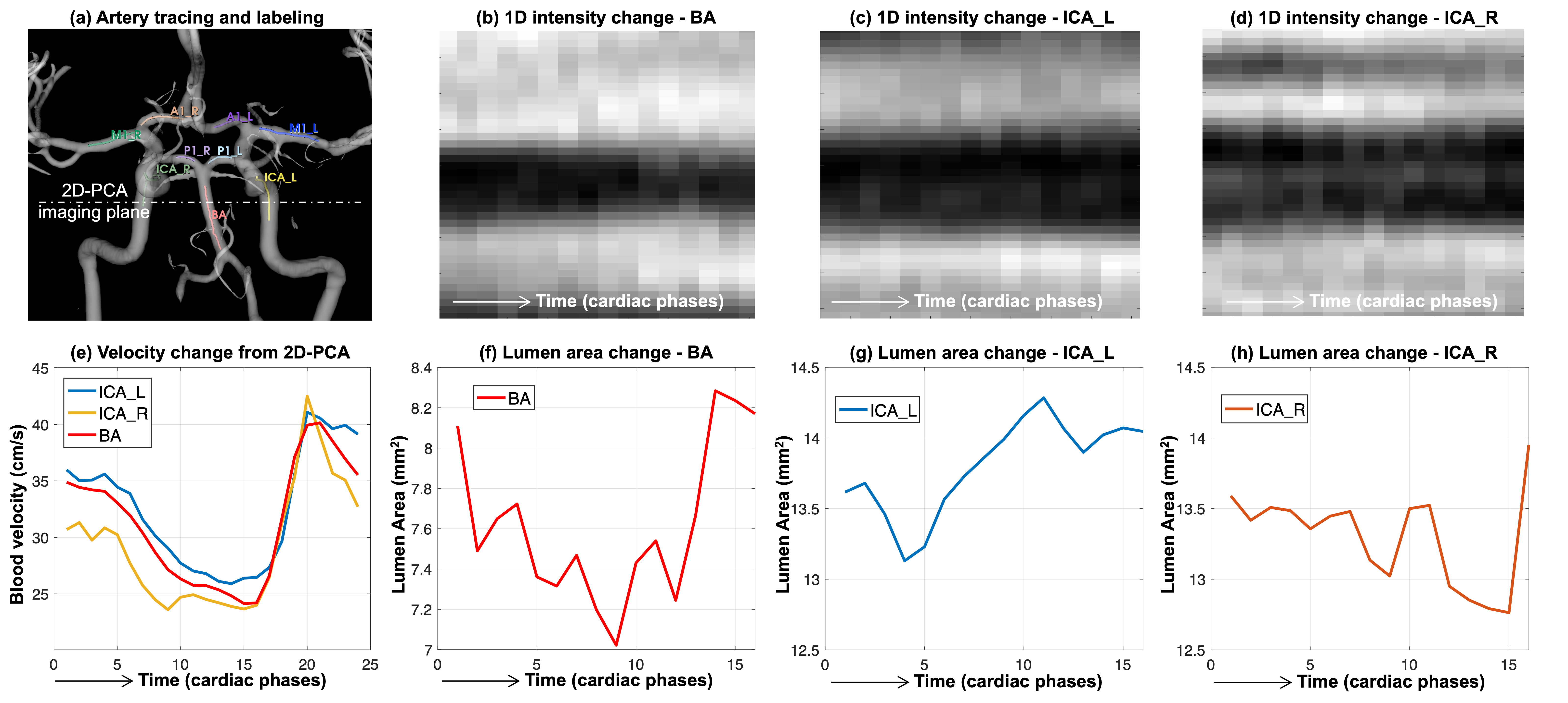
In vivo experiments: MRI data were collected on a healthy volunteer (Female, 21 years old) on a 3.0T Prisma-fit scanner (Siemens, Erlangen, Germany), using a 32-ch head coil. This experiment was approved by local IRB and written consent was obtained from the volunteer. Data were acquired using 3 sequences: 1) 3D TOF covering the Circle of Willis, 2) proposed time-resolved 3D black-bood cine covering the whole brain (0.83mm isotropic resolution, 16 phases, gated with finger pulse), and 3) 2D PCA with the imaging plane vertical to the internal carotid artery (ICA) and basilar artery (BA) (Fig. 4a). The imaging parameters can be found in Table 1.
The lumen changes among cardiac phases were obtained from black-blood images using the pipeline described above. The blood velocities in ICA and BA were calculated by averaging manually-drawn ROIs based on the 2D PCA data.
Results and Discussion
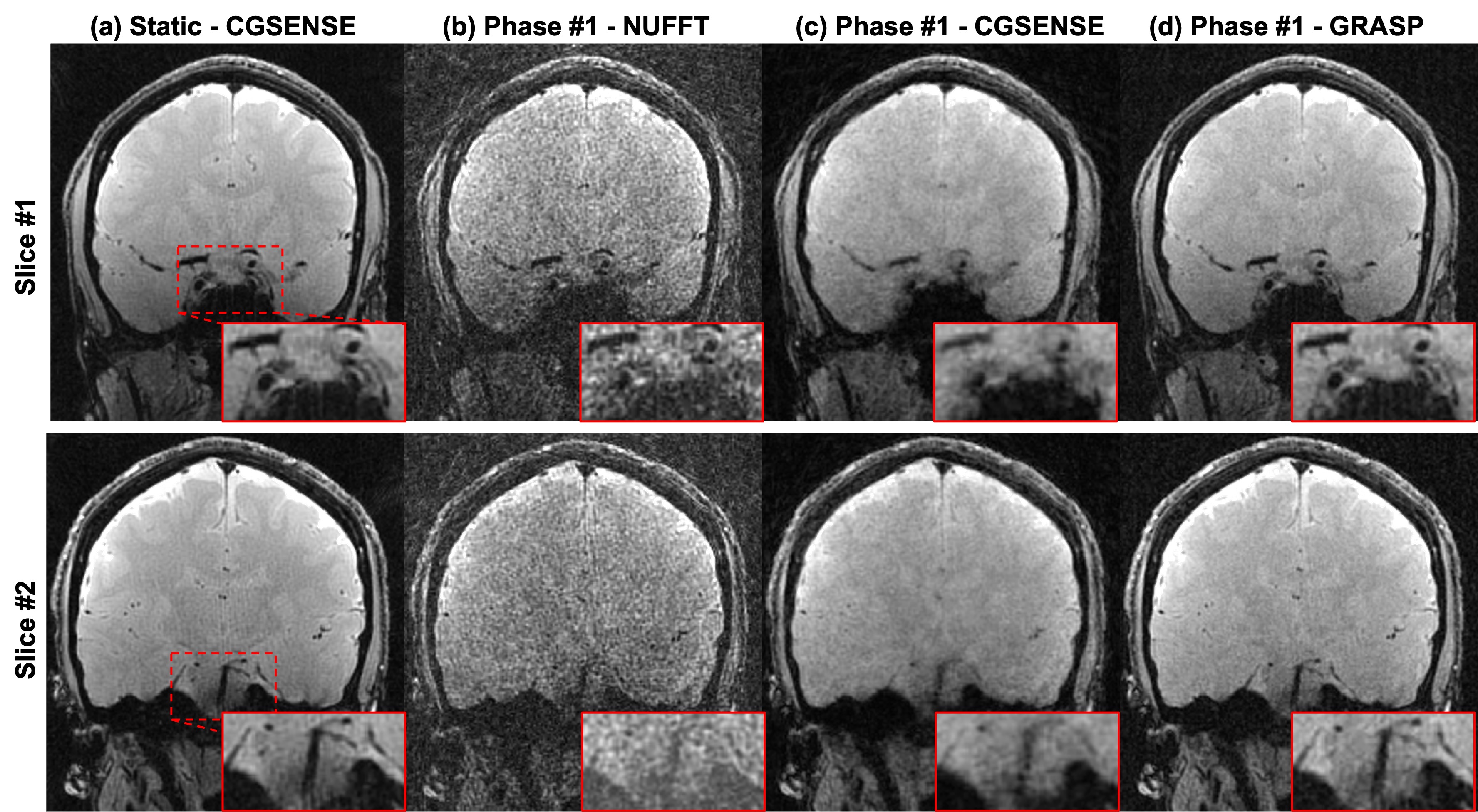
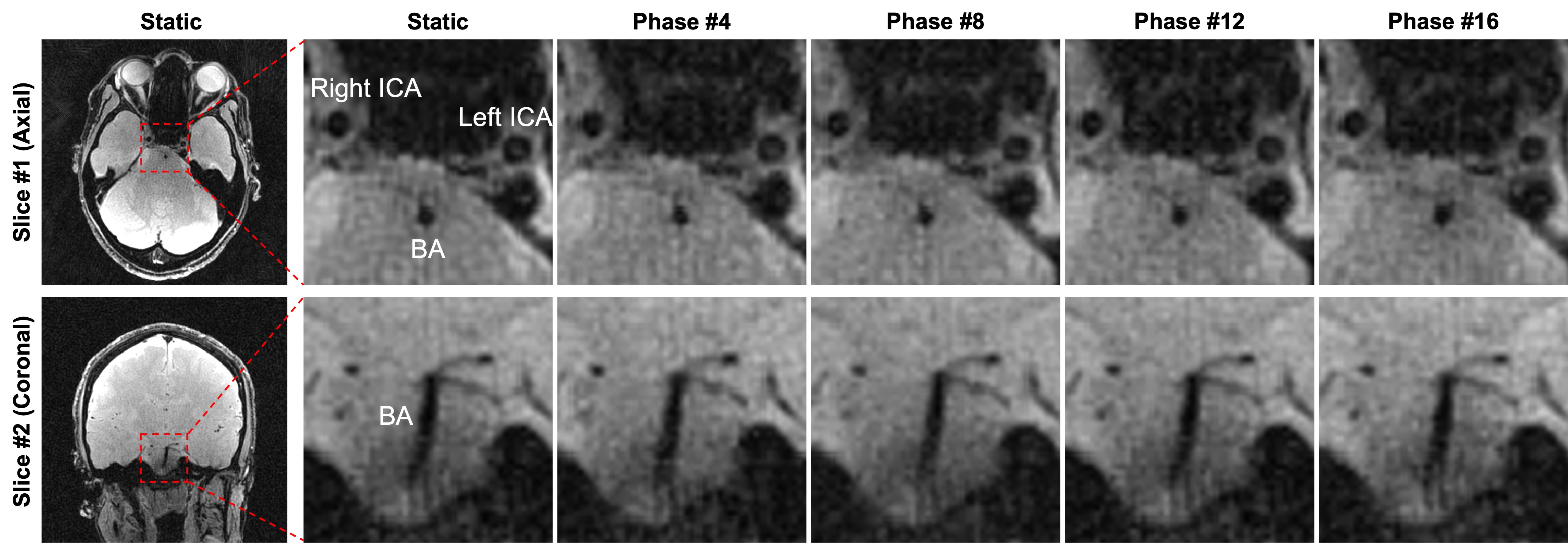
Both 3D static and dynamic black-blood images can be reconstructed from the proposed time-resolved 3D cine MRI, shown in Fig. 1. For the single-phase reconstruction, GRASP provides fewer aliasing artifacts and sharper lumen edges compared with NUFFT11 and CGSENSE12. Fig. 2 shows zoomed static and dynamic images of different cardiac phases, demonstrating that images of different cardiac phases have sharp lumen edges for major intracranial arteries, including ICA and BA.Fig. 3 shows the lumen changes of major intracranial arteries (ICA and BA) measured from the proposed 3D black-blood cine MRI. Both 1D signal intensities (b-d) and lumen areas (f-h) exhibited cardiac-driven changes, which are consistent with the blood velocity curves (e).
Our proposed 3D black-blood cine MRI can capture cardiac-driven lumen area changes (~15% in ICA and BA for this volunteer, equivalent to ~3 pixels). If the blood pressure can be measured, the local stiffness of each artery location can be assessed by calculating the vessel compliance: dividing the difference between maximum and minimum lumen areas by the pulse pressure.
Future work includes 1) reconstruction optimization by acquiring fully-sampled reference data on a flow phantom or a volunteer, 2) scan-rescan reproducibility test, and 3) validation of local stiffness measurement of intracranial arteries by acquiring data on volunteers in different age groups.
Conclusion
We have proposed a time-resolved 3D black-blood cine MRI technique that can generate whole-brain cine images with 0.83mm isotropic resolution and 16 cardiac phases in 8 mins. The images can capture cardiac-driven lumen area changes which are essential for local stiffness measurement.Acknowledgements
This study was supported by the 2023 radiology seed grant in our institute.References
1. Hughes TM, Wagenknecht LE, Craft S, Mintz A, Heiss G, Palta P, Wong D, Zhou Y, Knopman D, Mosley TH. Arterial stiffness and dementia pathology: Atherosclerosis Risk in Communities (ARIC)-PET Study. Neurology. 2018;90:e1248-e1256.
2. Poels MM, van Oijen M, Mattace-Raso FU, Hofman A, Koudstaal PJ, Witteman JC, Breteler MM. Arterial stiffness, cognitive decline, and risk of dementia: the Rotterdam study. Stroke. 2007;38:888-892.
3. Fu X, Huang C, Wong KS, Chen X, Gao Q. A new method for cerebral arterial stiffness by measuring pulse wave velocity using transcranial Doppler. Journal of atherosclerosis and thrombosis. 2016;23:1004-1010.
4. Rivera-Rivera LA, Cody KA, Eisenmenger L, Cary P, Rowley HA, Carlsson CM, Johnson SC, Johnson KM. Assessment of vascular stiffness in the internal carotid artery proximal to the carotid canal in Alzheimer’s disease using pulse wave velocity from low rank reconstructed 4D flow MRI. Journal of Cerebral Blood Flow & Metabolism. 2021;41:298-311.
5. Balu N, Yarnykh VL, Chu B, Wang J, Hatsukami T, Yuan C. Carotid plaque assessment using fast 3D isotropic resolution black‐blood MRI. Magnetic resonance in medicine. 2011;65:627-637.
6. Feng L, Grimm R, Block KT, Chandarana H, Kim S, Xu J, Axel L, Sodickson DK, Otazo R. Golden-angle radial sparse parallel MRI: combination of compressed sensing, parallel imaging, and golden-angle radial sampling for fast and flexible dynamic volumetric MRI. Magn Reson Med. 2014;72:707-717. doi: 10.1002/mrm.24980
7. Balu N, Zhang K, Hatsukami T, Yuan C. 3D Isotropic black-blood cine MRI of intracranial arteries. Paper/Poster presented at: Proc. Intl. Soc. Mag. Reson. Med; 2023;
8. Koktzoglou I. 4D Dark blood arterial wall magnetic resonance imaging: methodology and demonstration in the carotid arteries. Magnetic Resonance in Medicine. 2013;69:956-965.
9. Guo Y, Canton G, Chen L, Sun J, Geleri DB, Balu N, Xu D, Mossa‐Basha M, Hatsukami TS, Yuan C. Multi‐Planar, Multi‐Contrast and Multi‐Time Point Analysis Tool (MOCHA) for Intracranial Vessel Wall Characterization. Journal of Magnetic Resonance Imaging. 2022;56:944-955.
10. HashemizadehKolowri S, Nadin, Canton G, Balu N, Hatsukami T, Yuan C. Automated Localization of the Extracranial Carotid Artery in Black Blood Contrast MR Images Using a Deep Learning Approach. Paper/Poster presented at: Proc. Intl. Soc. Mag. Reson. Med; 2023;
11. Knoll F, Schwarzl A, Diwoky C, Sodickson DK. gpuNUFFT-an open source GPU library for 3D regridding with direct Matlab interface. Paper/Poster presented at: Proceedings of the 22nd annual meeting of ISMRM, Milan, Italy; 2014;
12. Pruessmann KP, Weiger M, Börnert P, Boesiger P. Advances in sensitivity encoding with arbitrary k‐space trajectories. Magnetic Resonance in Medicine: An Official Journal of the International Society for Magnetic Resonance in Medicine. 2001;46:638-651.
Figures