3346
Correlation Between Morphological Measurements of Intra- and Extracranial Vessel Wall and Whole-Body Fat Distribution Characteristics1Sanya Central Hosptial(Hainan Third People's Hospital), sanya, China, 2Lauterbur Research Center for Biomedical Imaging, Shenzhen Institute of Advanced Technology, Chinese Academy of Sciences, shenzhen, China, 3United Imaging Research Institute of Innovative Medical Equipment, shenzhen, China, 4Key Laboratory of Biomedical Imaging Science and System, Chinese Academy of Sciences, shenzhen, China
Synopsis
Keywords: Vessel Wall, Data Analysis, Vessel Wall Morphological Measurements; Whole-Body Fat Distribution
Motivation: Obesity is associated with cardiovascular diseases. However, its effect on cerebrovascular disease remains unclear.
Goal(s): To investigate the correlation between morphological measurements of intra- and extracranial arterial vessel walls and whole-body fat distribution characteristics.
Approach: A total of 98 volunteers underwent three-dimensional high-resolution MR vessel wall imaging and whole-body MR proton density fat fraction (PDFF) imaging on a 3.0T MRI scanner.
Results: The results indicate that the vessel wall thickness of both the extracranial carotid artery and vertebral artery is positively correlated with visceral adipose tissue and the liver-PDFF.
Impact: The assessment of vascular health in individuals with central obesity necessitates a comprehensive evaluation of both extracranial carotid arteries and vertebral arteries. This paradigm shift may have far-reaching implications for the prevention and treatment of cerebrovascular diseases associated with obesity.
Introduction
Atherosclerosis is the basis of cardiovascular disease. Studies have shown that whole body fat, especially visceral adipose tissue, is associated with atherosclerosis risk[1-3]. However, previous studies have focused on the extracranial carotid arteries and thoracic aorta, ignoring the intracranial arteries[4,5]. MRI is the preferred method for quantifying total body fat and assessing cardiovascular diseases, especially strokes. It offers accurate measurements of the arterial vessel wall and whole body fat distribution[6]. Therefore, the present study aims to investigate the correlation between morphological measurements of intra- and extracranial arterial vessel walls and body fat distribution characteristics.Methods
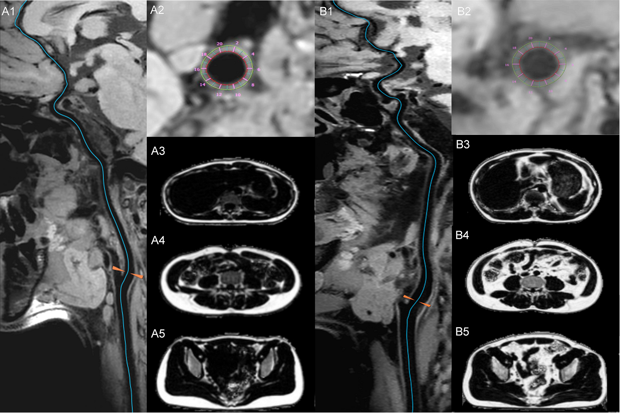
3D high-resolution intra- and extracranial arterial vessel wall images and whole-body MR proton density fat fraction (PDFF) images were acquired from 98 healthy volunteers aged between 21 and 78 years on a 3T MRI scanner (uMR790, United Imaging Healthcare, China). The morphological quantitative parameters of mean and maximum vessel wall thickness and mean vessel wall area were automatically calculated on seven vessel segments, including bilateral common carotid arteries (CCA), proximal internal carotid artery (ICA-C1), distal internal carotid artery (ICA-C4), A1 segment of the anterior cerebral artery (ACA), M1 segment of the middle cerebral artery (MCA), distal vertebral arteries (VA), and distal basilar arteries (BA), for each subject using dedicated plaque analysis software (uWS-MR). Furthermore, a deep learning-based fully automatic fat segmentation method was applied to segment and quantify total body fat, including total adipose tissue (TAT), internal adipose tissue (IAT), and subcutaneous adipose tissue (SAT). In addition, visceral adipose tissue (VAT), abdominal fat tissue (AFT), abdominal subcutaneous fat (ASF), and liver PDFF were also calculated. The whole body volume (WB) and abdominal volume (aWB) of the volunteers were also estimated. Subsequently, seven quantitative indices were normalized, comprising three representing characteristics of whole-body fat distribution (TAT/WB, IAT/WB, SAT/WB) and four representing characteristics of abdominal fat distribution (VAT/WB, AFT/aWB, VAT/aWB, ASF/aWB). All statistical analysis was performed using SPSS v25.0. Comparisons between two groups of quantitative data were conducted using either the independent samples t test or the Mann‒Whitney U test, with p < 0.05 considered statistically significant. Correlation analysis was carried out using Pearson or Spearman correlation analysis, with p < 0.01 considered statistically significant.Results
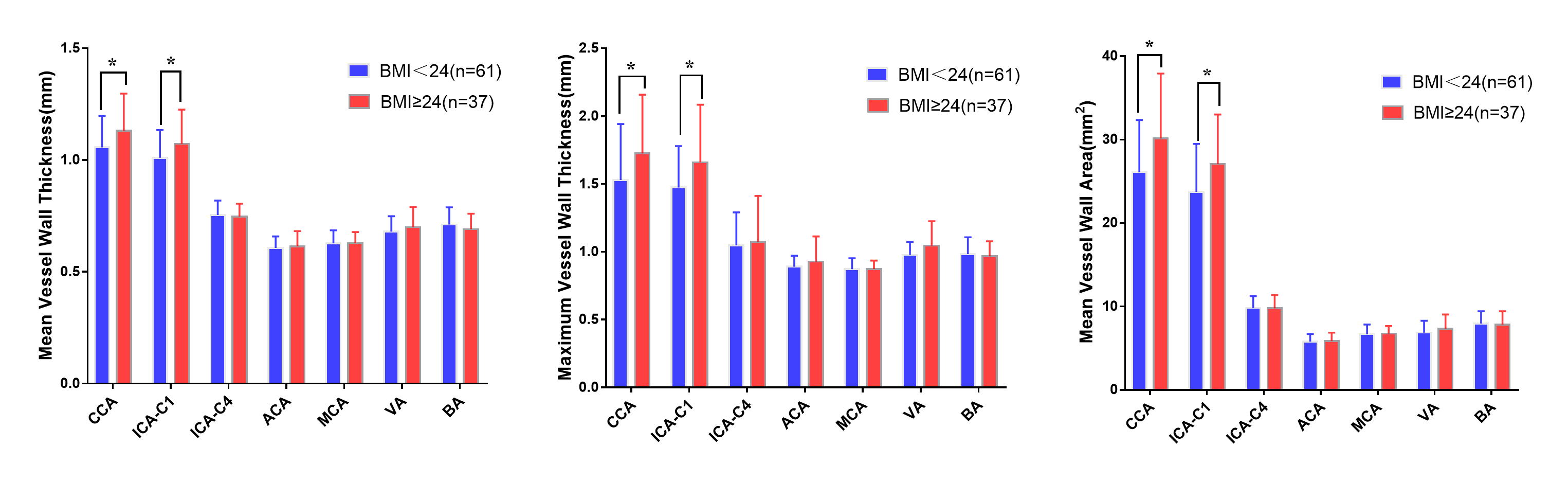
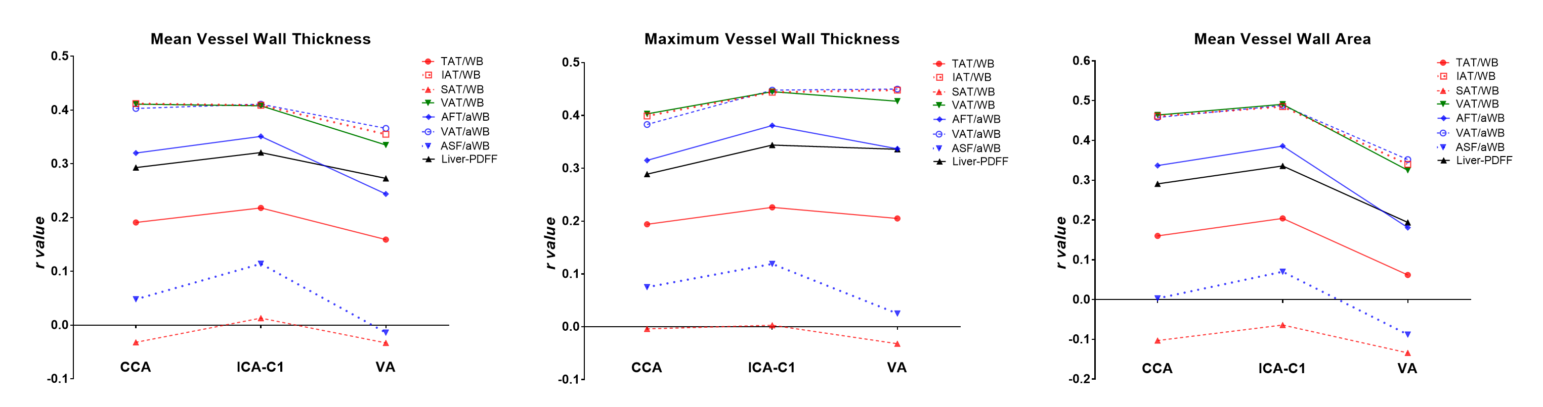
As shown in Figure 2, among these 98 volunteers, individuals with BMI≥24 exhibited significantly greater vessel wall thickness and vessel wall area in the CCA and ICA-C1 than individuals with BMI<24 (P<0.05). However, for the vascular segments of the ICA-C4, ACA, MCA, VA, and BA, there was no significant difference in the vessel wall morphological measurements between the two groups.For the ICA-V4, ACA, MCA, and BA segments, there was no significant correlation observed between the vessel wall morphological parameters and whole-body fat distribution characteristics. For CCA, ICA-C1, and VA, there was no significant correlation observed between the vessel wall morphological parameters and the TAT/WB, SAT/WB, and ASF/aWB volume ratios. However, a significant positive correlation (P < 0.01) was observed between these vessel wall morphological parameters and the volume ratio of IAT/WB, VAT/WB, AFT/aWB, VAT/aWB, Liver-PDFF except AFT/aWB and mean vessel wall thickness and area of VA, as well as PDFF and mean vessel wall area of VA (Figure 3). Among these, the VAT/WB volume ratio exhibited the highest correlation with vessel wall thickness, with an r-value of up to 0.491.
Discussion
In this study, the high BMI group exhibited significantly greater vessel wall thickness and area in the CCA and ICA-C1 than the low BMI group, further emphasizing the importance of BMI as a significant predictor of carotid artery wall thickness[7]. Unlike TAT and SAT, IAT, particularly VAT, shows a significant positive correlation with the vessel wall thickness and area of the CCA, ICA-C1, and VA. The accumulation of visceral adipose tissue around abdominal organs may lead to metabolic aberrations, inflammation, and atherosclerosis, consequently increasing arterial wall thickness.Furthermore, our study reveals a positive correlation between liver-PDFF and the thickness of extracranial carotid and intracranial vertebral artery walls. This indicates that hepatic fat deposition may have adverse effects on the health of these vascular walls[8].
In summary, IAT, especially VAT accumulation, and hepatic fat deposition may have an impact on the vessel wall thickness and area of extracranial carotid arteries and vertebral arteries.
Acknowledgements
The study was partially supported by the Natural Science Foundation of Guangdong Province-Outstanding Youth Project (2023B1515020002), National Key Technology Research and Development Program of China (2021YFF0501502), Key Laboratory for Magnetic Resonance and Multimodality Imaging of Guangdong Province (2023B1212060052), and Central Guidance for Local Science and Technology Development Project (ZYYD2023D02).References
[1] Després JP. Body fat distribution and risk of cardiovascular disease: an update. Circulation. 2012;126(10):1301-1313. doi:10.1161/CIRCULATIONAHA.111.067264
[2] Stranahan AM. Visceral adiposity, inflammation, and hippocampal function in obesity. Neuropharmacology. 2022;205:108920. doi:10.1016/j.neuropharm.2021.108920
[3] Tchernof A, Després JP. Pathophysiology of human visceral obesity: an update. Physiol Rev. 2013;93(1):359-404. doi:10.1152/physrev.00033.2011
[4] Lind L, Kullberg J, Ahlström H, Strand R. Relationships between carotid artery intima-media thickness and echogenicity and body composition using a new magnetic resonance imaging voxel-based technique. PLoS One. 2021;16(7):e0254732. Published 2021 Jul 23. doi:10.1371/journal.pone.0254732
[5] Lefferts WK, Sperry SD, Jorgensen RS, et al. Carotid stiffness, extra-media thickness and visceral adiposity in young adults. Atherosclerosis. 2017;265:140-146. doi:10.1016/j.atherosclerosis.2017.08.033
[6] Zhang Y, Guallar E, Malhotra S, et al. Carotid Artery Wall Thickness and Incident Cardiovascular Events: A Comparison between US and MRI in the Multi-Ethnic Study of Atherosclerosis (MESA). Radiology. 2018;289(3):649-657. doi:10.1148/radiol.2018173069
[7] De Michele M, Panico S, Iannuzzi A, et al. Association of obesity and central fat distribution with carotid artery wall thickening in middle-aged women. Stroke. 2002;33(12):2923-2928. doi:10.1161/01.str.0000038989.90931.be
[8] Peng H, Wang S, Wang M, et al. Nonalcoholic fatty liver disease and cardiovascular diseases: A Mendelian randomization study. Metabolism. 2022;133:155220. doi:10.1016/j.metabol.2022.155220
Figures