3344
T2-weighted imaging of the prostate with super-resolution deep learning reconstruction: impact on PI-QUAL assessment1Osaka University Graduate School of Medicine, Suita, Japan, 2Osaka University Hospital, Suita, Japan, 3Canon Medical Systems, Kawasaki, Japan, 4Canon Medical Systems, Otawara, Japan
Synopsis
Keywords: Prostate, Prostate
Motivation: Super-resolution deep learning reconstruction (SR-DLR) can simultaneously reduce noise and improve spatial resolution.
Goal(s): Our goal was to evaluate the usefulness of SR-DLR in prostate T2-weighted imaging (T2WI) with conventional and reduced acquisition times.
Approach: SR-DLR was applied to both conventional acquisition time T2WI and short acquisition time T2WI. Visibility of the anatomical structures of the prostate and image quality were evaluated.
Results: SR-DLR significantly improved image quality of prostate T2WI and visibility of detailed anatomical structures, especially in the small structures such as ejaculatory ducts.
Impact: SR-DLR improves T2WI image quality in prostate MRI and improves the visibility of detailed anatomical structures, and has the potential to reduce acquisition time while maintaining adequate image quality for diagnosis.
Introduction
T2-weighted imaging (T2WI) is an essential sequence for the morphological evaluation of the prostate and a dominant sequence in the diagnosis of transitional zone (TZ) cancers1. The Prostate Imaging Reporting and Data System (PI-RADS) recommends high spatial resolution imaging on T2WI, which requires a long time to obtain images with sufficient signal-to-noise ratio (SNR). Improving image quality, including T2WI, is essential in prostate MRI, and the recently proposed Prostate Imaging Quality (PI-QUAL) score2 has been reported to correlate with the PI-RADS-3 incidence, the positive predictive value in detecting clinically significant cancer, and the percentage of upgrades in postoperative pathology.Recently, deep learning reconstruction (DLR) has been introduced into clinical practice, and its usefulness in improving SNR and reducing acquisition time has been reported in prostate MRI. Super-resolution deep learning reconstruction (SR-DLR) is a newly developed technique that can simultaneously reduce noise and improve spatial resolution3. However, few reports have been published on the application of this technique to prostate MRI. The purpose of this study was to evaluate the usefulness of SR-DLR in prostate T2WI with conventional and reduced acquisition times.
Methods
This retrospective study included 30 patients who underwent prostate MRI including conventional acquisition time T2WI (c-T2WI) and short acquisition time T2WI (s-T2WI). The matrix sizes of c-T2WI and s-T2WI were 496 × 288 and 288 × 288, respectively. The acquisition times for c-T2WI and s-T2WI were 4 min 18 sec and 1 min 54 sec, respectively. Both T2WI were reconstructed using a conventional reconstruction technique (c-T2WIconv and s-T2WIconv) and SR-DLR (c-T2WIDLR and s-T2WIDLR).A radiologist placed regions of interest on the peripheral zone (PZ) and TZ of the prostate and the homogeneous area of the internal obturator muscle, and the signal intensity (SI) of the PZ and TZ and the standard deviation (SD) of the SI of the muscle were measured. The SNR was calculated by dividing the SI of the prostate by the SD of the muscle and compared between 4 images (c-T2WIconv, c-T2WIDLR, s-T2WIconv, and s-T2WIDLR) using Friedman’s test followed by post-hoc Wilcoxon signed rank test with Bonferroni correction (n = 6). As a qualitative analysis, two radiologists blinded to the acquisition and reconstruction methods reviewed the images and assigned scores on a 5-poin scale for visibility of the capsule, seminal vesicles, ejaculatory ducts, neurovascular bundles, and sphincter muscle, which are assessment items in PI-QUAL. They also rated image noise, artifact, and overall image quality on a 5-point scale.
Results
Quantitative Analysis:The results of the quantitative analysis are summarized in Figure 1. SR-DLR images showed significantly improved SNR compared to conventional images in both c-T2WI and s-T2WI (P < 0.01).
Qualitative Analysis:
The results of the assessment of the visibility of anatomical structures are shown in Figure 2. In Reader 1, the scores for SR-DLR images were significantly higher than conventional images for all assessments in both c-T2WI and s-T2WI (P < 0.01). In Reader 2, SR-DLR image scores for c-T2WI and s-T2WI were significantly higher than those for s-T2WI conventional images in the evaluation of the ejaculatory ducts (P < 0.05).
The results of the image quality assessment are shown in Figure 3. Both readers scored SR-DLR images higher than conventional images in all evaluations in both c-T2WI and s-T2WI (P < 0.05).
Representative cases are shown in Figures 4 and 5.
Discussion
Our results showed that SR-DLR reduced image noise and improved SNR in both c-T2WI and s-T2WI. In addition, SR-DLR images scored significantly higher in visual assessment than conventional reconstruction images. The visibility of anatomical structures tended to improve with SR-DLR, and in particular, the visibility of the ejaculatory ducts was significantly improved on c-T2WI and s-T2WI SR-DLR images compared to s-T2WI conventional images in both readers. This suggests that even in images with reduced spatial resolution due to reduced acquisition time, the use of SR-DLR improves the visibility of detailed anatomical structures, suggesting the possibility of reducing the acquisition time while maintaining the PI-QUAL scores. This is a preliminary study with a relatively small number of cases, and further evaluation of the clinical utility of this technique, including its detectability of prostate cancers, will be needed with a larger number of cases.Conclusion
SR-DLR improves T2WI image quality in prostate MRI and improves the visibility of detailed anatomical structures such as ejaculatory ducts. SR-DLR has the potential to reduce acquisition time while maintaining adequate image quality for diagnosis.Acknowledgements
No acknowledgement found.References
1. Turkbey B, Rosenkrantz AB, Haider MA, et al. Prostate Imaging Reporting and Data System Version 2.1: 2019 Update of Prostate Imaging Reporting and Data System Version 2. Eur Urol. 2019;76:340-351.
2. Giganti F, Allen C, Emberton M, Moore CM, Kasivisvanathan V; PRECISION study group. Prostate Imaging Quality (PI-QUAL): A New Quality Control Scoring System for Multiparametric Magnetic Resonance Imaging of the Prostate from the PRECISION trial. Eur Urol Oncol. 2020;3:615-619.
3. Matsuo K, Nakaura T, Morita K, et al. Feasibility study of super-resolution deep learning-based reconstruction using k-space data in brain diffusion-weighted images. Neuroradiology. 2023;65:1619-1629.
Figures
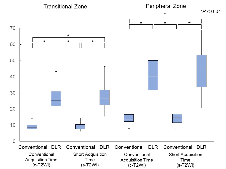
Signal-to-noise ratio of the peripheral and transitional zones of the prostate. In both zones the SNR was significantly improved by SR-DLR (P < 0.01).
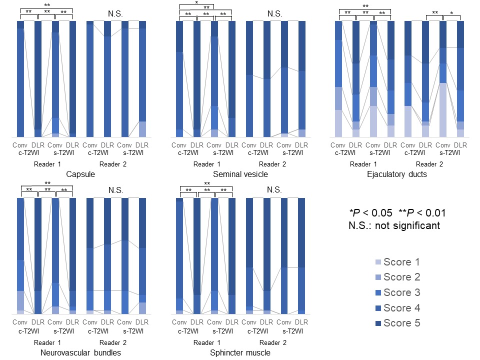
The graph shows the percentage of each score for the visibility of the capsule, seminal vesicles, ejaculatory ducts, neurovascular bundles, and sphincter muscle.
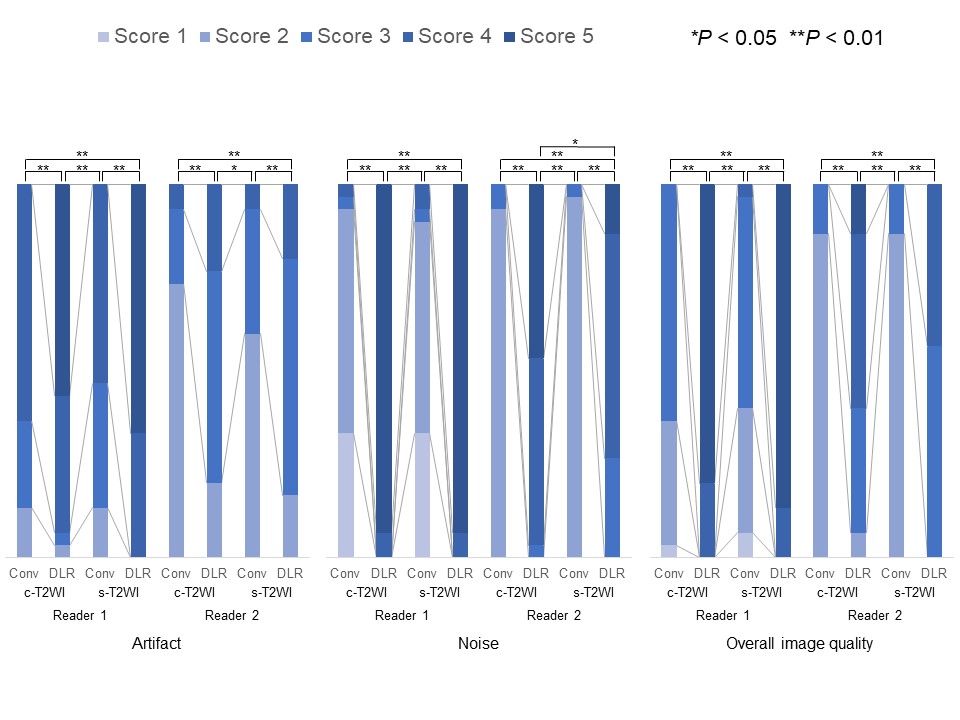
The graph shows the percentage of each score for the artifact, noise, and overall image quality.
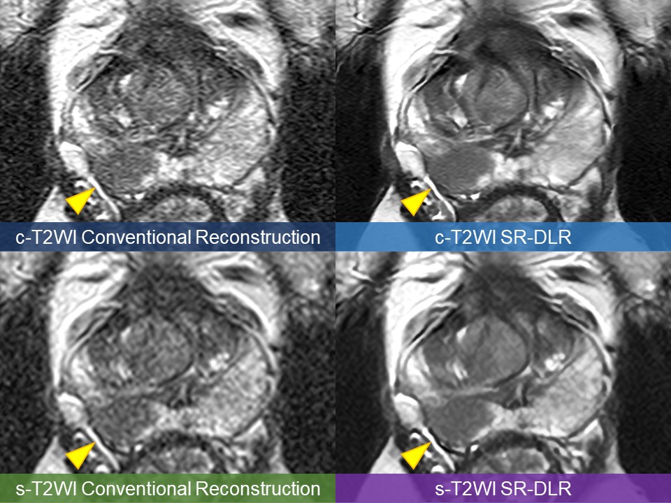
A case of prostate cancer (arrowheads). Image noise is reduced by SR-DLR in both c-T2WI and s-T2WI. Although the s-T2WI image is blurred due to the reduced spatial resolution, the SR-DLR image is sharpened, and the resolution is comparable to the c-T2WI with a conventional reconstruction technique.
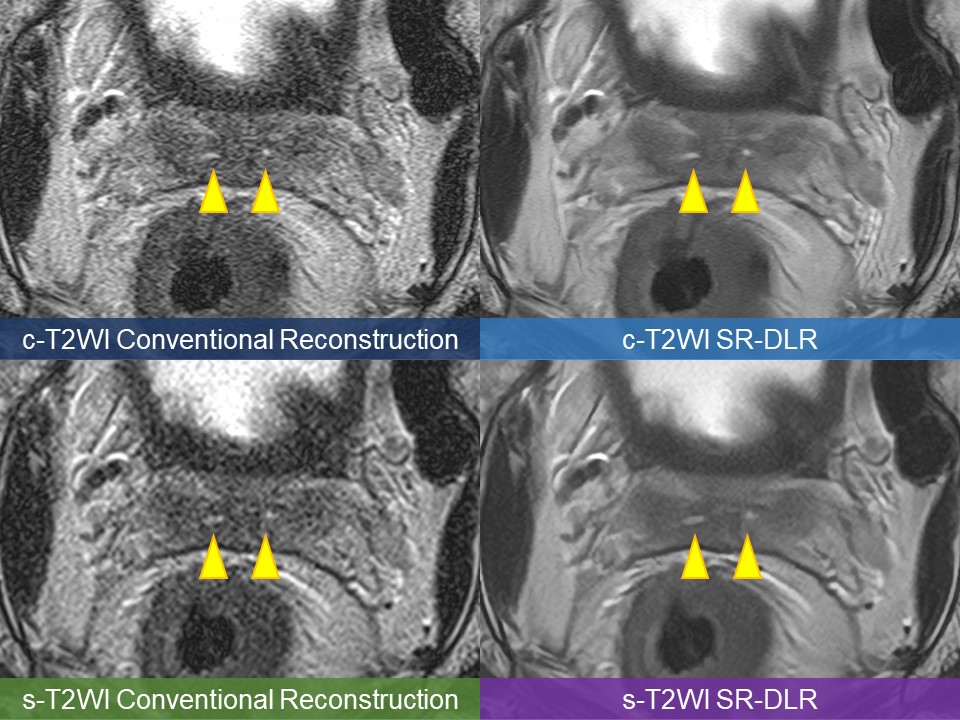
Although the ejaculatory ducts (arrowheads) are depicted on all images, the conventional reconstruction images are noisy and somewhat difficult to identify, especially on the s-T2WI image; the noise is reduced on the SR-DLR images, and the ejaculatory ducts are clearly depicted.