3329
BOLD fMRI responses to low and high frequency of sensory stimulation may reflect excitation and inhibition (im)balance1Center for Neuroscience Imaging Research (CNIR), Institute for Basic Science (IBS), Suwon 16419, Republic of Korea, Suwon, Korea, Republic of, 2Department of Biomedical Engineering, Sungkyunkwan University, Suwon, Korea, Republic of, 3Department of Intelligent Precision Healthcare Convergence, Sungkyunkwan University, Suwon, Korea, Republic of
Synopsis
Keywords: Task/Intervention Based fMRI, fMRI (task based), Excitation and Inhibition balance, Mouse fMRI, Autism
Motivation: The hyper- or hyporeactivity to sensory input is a common diagnostic criterion in autism, potentially influencing BOLD signals.
Goal(s): Our study was centered on unraveling the underlying mechanisms governing the positive and negative BOLD responses to low and high sensory stimulus frequencies. A ratio of BOLD responses of high frequency to low frequency may reflect E:I balance.
Approach: Our study delved into BOLD responses during varied sensory stimulation frequencies
Results: We observed increased neural activity and BOLD responses at lower frequencies, contrasting with suppressed cortical activity and subsequent negative BOLD responses at higher frequencies.
Impact: A ratio of BOLD responses of high frequency to low frequency may reflect E:I balance, necessary for the clinical utility of BOLD fMRI to hyper- or hypo-reactivity responses to sensory inputs in autism
Introduction
Brain dysfunctions in neurodevelopmental disorders such as autism spectrum disorder (ASD) and schizophrenia (SCZ) are supposed to associate with excitation and inhibition imbalance (E:I imbalance). This E:I ratio can be determined by electrophysiological recordings in brain slices. ASD individuals often exhibit either heightened (hyperreactivity) or reduced (hyporeactivity) responses to sensory inputs like sound, touch, or light. The abnormal E:I ratio can be due to abnormalities in PV interneurons (1), disrupting the balance between excitatory and inhibitory signals in the brain (2-5).The hemodynamic response to sensory stimuli results from the interplay between excitation and inhibition. Our hypothesis is that the sensory E:I ratio can be inferred by BOLD fMRI with low and high frequency stimulation. With low-frequency stimuli, excitatory neurons respond efficiently, experiencing reduced adaptation and less inhibition from nearby inhibitory neurons, thereby increasing the hemodynamic response. In contrast, higher-frequency stimuli prompt dominant inhibition due to the rapid firing of PV neurons and heightened adaptation in excitatory neurons, consequently reducing the hemodynamic response (6, 7). We evaluated our hypothesis by performing fMRI data with low and high frequency stimuli in anesthetized mice.
Method
Anesthesia: mouse strain & number? Initial injection with a mixture of ketamine (Ket: 100mg/kg) and xylazine (Xyl: 10mg/kg), and a supplementary dose (25mg/kg Ket and 1.25mg/kg Xyl) through IV injection.Stimulation: 40-s forepaw somatosensory stimulation with 5Hz or 40Hz, 2ms, and 0.5mA.
Functional MRI: BOLD-fMRI (156 × 156 × 500 μm3) with TE/TR of 11ms/1s on a 15.2T scanner.
Neural recording: A 16-channel electrode was perpendicularly inserted up to 1mm in the left forepaw somatosensory area.
Results
The initial examination was focused on measuring neural activity in response to 5Hz and 40Hz forepaw stimulation (Figure 1A). An electrode was inserted perpendicularly to a depth of 1mm into the cortex. Under Ketamine/Xylazine anesthesia, there was an increase in neural spiking in response to low-frequency stimuli, whereas the 40Hz stimuli resulted in a notable reduction in neural activity (Figure 1B). 5Hz stimuli generally led to an increased multi-unit activity (MUA), characterized by an initial peak, followed by a decay for the remainder of the stimulation period. Conversely, 40Hz stimuli initially increased MUA, quickly suppressed below the pre-stimulus spontaneous MUA, ending an instant rebound in MUA right after the end of stimulation.In our next investigation, we explored the effects of low and high-frequency stimuli on the BOLD response. During the 5Hz stimulation, a positive BOLD response was notably present in the thalamus, the left S1, and its related projection sites, including the ipsilateral S2 and contralateral areas (highlighted by red-yellow voxels in Figure 2B). In contrast, the 40Hz stimulation induced a negative BOLD response in the cortex, while a positive BOLD response persisted in the thalamic area. BOLD fMRI responses in S1 are closely related to MUA in S1, combined with neural and BOLD fMRI.
Discussion
Our study was centered on unraveling the underlying mechanisms governing the positive and negative BOLD responses to low and high stimulus frequencies, commonly observed in sensory-evoked BOLD studies. A ratio of BOLD responses of high frequency to low frequency (40Hz/5Hz) may reflect E:I balance. The 40Hz stimulation appears to suppress excitatory activity within the cortex by increased activation of nearby interneurons and the substantial adaptation of excitatory neurons, subsequently inducing a negative BOLD response. The observation of positive BOLD in the thalamus during the 40Hz stimulation implies a potential disparity in the ratio between excitation and inhibition compared to the cortex. Further studies are necessary in frequency-dependent BOLD fMRI of hypo- or hyper-sensitive ASD mice for evaluating the validity of our hypothesis and finding the clinical utility of BOLD fMRI.Acknowledgements
This study was supported by the Institute of Basic Science (IBS-R015-D1).References
- J. Yu, H. Hu, A. Agmon, K. Svoboda, Recruitment of GABAergic Interneurons in the Barrel Cortex during Active Tactile Behavior. Neuron 104, 412-427.e414 (2019).
- Q. Chen et al., Dysfunction of cortical GABAergic neurons leads to sensory hyper-reactivity in a Shank3 mouse model of ASD. Nat Neurosci 23, 520-532 (2020).
- E. Lee, J. Lee, E. Kim, Excitation/Inhibition Imbalance in Animal Models of Autism Spectrum Disorders. Biological Psychiatry 81, 838-847 (2017).
- N. Gogolla et al., Common circuit defect of excitatory-inhibitory balance in mouse models of autism. Journal of Neurodevelopmental Disorders 1, 172-181 (2009).
- O. Marín, Interneuron dysfunction in psychiatric disorders. Nat Rev Neurosci 13, 107-120 (2012).
- T. N. A. Dinh, W. B. Jung, H.-J. Shim, S.-G. Kim, Characteristics of fMRI responses to visual stimulation in anesthetized vs. awake mice. NeuroImage 226, 117542 (2021).
- H.-J. Shim et al., Mouse fMRI under ketamine and xylazine anesthesia: Robust contralateral somatosensory cortex activation in response to forepaw stimulation. NeuroImage 177, 30-44 (2018).
Figures
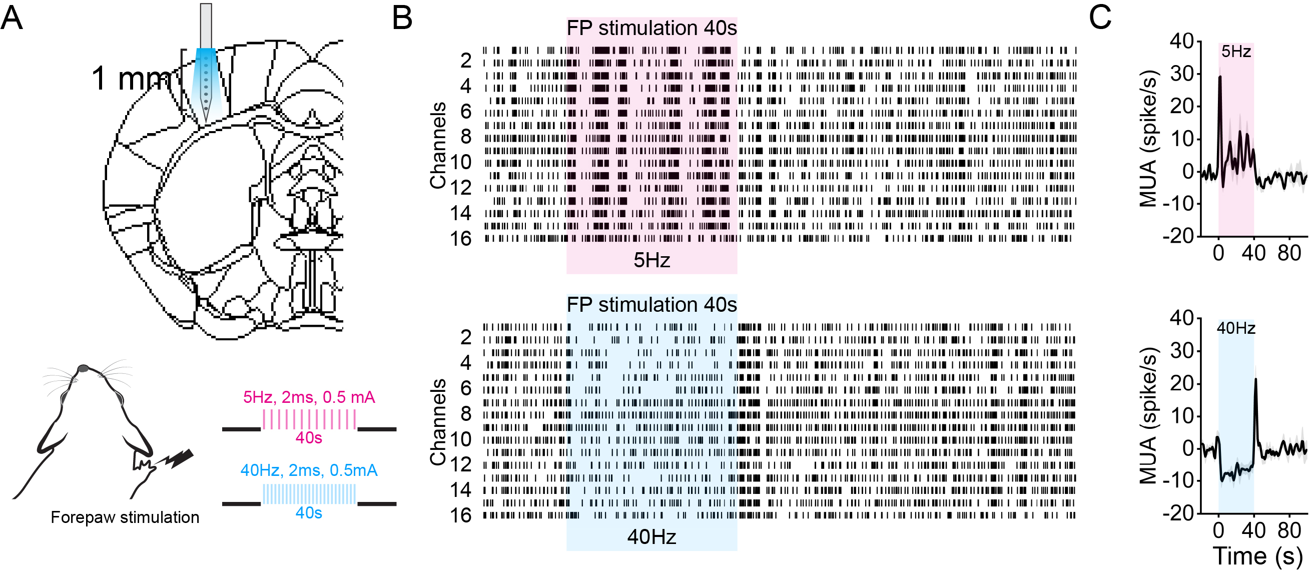
Figure 1. Neural activity responding to 5Hz and 40Hz forepaw stimulation in S1 of mice under Ket/Xyl condition.
A. Schematic of neural recording experiment.
B. Representative raster plot of 5Hz and 40Hz frequency stimulation, measured at left S1.
C. Multiunit activity changes (ΔMUA) corresponding to 5Hz and 40Hz stimulation.
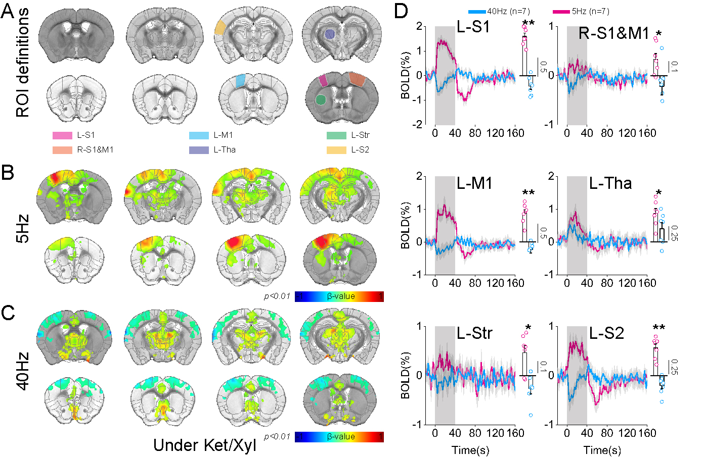
Figure 2. High-field BOLD fMRI responses to 5Hz and 40Hz forepaw stimulation of mice under Ket/Xyl condition.
A. ROIs related to L-S1 and its projection site, which is based on Allen mouse brain atlas.
B-C. BOLD fMRI activation map evoked by 5Hz and 40Hz forepaw stimulation, respectively.
D. Timecourses and mean BOLD changes for 5Hz and 40Hz forepaw stimulation for each ROI.