3328
Analyzing brain connectivity using a genetic reporter for MRI1Massachusetts Institute of Technology, Boston, MA, United States, 2Massachusetts Institute of Technology, Cambridge, MA, United States
Synopsis
Keywords: fMRI Acquisition, Brain, DIANA, Steady-State
Motivation: Anatomical connectivity underlies brain function, but currently cannot be traced robustly in living animals.
Goal(s): We sought to understand how the structure and plasticity of neuronal projections contribute to functional relationships.
Approach: A genetic reporter for MRI was delivered using viral injections and visualization of the reporter was compared with histology, diffusion tractography, and resting state fMRI results.
Results: We validated the new tracing tool in mice, showed that some but not all projection strengths correlate with functional measures, and demonstrated that connection strength changes relate variably to functional connectivity changes in a longitudinal model of opioid exposure.
Impact: This work validates and applies a genetic reporter for fMRI that permits neuronal projections from viral injection sites to be visualized longitudinally with fidelity in individual animals. Our results help explain functional connectivity relationships in terms of circuit-level contributions.
Introduction
The ability to map defined connectivity patterns by magnetic resonance imaging (MRI) could be enormously beneficial for explaining complementary structural and functional measures, but reliable mapping of neural connections in living mammalian brains using MRI is challenging. We have developed a novel genetic technique that allows virally transduced neural projections to be mapped longitudinally throughout the entire brains of individual animals using hemodynamic MRI contrast. Here we apply this method to analyze the connectivity and plasticity of the rodent striatum (Str), a subcortical hub for integration of motivational and cognitive signals. We investigate the correspondence between retrograde and anterograde striatal projection densities, non-directional diffusion tractography, and resting state functional connectivity assessed using functional magnetic resonance imaging (fMRI). We examine the extent to which anatomical connectivity predicts the other measures at individual and group levels, and we use an opioid exposure model to probe how anatomical plasticity underlies functional changes over time.Methods
Imaging of virally transduced cells was accomplished using a technique called bioluminescence imaging using hemodynamics (BLUsH). BLUsH provides a means for detecting bioluminescent reporters in deep tissue, following systemic photosensitization of the vasculature by expression of a light-dependent adenylate cyclase. To implement BLUsH at a brain-wide scale in mice, we injected a Cre-dependent adeno-associated virus (AAV) encoding the adenylate cyclase into the ventricles of transgenic mice that express Cre recombinase under control of the smooth muscle-specific promoter sm22a. Retrograde and anterograde connections to striatal sites were probed using AAV2-retro and AAV1 viruses encoding a Gaussia luciferase (GLuc) variant. Hemodynamic imaging was performed using echo planar imaging (EPI) on a 9.4 T Bruker Biospec scanner; anatomical RARE scans were also obtained. For BLUsH, data were acquired for 90 minutes, with the GLuc substrate CTZ injected at t = 30 min. Diffusion tensor imaging data were acquired using EPI spatial encoding with 20 diffusion directions at b = 750 s/mm2 and reference volumes at b = 0 s/mm2. Tractography was performed using the ProbtrackX package in FSL.Results and discussions
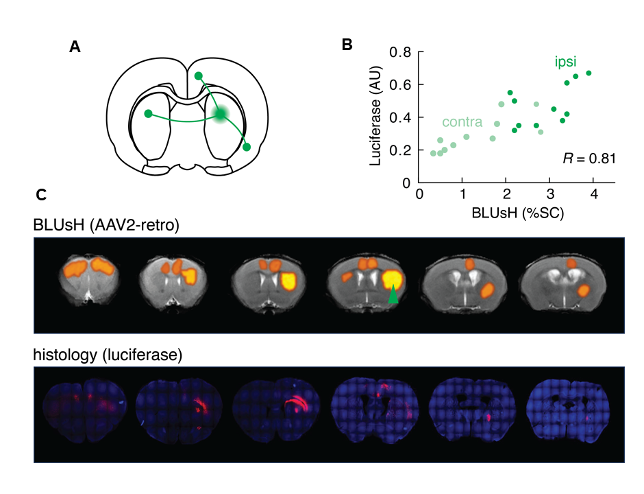
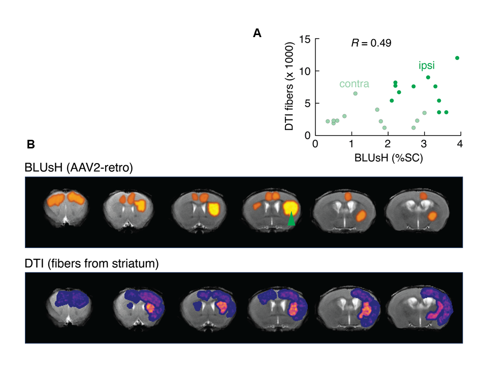
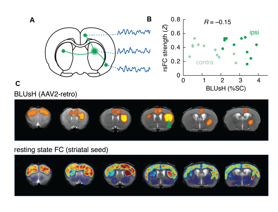
To validate our approach for anatomical tract tracing using viral vectors, BLUsH results were compared with histological analysis of animals injected with retrograde (n = 17) and anterograde (n = 8) vectors encoding GLuc. Spatial correspondence of aligned data was good, with peaks of BLUsH signal aligning well with postmortem visualization of GLuc expression on a regional basis. Spatial correlation coefficients were computed over regions of interest (ROIs) from individual animals, with a mean correlation of 0.81 (p-value<0.001, n=17) over retrograde tracing experiments and 0.85 (p-value<0.001, n=8) over anterograde tracing trials. We also found that diffusion-based measures correlated significantly with retrograde (R=0.49, p-value=0.018) but not anterograde (R=0.32, p-value<0.132) BLUsH results, likely reflecting the greater cellular specificity provided by the BLUsH approach. We next examined the ability of BLUsH-based anatomical connectivity to predict fMRI measures by comparing BLUsH tracing results to resting state functional connectivity (rsFC) strengths between Str and distal regions. We found that rsFC results were poorly correlated with BLUsH-based anatomical tracing in general (retrograde: R=-0.15, p-value<0.50, anterograde R=-0.06, p-value=0.7984), particularly between striatal sites and ROIs in the contralateral hemisphere. Interestingly, however, specific anatomical and functional connections were significantly correlated with each other over individuals (PFC R=0.51, p-value=0.0368). This suggests specific monosynaptic connections that play a substantial role in determining rsFC phenotypes and highlights the importance of being able to measure structural and functional parameters within individual living animals. We sought to determine whether plasticity of structural connections over time could be related to corresponding functional changes in an opioid exposure model. Eight animals were injected with BLUsH vectors for striatal connectivity mapping and scanned before and after a 10-day treatment with daily fentanyl doses. Comparison of BLUsH viral tracing data before and after the treatment revealed that some but not all striatal projections were significantly attenuated by exposure (t-test M1/2 p-value=0.042, PFC p-value=0.004: n=5 for both). These reductions were not observed in drug-free control animals. Some but not all of the fentanyl-dependent reductions in striatal anatomical connectivity were accompanied by parallel reductions in rsFC. Remarkably, a significant reduction in the ipsilateral PFC→Str anatomical connectivity strength was accompanied by an increase in rsFC between these regions (t-test p-value=0.04, n=5).Conclusion
BLUsH contrast with viral tract tracers enables mapping of cellular-level projection patterns on a brain-wide scale within individual animals over time. Projection patterns measured in vivo using BLUsH can be dissociated from functional connectivity measures, distinguishing the contributions of specific interregional connections to circuit-wide activity patterns in the brain.Acknowledgements
No acknowledgement found.References
No reference found.Figures