3324
How Variable Are Our Rat Sensory-Evoked Functional MRI Datasets?1Donders Institute for Brain, Behaviour, and Cognition, Radboud University, Nijmegen, Netherlands, 2Department for Medical Imaging, Radboud University Medical Center, Nijmegen, Netherlands, 3Center for Animal MRI, The University of North Carolina at Chapel Hill, Chapel Hill, NC, United States, 4The University of North Carolina at Chapel Hill, Chapel Hill, NC, United States, 5High-Field Magnetic Resonance, Max Planck Institute for Biological Cybernetics, Tuebingen, Germany, 6Cerebral Imaging Centre, Douglas Mental Health University Institute, Verdun, QC, Canada, 7Integrated Program in Neuroscience, McGill University, Montreal, QC, Canada, 8Department of Radiology, Lausanne University Hospital, Lausanne, Switzerland, 9Laboratory for Functional and Metabolic Imaging, Ecole Polytechnique Fédérale de Lausanne, Lausanne, Switzerland, 10Champalimaud Research, Champalimaud Centre for the Unknown, Lisbon, Portugal, 11Institute of Experimental and Clinical Pharmacology and Toxicology, FAU Erlangen-Nürnberg, Erlangen, Germany, 12Department of Neuroimaging, King's College London, London, United Kingdom, 13Clinic of Radiology, University Hospital Münster, Muenster, Germany, 14Brainnetome CenterBrainnetome Center, Institute of Automation, Chinese Academy of Sciences, Brainnetome CenterBrainnetome Center, China, 15Max-Delbrück Center for Molecular Medicine in the Helmholtz Association, Berlin, Germany, 16NeuroSpin, CEA Saclay, Paris, France, 17Functional Imaging Laboratory, German Primate Center - Leibniz Institute for Primate Research, Göttingen, Germany, 18Faculty of Biology and Psychology, Georg-August University of Göttingen, Göttingen, Germany, 19DFG Research Center for Nanoscale Microscopy and Molecular Physiology of the Brain (CNMPB), Göttingen, Germany, 20Institute of Development, Aging and Cancer, Tohoku University, Sendai, Japan, 21National Institutes for Quantum Science and Technology, Chiba, Japan, 22Radiology, Brigham and Women's Hospital, Boston, MA, United States, 23Faculty of Biology and Psychology, German Primate Center - Leibniz Institute for Primate Research, Göttingen, Germany, 24Georg-August University of Göttingen, Göttingen, Germany, 25Biological and Biomedical Engineering, McGill University, Montreal, QC, Canada, 26Department of Psychiatry, McGill University, Montreal, QC, Canada, 27Queensland Brain Institute and Centre for Advanced Imaging, University of Queensland, St Lucia, Australia, 28Instituto de Investigaciones Biomédicas de Barcelona (IIBB), Instituto de Investigaciones Biomédicas de Barcelona (IIBB), Barcelona, Spain, 29Institute of Development, Aging and Cancer, Tohoku University, Sendai, Korea, Republic of, 30Experimental and Clinical Research Center, A Joint Cooperation Between the Charité Medical Faculty and the Max-Delbrück Center for Molecular Medicine in the Helmholtz Association, Berlin, Germany, 31Erwin L. Hahn Institute for MR ImagingErwin L. Hahn Institute for MR Imaging, University of Duisburg-Essen, Essen, Germany, 32Institute of Neuroscience, University of Barcelona, Barcelona, Spain, 33Athinoula A. Martinos Center for Biomedical Imaging, Massachusetts General Hospital and Harvard Medical School, Charlestown, MA, United States, 34Brainnetome CenterBrainnetome Center, Institute of Automation, Chinese Academy of Sciences, Beijing, China
Synopsis
Keywords: fMRI Acquisition, Brain, rats, multicenter, sensory-evoked, heterogeneity
Motivation: We address the need for standardization and collaboration in rat sensory-evoked fMRI by providing evidence-based recommendations and open-access datasets, fostering community growth.
Goal(s): We aim to assess inter- and intra-datasets variability, focusing on image acquisition and experimental protocols, and to optimize analysis by comparing hemodynamic response functions and denoising methods.
Approach: We collected 17 rat datasets from 10 centers, applied standardized preprocessing, and analyzed the sensory-evoked responses at individual and groups levels. Project code is openly available.
Results: Our study revealed significant diversity in rat attributes, anesthesia protocols, and imaging acquisition parameters across datasets. We are currently optimizing analyses to strengthen protocol robustness.
Impact: We present evidence for the substantial heterogeneity intra- and inter-datasets of rat sensory-evoked fMRI. We will provide guidelines to enhance reproducibility, facilitate cross-laboratory comparisons, collaborations in neuroimaging research, and encourage more robust findings with potential translational applications.
Purposes
Rat sensory-evoked fMRI is used to study the physiology of the blood-oxygenation level dependent (BOLD) contrast, to study animal models, among many other applications. Here, we examine how rat task fMRI protocols compare between imaging sites and centers to identify evidence-based recommendations. We are in the process of comparing the implementation of different hemodynamic response functions, and to examine denoising methods. We promote and strengthen collaborations and discussion within the community by providing open access datasets and code [1].Methods
We collected rat sensory-evoked fMRI datasets without restrictions on strain, sex, age, weight, anesthesia, acquisition system or imaging sequence (Fig. 1). Datasets were converted to BIDS [2], preprocessed using RABIES (version: 0.4.8, [3]), and visually inspected for registration quality. Exclusion criteria included poor raw data and misregistration during preprocessing steps. We performed first-level analysis on individual scans, and a second-level group analysis analysis of the n=10 scans within each dataset. These analyses were performed in Nilearn (version: 0.10.0, [4]), motion parameters were included as regressors in the General Linear Model (GLM) to account for motion-related artifacts, and we applied smoothing of 0.45 millimeters. The stimulation paradigms were specified by data acquisition authors. We extracted timeseries and residuals from Regions of interests (ROIs) drawn based on the stimulation location (primary somatosensory cortex forelimb or hindlimb, primary somatosensory cortex barrel field). The SIGMA rat template was used to display individual-level and dataset-level statistical maps, and compare the activation cluster across datasets and between individuals within datasets. We are currently implementing different hemodynamic response functions in the analysis, namely the nilearn’s defaults: Glover and SPM without derivatives [4], and two custom rat functions [5, 6]. We are also adding new datasets to the analysis. Results are yet to be processed. The code for this project is available under terms of the Apache-2 license [1].Results
We gathered 17 rat datasets of 10 functional scans each, from 10 centers, totalling 161 scans. We excluded 5 scans, due to missing functional images (4/181), and failure in the functional-to-anatomical registration evident in the quality check after RABIES preprocessing (1/181). Expectedly, we find heterogenous rat attributes, image acquisition and experimental protocols across different laboratories, Thus, it is challenging to identify consistent patterns or effects and it limits their generalization to a broader context or population. We observe a sex bias (64% male, 36% females) across three different strains (Fig. 1.a). The anesthesia protocols and magnetic field strength distribution also align with the current trend in the field [7] with primarily Isoflurane and Medetomidine, using mostly 9 T and 14 T or larger field strength (Fig. 1.a,b). The datasets consist of somatosensory (either front- , hindpaw, or whiskers), stimulations. Group analysis revealed distinct activation clusters in 12 of the 17 datasets, but intensity and distribution varied considerably, even within similar stimulation types and field strengths (Fig. 2). This diversity hindered the identification of optimal experimental parameters. Individual-level analysis showed substantial variability, even within datasets demonstrating group-level clusters. We observed an enhanced consistency across individual scans associated with clearer group-level activation clusters, underscoring the impact of individual responses. Optimization of the analysis is ongoing, including the comparison of different hemodynamic response functions, and denoising methods.Discussion
We attribute the large differences between datasets to the substantial diversity of rat characteristics and individual responses, as well as image acquisition and experimental protocols dependent on the source laboratory. The diversity of the datasets will make it challenging to exactly pinpoint the reasons for robust group-level activation clusters supported by reduced heterogeneity at the individual level. We questioned the reliability of results based solely on group-level findings, as we observed considerable heterogeneity at the individual level. To address this, we propose combining various physiological readouts and encourage researchers to inspect their data and adopt greater quality assessment. An idea could be to adapt human fMRI quality assessment guidelines for use in rodent studies. On the other hand, we preprocessed datasets through RABIES pipeline [3], although we do not have evidence against using it on sensory-evoked fMRI on rodents, it could be an incomplete preprocessing method. We support further research into these concerns to establish standardized protocols. We are comparing different hemodynamic response function models which are expected to enhance some of the results. Finally, we anticipate that NORDIC denoising [8] will further enhance signal detection. In summary, our work describes the large variability in sensory-evoked fMRI datasets in the rat in line with what we observed with task-free conditions [7]. We can draw lessons from this to build more robust protocols.Acknowledgements
We extend our sincere appreciation to all researchers and laboratories who collaborated by providing the crucial rat sensory evoked fMRI datasets for this study.References
[1] https://github.com/grandjeanlab/multirat_se.git
[2] Gorgolewski, K. J., Auer, T., Calhoun, V. D., Craddock, R. C., Das, S., Duff, E. P., Flandin, G., Ghosh, S. S., Glatard, T., Halchenko, Y. O., Handwerker, D. A., Hanke, M., Keator, D., Li, X., Michael, Z., Maumet, C., Nichols, B. N., Nichols, T. E., Pellman, J., Poline, J. B., … Poldrack, R. A. (2016). The brain imaging data structure, a format for organizing and describing outputs of neuroimaging experiments. Scientific data, 3, 160044. https://doi.org/10.1038/sdata.2016.44
[3] Desrosiers-Grégoire, G., Devenyi, G. A., Grandjean, J., & Chakravarty, M. M. (2022). Rodent Automated Bold Improvement of EPI Sequences (RABIES) : a standardized image processing and data quality platform for rodent FMRI. bioRxiv (Cold Spring Harbor Laboratory). https://doi.org/10.1101/2022.08.20.504597
[4] Abraham, A., Pedregosa, F., Eickenberg, M., Gervais, P., Mueller, A., Kossaifi, J., Gramfort, A., Thirion, B., & Varoquaux, G. (2014b). Machine learning for Neuroimaging with SciKit-Learn. Frontiers in Neuroinformatics, 8. https://doi.org/10.3389/fninf.2014.00014
[5] Lambers, H. et al. (2020). A cortical rat hemodynamic response function for improved detection of BOLD activation under common experimental conditions. NeuroImage 208, 116446.
[6] Silva, A. C., Koretsky, A. P. & Duyn, J. H. (2007). Functional MRI impulse response for BOLD and CBV contrast in rat somatosensory cortex. Magn. Reson. Med. 57, 1110–1118
[7] Mandino, F. et al. Animal Functional Magnetic Resonance Imaging: Trends and Path Toward Standardization. Front. Neuroinformatics 13, 78 (2020).
[8] Tellez Ceja, I., Starke, S., Gladytz, T., Tabelow, K., Niendorf, T., Reimann, H. (2023). En Route to Fine-Grained Neurosignatures in the Individual Brain: Evaluating Methodology to Boost Spatial Accuracy & Sensitivity of BOLD fMRI. ISMRM annual meeting (Toronto), Oral Session: Novel Methods in fMRI.
Figures
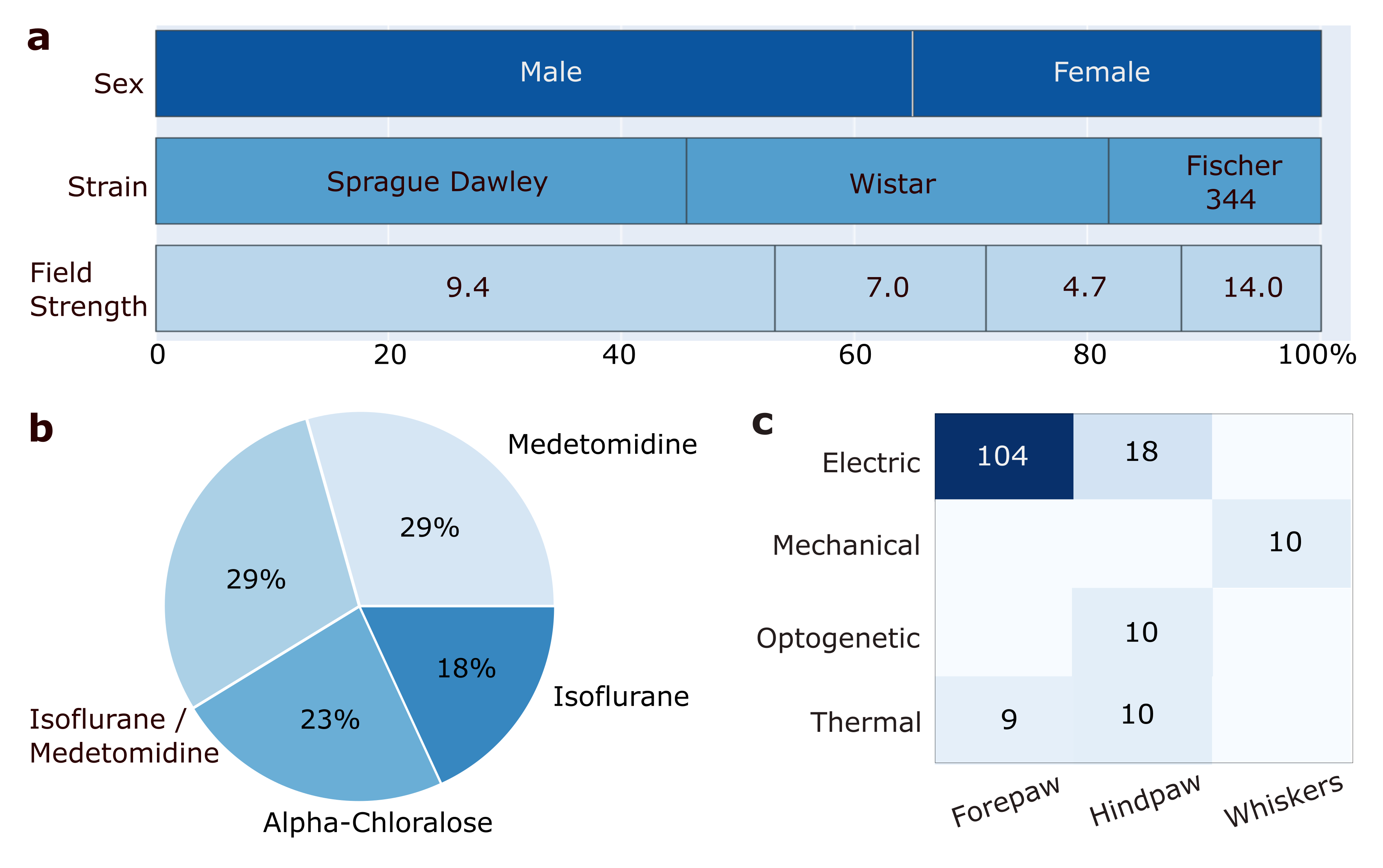
Figure 1: Dataset description in percentage of scans, a. sex, strain and field strength, b. anesthesia, c. stimulation type and location.
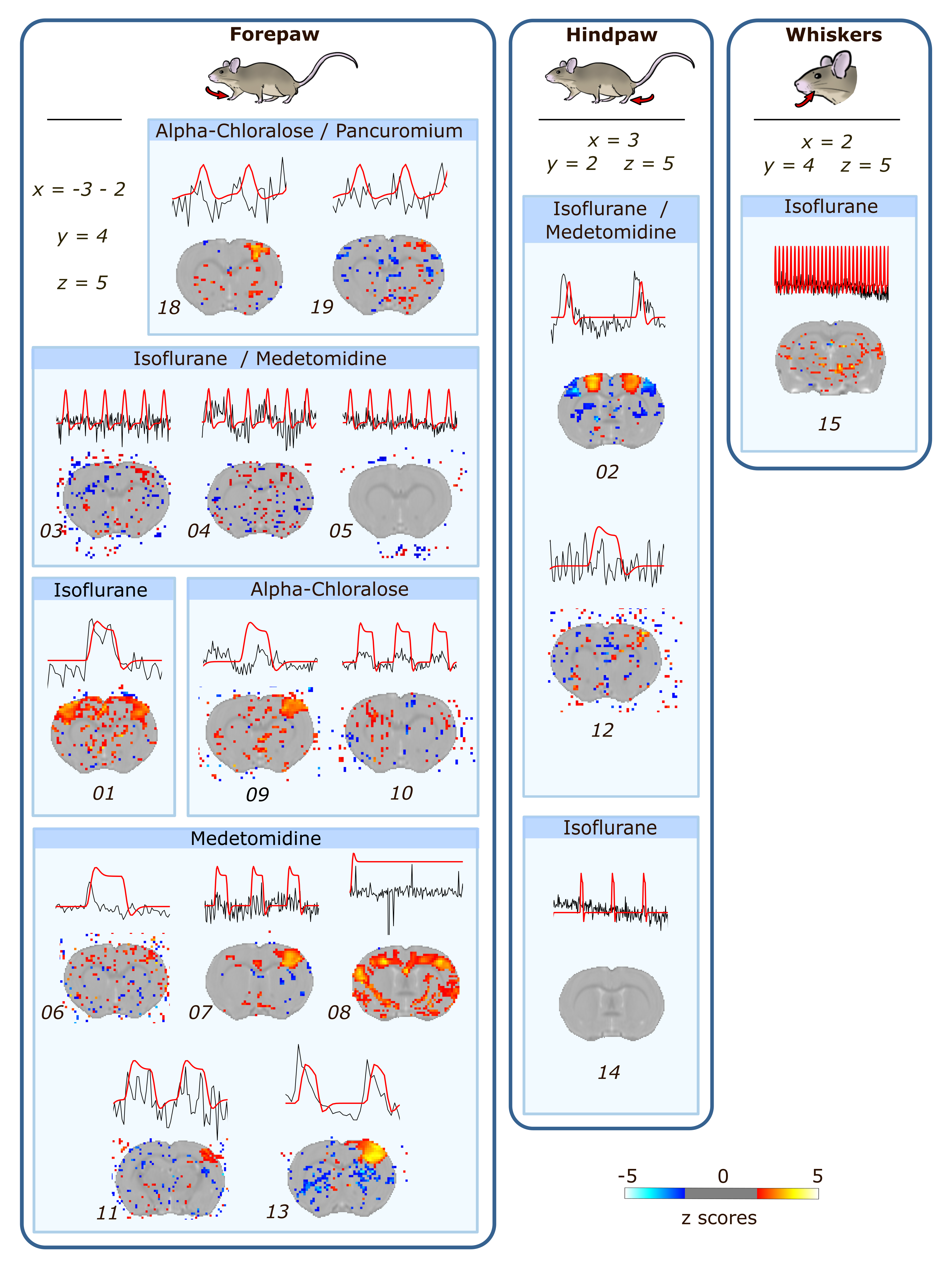
Figure 2: Second-level analysis statistical (Z scores) maps accompanied by the Glover model depiction of the hemodynamics response function (red) juxtaposed alongside the recorded timeseries (black). Thresholds of activity were set to 1.7, and 1.5 for datasets 03, 04 and 05 only. Coordinates are given for all scans within the stimulation site box. Spatial arrangement of maps according to stimulation location, anesthesia of maintenance, and dataset.
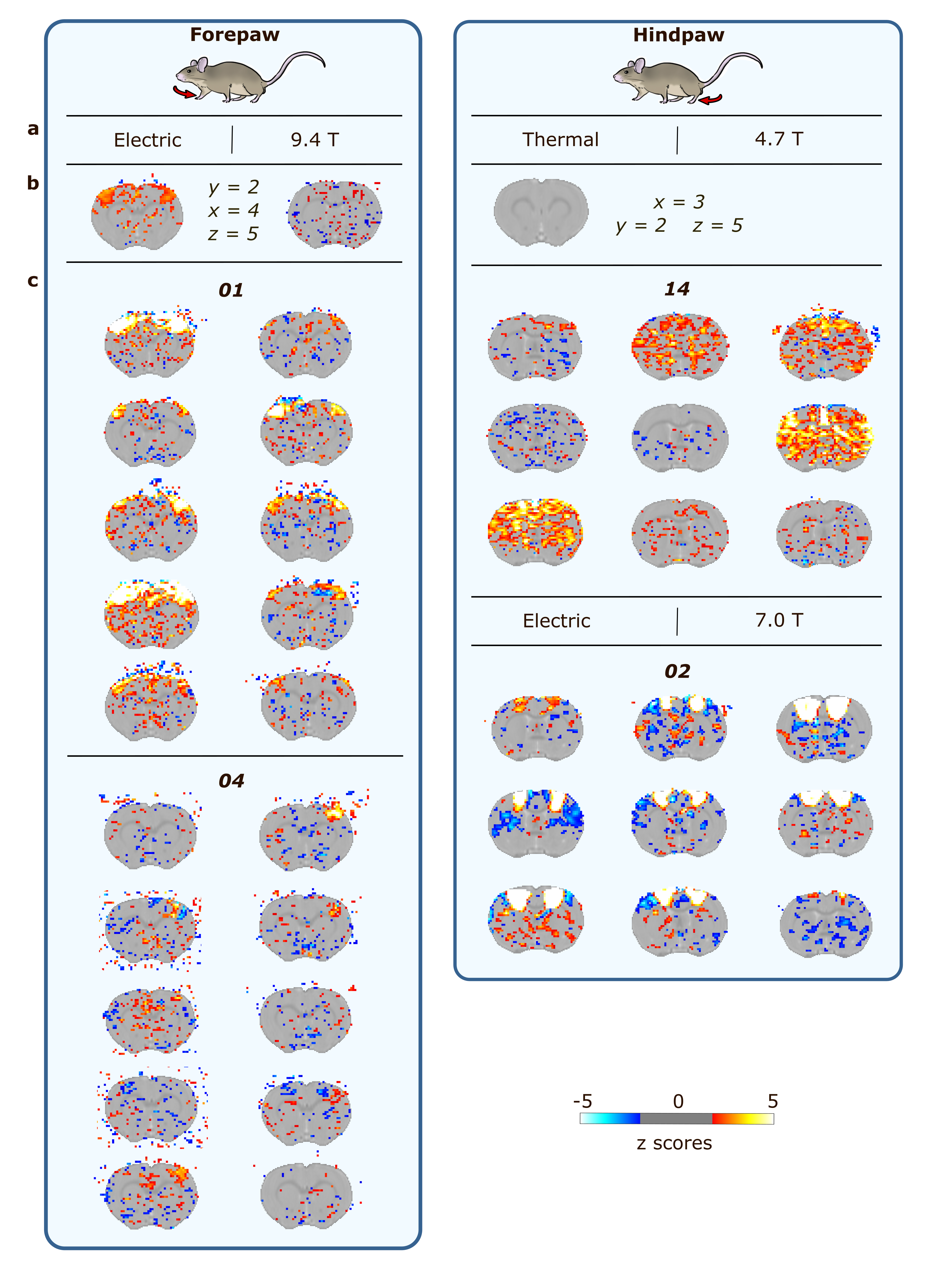
Figure 3: Example of the heterogeneity at the individual level for 4 datasets, specifying the a. stimulation type and field strength, b. second-level statistical maps with coordinates, c. first-level statistical maps. Maps are arranged according to stimulation location.