3323
Protocol for awake task free fMRI using freely behaving head fixed mice.1Donders Institute for Brain, Cognition and Behaviour, Nijmegen, Netherlands, 2Radboud University Medical Center, Nijmegen, Netherlands
Synopsis
Keywords: fMRI Acquisition, fMRI (resting state), Mice, Awake Imaging
Motivation: To increase the translatability of preclinical imaging data, awake protocols have to be developed.
Goal(s): Here we outline a protocol for awake task free fMRI using freely behaving head fixed mice.
Approach: After headplate surgery and habituation to a holder containing a treadmill, mice are scanned up to 6 times.
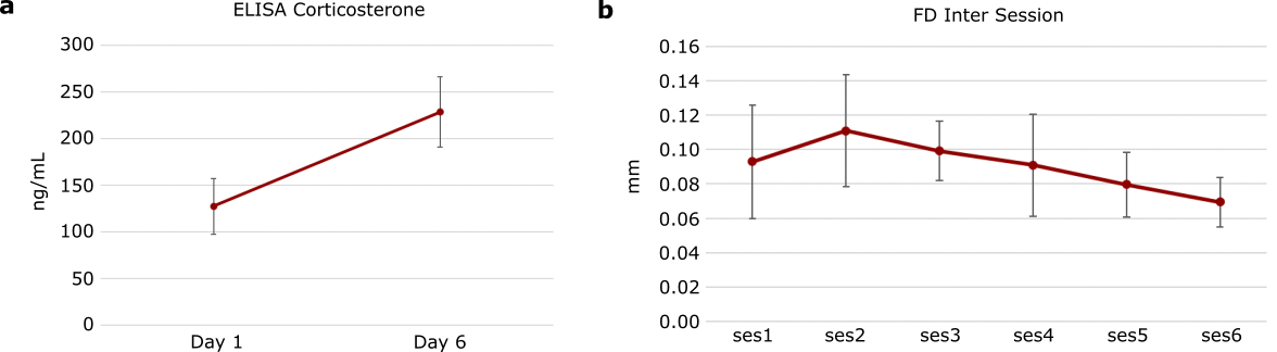
Results: The corticosterone and framewise displacement show that habituation seems to continue during scanning, suggesting that the habituation protocol needs to be lengthened. However, after dual regression analysis, activity within task free networks can be seen in the data, meaning that this protocol takes steps in the improvement of translatability of data.
Impact: To increase the translatability of functional MRI data, development of awake protocols is necessary. This allows for removal of the confound of anesthesia as well as opening up the option for behavioral paradigms during scanning.
Introduction
Functional Magnetic Resonance Imaging remains the mainstay technique for studying whole brain networks in human subjects. However, due to the lack of genetic control and experimental invasiveness, animals such as mice and rats are often used as models for human psychopathology. Currently, most preclinical fMRI is performed using anesthesia to prevent movement of the animals in the scanner1,2. As anesthesia influences the activity of brain networks, this hampers the translation of data between animals and humans3. For this reason, the development of awake animal scanning protocols is necessary. Here we outline a protocol for awake task free fMRI using freely behaving head fixed mice.Methods
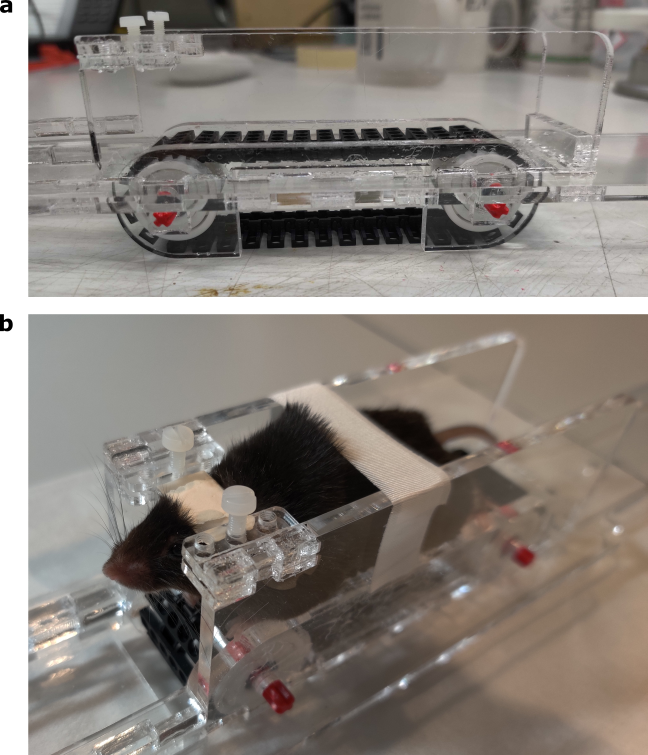
Mice (N=10) underwent surgery to fit a plastic headplate to the skull. They were then habituated to being head fixed in a specialized holder. This holder cut from PLA using a laser cutter and contains a treadmill which allows the mice to walk freely (Figure 1). MRI background noise (80 dB) was playing during the habituation which was a stepwise increase from 10 to 60 minutes over the course of 6 days. On the first and last day, blood was taken after habituation for corticosterone measurement with ELISA. After habituation, mice were scanned up to 6 times using a Bruker 11.7T scanner and a surface coil. Data was preprocessed and analyzed using Rodent Automated Bold Improvement of EPI Sequences (RABIES)4. The data was analyzed using dual regression analysis.Results/Discussion
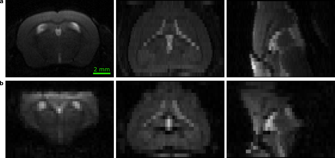
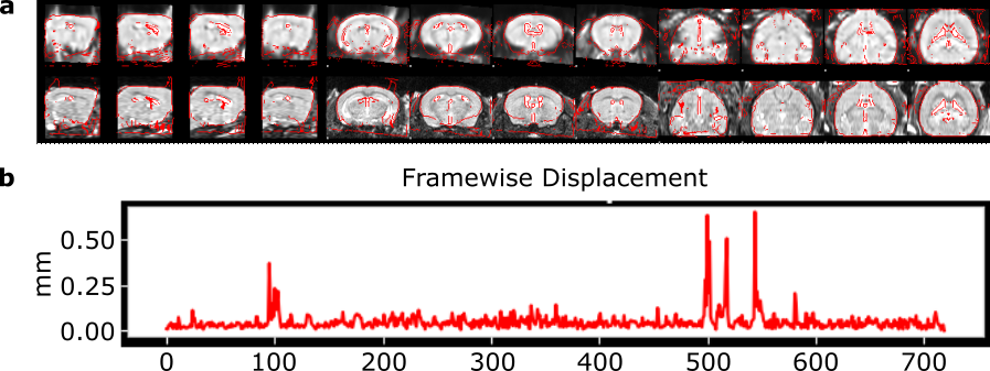
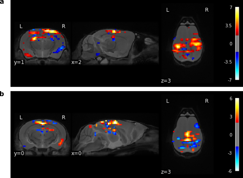
Examples of structural and functional scans can be seen in Figure 2. After preprocessing, RABIES provides quality control information showing for instance whether registration was successful and the framewise displacement, which is related to movement during scanning (Figure 3). Corticosterone measurements were used as a proxy for stress during habituation, while movement was used as a proxy for stress during scanning. As can be seen in Figure 4, corticosterone rises during habituation, but movement goes down over scanning sessions. This suggests that the habituation was still taking place during the scanning period and that the habituation protocol should be lengthened. Dual regression analysis uses networks derived from an Independent Component Analysis to derive individual level activity maps for the ICA networks, in this case from the DSURQE templates5. Here we show two example maps, corresponding to somatosensory and retrosplenial areas, showing that this protocol allows for the detection of task free networks in awake freely behaving mice (Figure 5). Although the habituation protocol needs to be optimized further, this protocol takes steps in the improvement of translatability of data and provides the option for behavioral paradigms inside the scanner, thereby increasing the reach and scope of preclinical fMRI studies.Acknowledgements
We would like to thank NWO for funding this researchReferences
- Grandjean, J. et al. A consensus protocol for functional connectivity analysis in the rat brain. Nat. Neurosci. 26, 673–681 (2023).
- Mandino, F. et al. Animal Functional Magnetic Resonance Imaging: Trends and Path Toward Standardization. Front. Neuroinformatics 13, 78 (2019).
- Tsurugizawa T, Yoshimaru D. Impact of anesthesia on static and dynamic functional connectivity in mice. Neuroimage. 2021;241:118413.
- Desrosiers-Gregoire, G., Devenyi, G. A., Grandjean, J. & Chakravarty, M. M. Rodent Automated Bold Improvement of EPI Sequences (RABIES): A standardized image processing and data quality platform for rodent fMRI. 2022.08.20.504597
- Zerbi V, Grandjean J, Rudin M, Wenderoth N. Mapping the mouse brain with rs-fMRI: An optimized pipeline for functional network identification. Neuroimage. 2015;123:11-21.
Figures