3322
AwakeRodent, the multi-center, multi-species, multi-modality study!1Donders Institute for Brain, Behaviour, and Cognition, Radboud University, Nijmegen, Netherlands, 2CAS Center for Excellence in Brain Sciences and Intelligence Technology, Institute of Neuroscience, Chinese Academy of Sciences, Shanghai, China, 3iHuman Institute, ShanghaiTech University, Shanghai, China, 4ShanghaiTech University, Shanghai, China, 5Center for Excellence in Molecular Cell Science, Shanghai Institute of Biochemistry and Cell Biology, Chinese Academy of Sciences, Shanghai, China, 6University of Chinese Academy of Sciences, Beijing, China, 7Instituto de Neurociencias, CSIC-UMH, San Juan de Alicante, Spain, 8Molecular Biology and Genetics, Aarhus University, Aarhus, Denmark, 9Center for Neuroscience and Cognitive systems, Istituto Italiano di Tecnologia, Rovereto, Italy, 10Amsterdam UMC, University of Amsterdam, Amsterdam, Netherlands, 11A.I.V. Institute for Molecular Sciences, University of Eastern Finland, Kuopio, Finland, 12School of Biomedical Engineering, ShanghaiTech University, Shanghai, China, 13Center for Excellence in Brain Science and Intelligence Technology, Chinese Academy of Sciences, Shanghai, China
Synopsis
Keywords: fMRI Acquisition, Brain, awake imaging, rodents, multi-center
Motivation: To bring awareness on the heterogeneity in awake rodent functional imaging. We aim to identify protocol differences that optimize awake functional connectivity, reproducibility and collaboration.
Goal(s): To aggregate awake rodent functional datasets, establish population parameters, provide evidence-based recommendations, compare functional Magnetic Resonance Imaging and functional Ultrasound, foster collaborations.
Approach: We collect datasets from mice and rats, preprocess and run a seed-based analysis at the individual-level using RABIES. Group-level analysis is performed in Python, resulting in functional connectivity specificity maps.
Results: We have gathered 5 mice-datasets and 4 rat-datasets totaling 122 scans with great variability in rodent characteristics, imaging methods, and experimental designs.
Impact: Findings will empower researchers to refine awake rodent functional imaging, enhancing investigations into cognitive and behavioral processes, without the anesthesia confounds. This approach closely parallels human brain imaging, enhancing translational relevance and providing an accurate representation of conscious brain function.
Introduction
Awake whole-brain functional imaging in rodents presents an emerging frontier in translational and biomedical research. Previous comparisons between task-free functional MRI (fMRI) datasets in mice [1] and rats [2] demonstrated that awake data exhibit fewer plausible functional connectivity patterns compared to anesthetized counterparts. We hypothesize that variations in laboratory practices and protocols contribute to these disparities. We aim to exploit this variability to identify approaches that enhance the detection of functional networks in awake rodents. Our ultimate goal is to establish best practices, foster collaboration, and potentiate our research community.Methods
We invite laboratories to participate by contributing one or more datasets. The inclusion criteria encompass datasets with a minimum of four task-free functional MRI or functional ultrasound scans in either mice or rats, without genetic or experimental manipulation, any strain, gender, age, or weight, provided they meet a temporal resolution of 0.25 Hz or above and specific field-of-view dimensions. We exclude datasets that fail the quality assessment after RABIES preprocessing. We convert the raw data in accordance with Brain Imaging Data Structure standards. Preprocessing involves co-registration to relevant spaces (DSURQE for mice, SIGMA for rats), denoising via motion regression, and temporal filtering. Two seeds were positioned symmetrically in the primary somatosensory cortex. We generated seed-based maps at the individual scan level, within RABIES. Subsequently, we computed a group-level analysis for each dataset, using Python with nilearn. The primary outcome is a statistical (Z score) maps of functional connectivity, with a statistical threshold set to 1.9 (Fig.2). We are committed to transparency and reproducibility, we adhere to the Brain Imaging Data Structure format and we provide diagnostic outputs for quality assurance. The code for this project is available under terms of the Apache-2 license (https://github.com/grandjeanlab/awake).Results
We have gathered 5 mice-datasets and 4 rat-datasets totaling 122 functional scans. Datasets exhibit notable variability in terms of rodent characteristics, imaging methods, and experimental designs, underscoring the need for harmonization in the field (Fig. 1). Preliminary analyses have revealed different levels of plausible functional connectivity patterns (Fig. 2). These findings highlight the complexities to optimize protocols of task-free fMRI experiments in rodents and the necessity to establish standardized practices for reproducibility. We are in the process of collecting and analyzing data.Discussion: Our results underscore the significant variability within and between datasets of awake rodent functional imaging. While we acknowledge the challenges posed by this heterogeneity, it also provides us with a unique opportunity to identify best practices. Therefore, we encourage collaborative efforts, including transparency and open access to data for peer scrutiny, deeper insights, and constructive feedback.Acknowledgements
We extend our sincere appreciation to all researchers and laboratories who collaborated by providing the crucial rodent resting state fMRI datasets for this study.References
[1] Grandjean, J., Canella, C., Anckaerts, C., Ayrancı, G., Bougacha, S., Bienert, T., Buehlmann, D., Coletta, L., Gallino, D., Gass, N., Garin, C. M., Nadkarni, N. A., Hübner, N. S., Karatas, M., Komaki, Y., Kreitz, S., Mandino, F., Mechling, A. E., Sato, C., Sauer, K., … Gozzi, A. (2020). Common functional networks in the mouse brain revealed by multi-centre resting-state fMRI analysis. NeuroImage, 205, 116278. https://doi.org/10.1016/j.neuroimage.2019.116278
[2] Grandjean, J., Desrosiers-Gregoire, G., Anckaerts, C., Angeles-Valdez, D., Ayad, F., Barrière, D. A., Blockx, I., Bortel, A., Broadwater, M., Cardoso, B. M., Célestine, M., Chavez-Negrete, J. E., Choi, S., Christiaen, E., Clavijo, P., Colon-Perez, L., Cramer, S., Daniele, T., Dempsey, E., Diao, Y., … Hess, A. (2023). A consensus protocol for functional connectivity analysis in the rat brain. Nature neuroscience, 26(4), 673–681. https://doi.org/10.1038/s41593-023-01286-8Figures
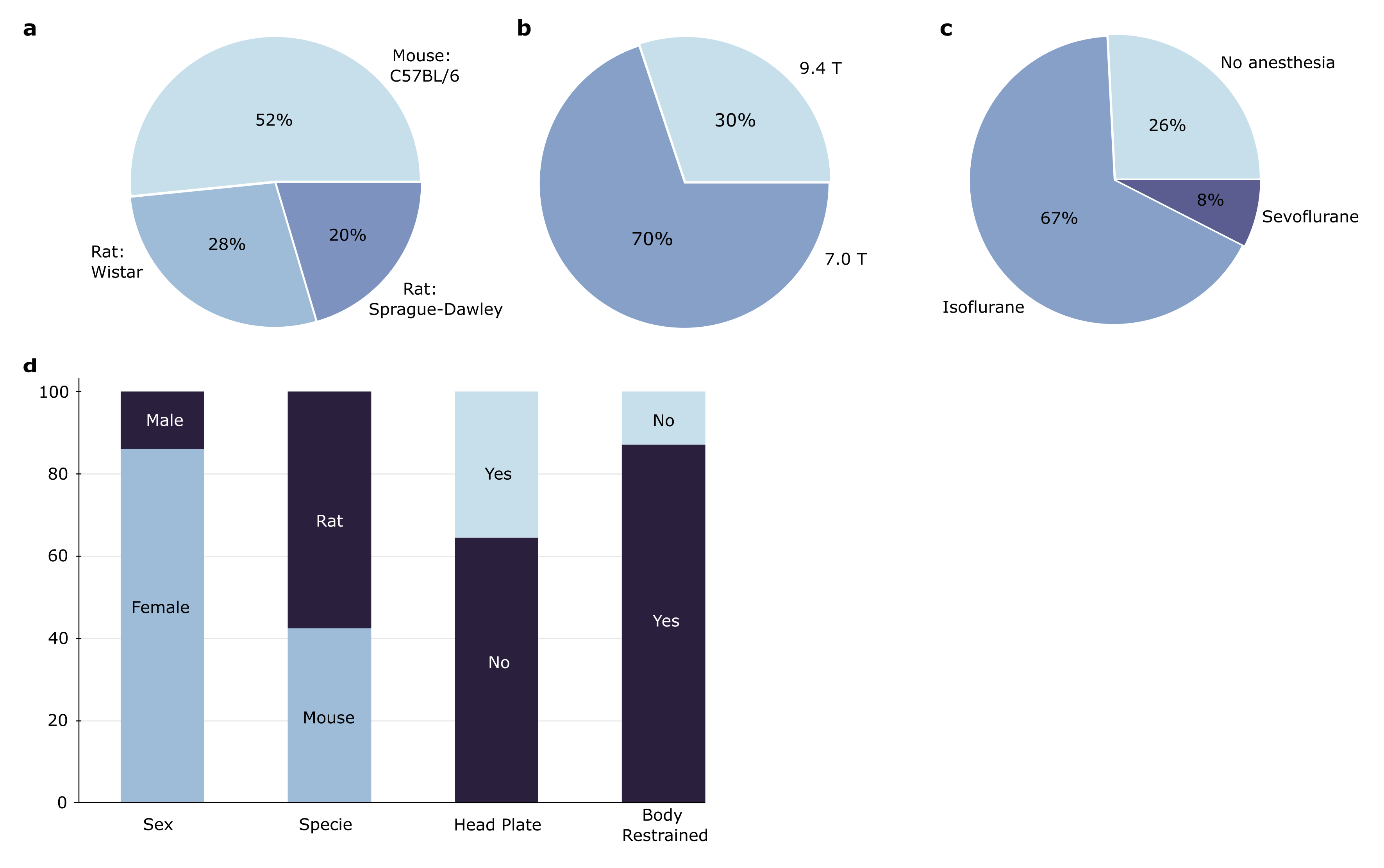
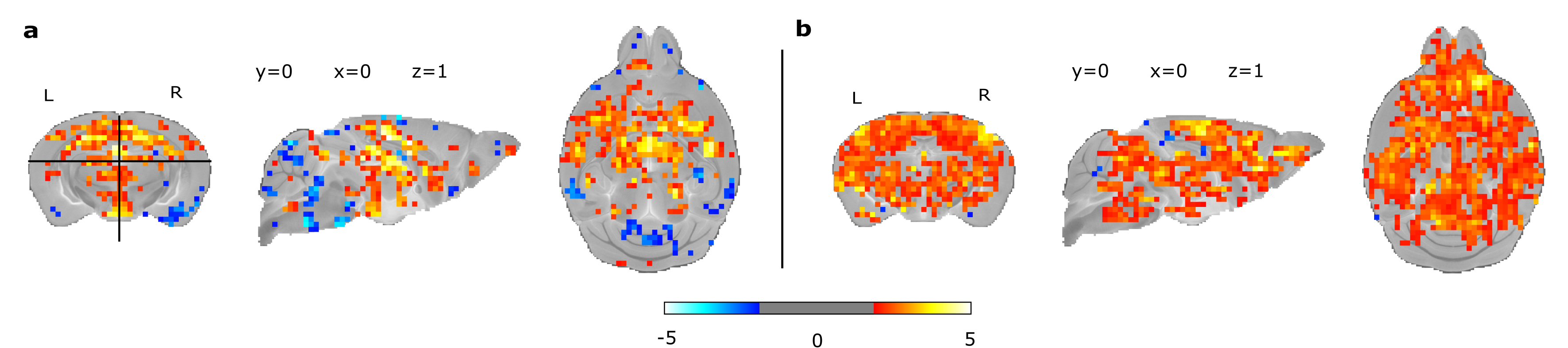
Figure 2: Group level analysis statistical (Z scores) maps, showing functional connectivity for seeds symmetrically based in the primary somatosensory cortex. The thresholds of activity were set at 1.9. Example of 2 mice datasets, a. dataset 01, n=4 mice, b. dataset 02, n=8 mice.