3321
Comparing responses to visual and auditory stimuli in anesthetized and minimally restrained awake mice using quiet zero echo time MB-SWIFT fMRI1A.I. Virtanen Institute for Molecular Sciences, Kuopio, Finland, 2Center for Magnetic Resonance Research, Minneapolis, MN, United States, 3Roche Innovation Center Basel, Basel, Switzerland
Synopsis
Keywords: Task/Intervention Based fMRI, fMRI (task based), Awake, fMRI, mouse, MB-SWIFT, ketamine, xylazine
Motivation: Anesthesia is a major confounding factor for pre-clinical fMRI and thus awake fMRI protocols with minimal body restraints are needed.
Goal(s): Our goal was to develop a novel motion-tolerant fMRI approach for awake mice and to compare sensory responsiveness between awake and anesthetized mice.
Approach: Following a 14-day habituation protocol, mice were imaged using zero-echo-time MB-SWIFT fMRI with visual and auditory stimulation schemes in awake state and under ketamine-xylazine anesthesia.
Results: In awake mice, the activation of all key nodes was detected after both sensory stimuli. However, anesthesia suppressed particularly the auditory responses and affected the fMRI response shapes.
Impact: Awake animal imaging has gained much popularity in recent preclinical studies. Together with quiet and motion-tolerant MB-SWIFT imaging our approach has potential for more complex behavioral fMRI designs that improve our understanding of neuroscience and support translational drug discovery.
Introduction
In preclinical fMRI, animals are routinely imaged under anesthesia, a circumstance that can markedly influence their responsiveness to stimuli1,2. Recognizing this limitation, various awake or behavioral imaging strategies1-5, have been introduced. However, the prevalent echo planar imaging based awake imaging approaches often grapple with challenges such as artifacts caused by body motion, and high acoustic noise that introduces additional stress to animals. The recently developed zero echo time MB-SWIFT sequence6 has emerged as a promising alternative for awake rat fMRI because it is inherently motion-tolerant and silent7. These features make this imaging sequence ideally suited for behavioral rodent imaging8, where body motions are expected or even desired as a presentation of behavior. While head-fixed setup with minimal body restraint has been used before in rats with MB-SWIFT8, it has not been implemented in mice so far. Therefore, we first developed a novel method for minimally restraint awake mice and then validated the method’s performance by studying visual and auditory responsiveness. Results from awake mice were compared with the sensory responses under the widely used ketamine-xylazine anesthesia protocol.Methods
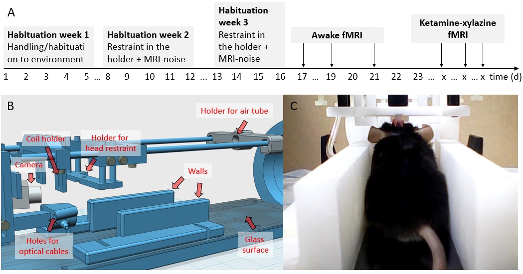
All mice (n=8) underwent surgery where head posts designed to fit to the awake MRI holder (Figure 1) were implanted. Thereafter, animals underwent a 14-day habituation protocol to mitigate stress and extensive motion in the subsequent awake fMRI procedures. The habituation protocol included a rigid head fixation by the implant while the rest of the body and legs were freely movable on top of a slightly slippery glass surface.Functional imaging was performed 2-3 times on each mouse either in awake (n=23 sessions) or ketamine-xylazine (ketamine 100mg/kg, xylazine 10mg/kg i.p.) anesthesia (n=21 sessions) conditions by employing a radial 3D MB-SWIFT sequence on a 9.4T magnet (Agilent DirectDrive), with the following parameters: a repetition time of 0.82ms, acquisition time per volume 1.7s, flip angle 3°, transmission bandwidth of 125kHz, a matrix size of 643, field of view 243mm³, isotropic resolution of 375μm. Average and peak acoustic sound pressure of MB-SWIFT was 64 and 85dB, respectively.
During imaging, either blue LED light flashing at a frequency of 5Hz, or air-pressure-induced sound pulses at a frequency of 13Hz, were delivered 5 times each in a 10s ON/50s OFF pattern with randomized order.
Results
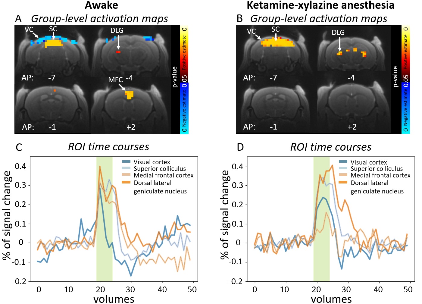
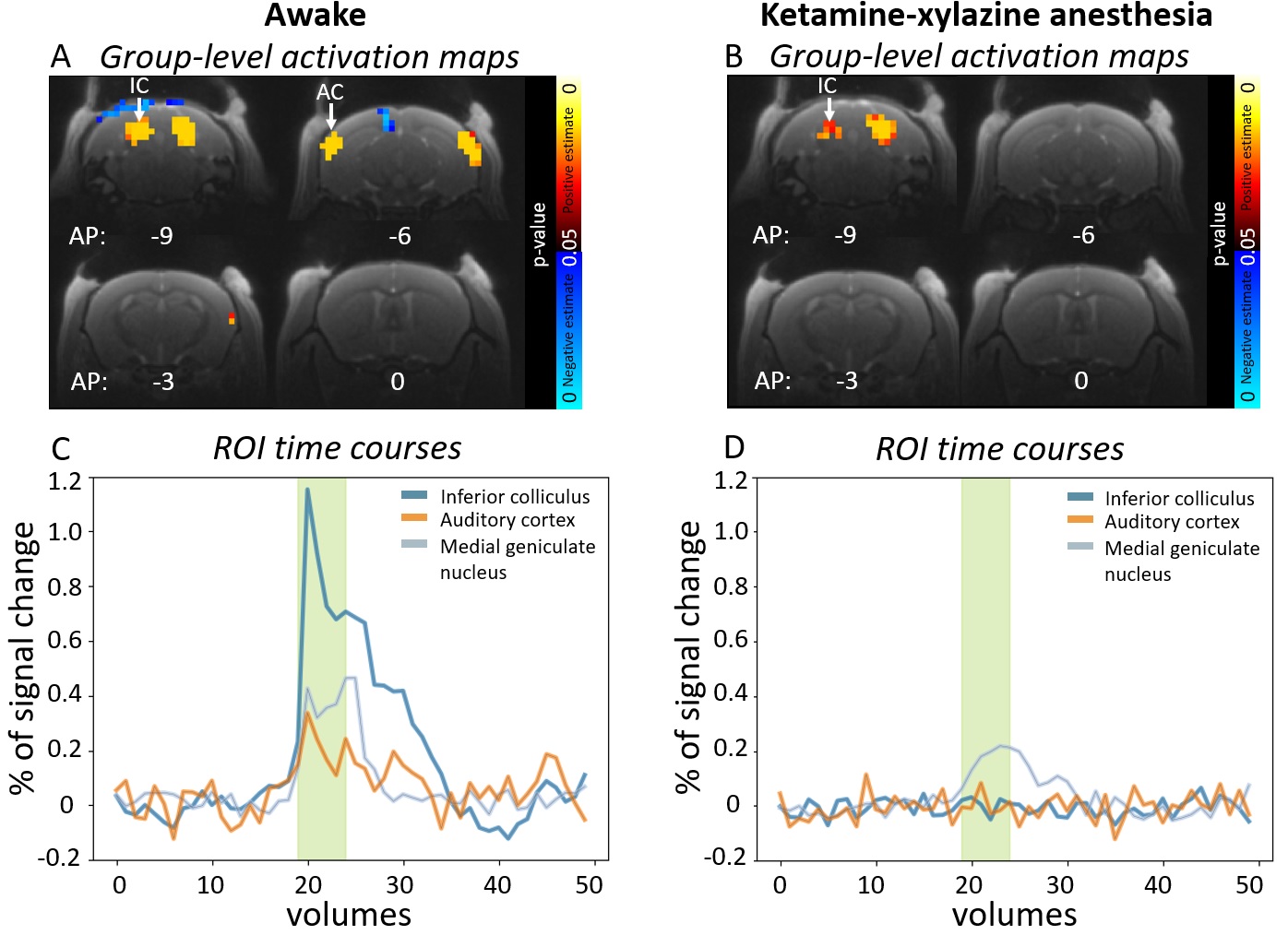
For visual stimulation, visual cortex (VC), superior colliculus (SC) and dorsal lateral geniculate nucleus (DLG) exhibited the strongest responses in both the awake and anesthetized groups (Figure 2). Medial frontal cortex (MFC) was more clearly activated in the awake group. Additionally, in the awake group, response shape in VC altered from response shape in anesthetized group by having a shorter duration, slightly stronger amplitude (0.3%, awake vs 0.25%, anesthetized) and a negative undershoot, thus resulting to negative parameter estimates.For auditory stimulation, the strongest responses were seen in the auditory cortex (AC), inferior colliculus (IC) and medial geniculate nucleus (MGN) in the awake group (Figure 3). However, in the anesthetized group, the responses were substantially blunted in all analyzed ROIs and only the inferior colliculus exhibited a weak response (0.2%, anesthetized vs 0.4%, awake).
Discussion
The MB-SWIFT approach allowed distortion-free and well-localized activation mapping in response to visual and auditory stimuli in minimally restrained awake mice. Silent scanning, together with the habituation protocol, translated into only modest levels of animal motion, and awake mice were able to distinguish auditory stimuli from the low background scanning noise.The imaging protocol allowed effective exploration of basic visual systems in both the awake and anesthetized animals. However, activation in the frontal cortex was more strongly detectable in the awake group, illustrating a better suitability of awake animals to study higher order cortical information processing that is blunted under anesthesia. Furthermore, changed response shape in VC in awake animals is an interesting phenomenon that needs further investigations.
Auditory responses were markedly suppressed in the anesthetized group, illustrating blunted processing of sound under the anesthesia protocol used. Here, the acoustic sound pressure (90dB) used for stimulation enabled activation of the inferior colliculus, but further ascending brain nodes including the auditory system were not detectable in anesthetized animals. The underlying mechanism is elusive but ketamine-xylazine may block ascending connections from the inferior colliculus which can possibly be related to an increased threshold of sound pressure needed in anesthetized mice.
Conclusion
We have pioneered a novel fMRI approach tailored for awake mice utilizing acoustically quiet and artefact-free MB-SWIFT sequence and demonstrated its utility by detecting robust whole-brain sensory stimulation responses. Ketamine-xylazine anesthesia was found to be an alternative to awake imaging for studying the visual system but showed strongly suppressed auditory system responses.Acknowledgements
This work was supported by F. Hoffmann-La Roche Ltd and The Finnish Cultural Foundation (grant no. 00230292).References
1. Paasonen J. et al. Functional connectivity under six anesthesia protocols and the awake condition in rat brain. Neuroimage. 2018.
2. Dinh T.N.A. et al. Characteristics of fMRI responses to visual stimulation in anesthetized vs. awake mice. NeuroImage. 2021.
3. Lahti K.M. Comparison of evoked cortical activity in conscious and propofol- anesthetized rats using functional MRI. Magn. Reson. 1999.
4. Sakurai, K. et al. Hyper BOLD Activation in Dorsal Raphe Nucleus of APP/PS1 Alzheimer’s Disease Mouse during Reward-Oriented Drinking Test under Thirsty Conditions. Sci Rep 2020.
5. Zhe H. et al. Awake and behaving mouse fMRI during Go/No-Go task, NeuroImage, 2019.
6. Idiyatullin D. et al. Multi-Band-SWIFT. J Magn Reson. 2015.
7. Paasonen J. Multi-band SWIFT enables quiet and artefact-free EEG-fMRI and awake fMRI studies in rat. Neuroimage. 2020.
8. Paasonen J. et al. Whole-brain studies of spontaneous behavior in head-fixed rats enabled by zero echo time MB-SWIFT fMRI. Neuroimage. 2022.
Figures