3320
9.4T MR based rs-fMRI of anesthetized mice after passive hyperthermia1Wuhan United Imaging Life Science Instruments Ltd.,, Wuhan, China, 2960th Hosp Joint Logist Support Force PLA, Dept Radiology, Jinan, China
Synopsis
Keywords: Functional Connectivity, Neuroscience
Motivation: The effects of passive hyperthermia on brain function in resting-state mice are unknown.
Goal(s): To expiore the rs-fMRI in hyperthermia treated and normal mice brain.
Approach: The ICA network and ROI-ROI FC in passive hyperthermia mice brain were compared with normal control.
Results: The ICA networks changed and the overall ROI-ROI FC of the passive hyperthermia mice decreased.
Impact: After prolonged exposure to high temperature has a greater impact on the overall perception and cognitive level of mice, which might help understand the relationship between neuronal activities and physiological thermal sensation and regulation as well as behavioral changes.
Introduction
Exposure to hyperthermia poses a challenge to physiological homeostasis and can potentially result in heat-related illnesses and mental health, causing a greater threat to people's lives. For instance, environmental high temperature decreases vigilance, as well as other negative effects on the physiological state of the human body, cognitive thinking, and behavioral ability. In terms of the effects of heat stress on attention and cognition, large amount of human studies have been explored [1-4] but remain limited to behaviors etc.Given heat stress has harmful effects on the brain structure and synaptic density [5] and mild warming in hyperthermia affects both exercise performance and cognitive function [6-8], changes in intrinsic networks and functional connection strength in different brain regions across the whole brain after a certain period of passive hyperthermia exposure have not been explored. Thus, resting-state functional magnetic resonance imaging (rs-fMRI) provides an important experimental approach to understanding the nature of the brain’s intrinsic functional activity in the absence of controlled stimuli or any observed behaviors [9] [10].
Herein, we performed independent component analysis (ICA) [11] of synchronous low frequency BOLD rs-fMRI signal recorded from lightly anesthetized mice for identifying the functional connectivity (FC) of spatially distinct regions of the brain and thus seeking insight into its overall organization. We further studied the variation of ICA networks and functional connection strength between the cotex network (CN), the subcortical network (SN) and DMN in passive hyperthermia exposed and normal control mice.
Methods
MR Instruments: MRI measurements were carried out on a 9.4T uMR scanner (United Imaging, Wuhan, China), equipped with a gradient system capable of producing up to 1000mT/m in all directions. An 86 mm quadrature resonator was used for, transmission, while a single loop surface coil was used for reception.Animals: All animal experiment were preapproved by the Chinese PLA, the 960th Hospital of Joint Hospital of Joint Logistics Support Force of PLA, Shandong Province, P. R. China (permit number: 2019e12).Twelve C56BL/6J male mice which weighing about 24~29g and aged 12~16 weeks old were used in the experiment, subdivided into two groups: Hyperthermia group (HT, n=6) and Normal control group (NC, n=6).
Hyperthermia treatment: We designed a heating box to enable mice to undergo 40 min of 40 ± 0.5 ℃ hyperthermia treatment before scanning. Ambient humidity ranged from 40% to 50% during the experiment.
Anesthesia and preparation for MRI experiments: Monitoring of physiological parameters was achieved with a 1030 system. Body temperature was measured with a fiber optic rectal probe and kept at 37℃±1℃ by a water circulating system. The standard anesthesia protocol for rsfMRI of mouse, i.e. isoflurane in combination with dexmedetomidine[12], was used .
MRI scans: An anatomical reference scan was acquired using a FSE-2D T2W sequence, with the following parameters: TE/TR: 48.8/3700ms, FOV: 14mm×19mm, Slices: 40×0.4mm, Matrix Size: 177×240, bandwith=30Hz/Pixel, ETL: 7, Voxel Size:0.08×0.08×0.40mm3, Average: 4, Scan time: 6min40s.The fMRI experiments were performed using the EPI sequence , images were acquired using the following parameters: TE/TR: 13.5/2000ms, FOV: 14mm×19mm, Slices: 40×0.4mm, Matrix Size: 56×76, bandwith: 4000Hz/Pixel, Voxel Size: 0.25×0.25×0.4mm3, Scan time: 10min0s. The slices were acquired in the same position as in the anatomical scans.
Data analysis: Refer to the previous article[11][13].
Results and Discussion
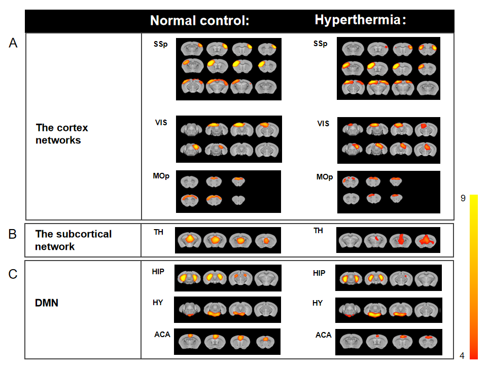
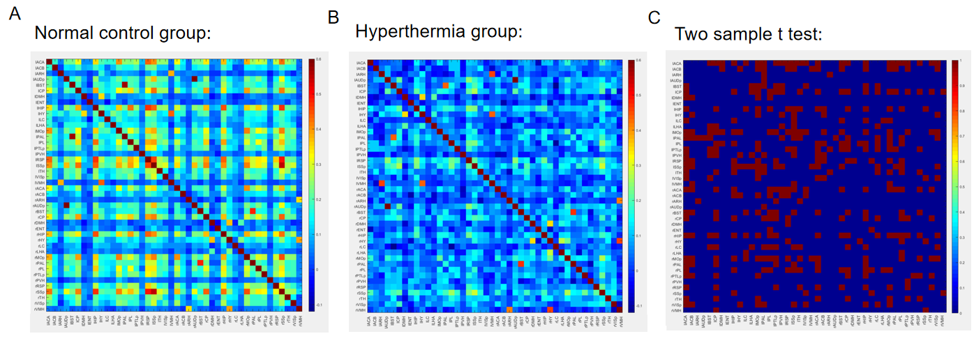
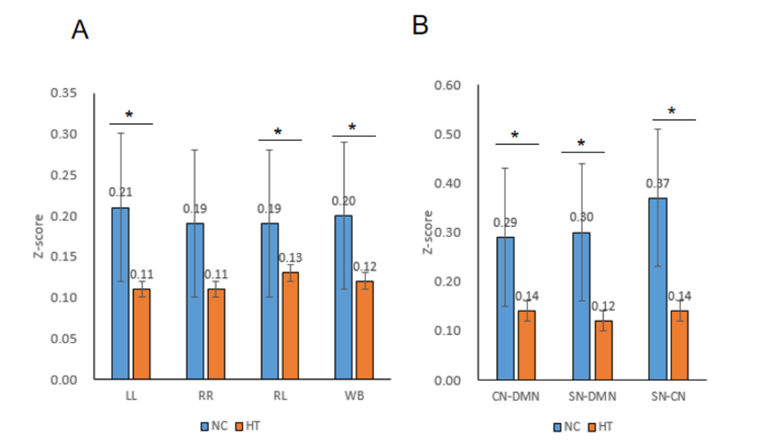
Group ICA analysis results (Figure 1) from the obtained rsfMRI GRE EPI images showed 7 intrinsic connectivity networks (ICNs) (CN: SSp, VIS, MOp; SN: TH; DMN: HIP,HY, ACA) in both HT and NC. The intensity and region size of ICNs changed in all networks. Color-coded matrices of NC (Figure 2A) and HT (Figure 2B) showed the z-scores corresponding to interactions between different brain regions, which were analyzed both left and right hemisphere separately. Further two-sample t-test results (Figure 2C) showed that some of the intensity of FCs is noticeably significant different between HT and NC. In addition, averaged z-score values of left and right intra-, inter-hemispheric, whole-brain (A) and inter-network functional connectivities (FCs)(B) showed that the power of FCs of the HT are substantially reduced when comparing to with the NC.As the sensorimotor network and DMN are often relevant functional networks in studies of functional deficits during brain disease and functional improvements in treatment strategies[13], and the previous studies found that communication enhanced between the brain's hemispheres during heat stress affects cognition [3], indicating that after prolonged exposure to high temperature has a greater impact on the overall perception and cognitive level of mice, which might help understand the relationship between neuronal activities and physiological thermal sensation and regulation as well as behavioral changes.
Acknowledgements
This work was supported by the no. 2019-JCJQ-ZNM-02 foundation.References
1. Hancock PA: Sustained attention under thermal stress. Psychological bulletin 1986, 99(2):263-281.
2. Hancock PA, Vasmatzidis I: Effects of heat stress on cognitive performance: the current state of knowledge. International journal of hyperthermia : the official journal of European Society for Hyperthermic Oncology, North American Hyperthermia Group 2003, 19(3):355-372.
3. McKiernan KA, D'Angelo BR, Kaufman JN, Binder JR: Interrupting the "stream of consciousness": an fMRI investigation. NeuroImage 2006, 29(4):1185-1191.
4. Han W, Qian S, Jiang Q, Liu K, Li B, Sun G: Regional and long-range neural synchronization abnormality during passive hyperthermia. Behavioural brain research 2018, 341:9-15.
5. Erfani M, Ghazi Tabatabaei Z, Sadigh-Eteghad S, Farokhi-Sisakht F, Farajdokht F, Mahmoudi J, Karimi P, Nasrolahi A: Rosa canina L. methanolic extract prevents heat stress-induced memory dysfunction in rats. Experimental physiology 2019, 104(10):1544-1554.
6. Raymann RJ, Van Someren EJ: Time-on-task impairment of psychomotor vigilance is affected by mild skin warming and changes with aging and insomnia. Sleep 2007, 30(1):96-103.
7. Romeijn N, Van Someren EJ: Correlated fluctuations of daytime skin temperature and vigilance. Journal of biological rhythms 2011, 26(1):68-77.
8. Ramautar JR, Romeijn N, Gómez-Herrero G, Piantoni G, Van Someren EJ: Coupling of infraslow fluctuations in autonomic and central vigilance markers: skin temperature, EEG β power and ERP P300 latency. International journal of psychophysiology : official journal of the International Organization of Psychophysiology 2013, 89(2):158-164.
9. Fox MD, Snyder AZ, Vincent JL, Corbetta M, Van Essen DC, Raichle ME: The human brain is intrinsically organized into dynamic, anticorrelated functional networks. Proceedings of the National Academy of Sciences of the United States of America 2005, 102(27):9673-9678.
10. Raichle ME: The brain's dark energy. Scientific American 2010, 302(3):44-49.
11. Beckmann CF, DeLuca M, Devlin JT, Smith SM: Investigations into resting-state connectivity using independent component analysis. Philosophical transactions of the Royal Society of London Series B, Biological sciences 2005, 360(1457):1001-1013.
12. Blazquez Freches G, Chavarrias C, Shemesh N: BOLD-fMRI in the mouse auditory pathway. NeuroImage 2018, 165:265-277.
13. Egimendia, A. et al. Aging Reduces the Functional Brain Networks Strength-a Resting State fMRI Study of Healthy Mouse Brain. Frontiers in aging neuroscience 11, 277, doi:10.3389/fnagi.2019.00277 (2019).
Figures